
Hyper Gram EKG Programs Yellow Card The Hyper

- Slides: 4
Hyper. Gram* EKG Programs “Yellow Card” The Hyper. Gram EKG programs are designed to help guide Third Year Medical Students in using a 10 item system for rapidly interpreting “real” EKGs using the “Yellow Card”. This card is based on a simple approach to EKG Interpretation that traces the sequences of depolarization and repolarization of the heart and asks: 1) “Is this step normal? ” If the answer is no, It asks “What is the abnormality? ” Because of its simplicity it is of necessity incomplete. However it covers all the common important problems. More importantly, it helps the student avoid missing obvious abnormalities. As it is used, the students develop confidence in their ability to interpret EKGs systematically, and they begin to fill in gaps not covered on the Yellow Card. To learn EKG interpretation, nothing is a substitute for practice. The Yellow Card makes practice easier. The more EKGs a student interprets, the more proficient he/she becomes. The card is laminated and designed to fit easily into a shirt/jacket pocket. If one is not available, slides 3 and 4 can be printed as a handout, 2 slides per page, cut and folded. This should produce a compact paper version. The 3 second lines on the original may not be correct on a printed version. To correct for this, make two marks, 75 mm apart (@25 mm/sec = 3 sec along the x axis). Abbreviations from the card are defined on the next slide. 3/14/2010 *Hyper. Gram© is a teaching program created originally by Robert Zelis, M. D. for the Macintosh computer using the Hyper. Card program. Copyright 1996 & 2010.
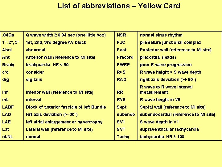
List of abbreviations – Yellow Card . 04 Qs Q wave width ≥ 0. 04 sec (one little box) NSR normal sinus rhythm 1°, 2°, 3° 1 st, 2 nd, 3 rd degree AV block PJC premature junctional complex Abnl abnormal Posterior wall (reference to MI site) Anterior wall (reference to MI site) Precord precordial (leads) Brady bradycardia. HR < 50 PWRP poor R wave progression c/o consider R>S R wave height > S wave depth digitalis RAD right axis deviation (>+ 90°) Inferior wall (reference to MI site) RR R wave to R wave interval measurement interval RV 6 R wave height in V 6 LABF Block of anterior fascicle of left Bundle Septal wall (reference to MI site) LAD left axis deviation (>- 30°) subendocardial (reference to MI site) LAE left atrial enlargement or hypertrophy SV 1 S wave depth in V 1 Lateral wall (reference to MI site) SVT supraventricular tachycardia nl/NL normal Tachy tachycardia. HR ≥ 100
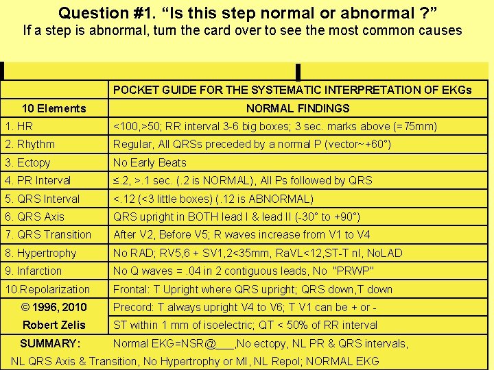
Question #1. “Is this step normal or abnormal ? ” If a step is abnormal, turn the card over to see the most common causes POCKET GUIDE FOR THE SYSTEMATIC INTERPRETATION OF EKGs 10 Elements NORMAL FINDINGS 1. HR <100, >50; RR interval 3 -6 big boxes; 3 sec. marks above (=75 mm) 2. Rhythm Regular, All QRSs preceded by a normal P (vector~+60°) 3. Ectopy No Early Beats 4. PR Interval ≤. 2, >. 1 sec. (. 2 is NORMAL), All Ps followed by QRS 5. QRS Interval <. 12 (<3 little boxes) (. 12 is ABNORMAL) 6. QRS Axis QRS upright in BOTH lead I & lead II (-30° to +90°) 7. QRS Transition After V 2, Before V 5; R waves increase from V 1 to V 4 8. Hypertrophy No RAD; RV 5, 6 + SV 1, 2<35 mm, Ra. VL<12, ST-T nl, No. LAD 9. Infarction No Q waves =. 04 in 2 contiguous leads, No "PRWP" 10. Repolarization Frontal: T Upright where QRS upright; QRS down, T down © 1996, 2010 Precord: T always upright V 4 to V 6; T V 1 can be + or - Robert Zelis ST within 1 mm of isoelectric; QT < 50% of RR interval SUMMARY: Normal EKG=NSR@___, No ectopy, NL PR & QRS intervals, NL QRS Axis & Transition, No Hypertrophy or MI, NL Repol; NORMAL EKG
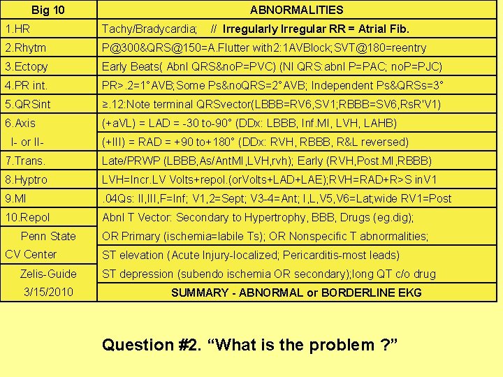
Big 10 ABNORMALITIES 1. HR Tachy/Bradycardia; 2. Rhytm P@300&QRS@150=A. Flutter with 2: 1 AVBlock; SVT@180=reentry 3. Ectopy Early Beats( Abnl QRS&no. P=PVC) (Nl QRS: abnl P=PAC; no. P=PJC) 4. PR int. PR>. 2=1°AVB; Some Ps&no. QRS=2°AVB; Independent Ps&QRSs=3° 5. QRSint ≥. 12: Note terminal QRSvector(LBBB=RV 6, SV 1; RBBB=SV 6, Rs. R'V 1) 6. Axis (+a. VL) = LAD = -30 to-90° (DDx: LBBB, Inf. MI, LVH, LAHB) I- or II- // Irregularly Irregular RR = Atrial Fib. (+III) = RAD = +90 to+180° (DDx: RVH, RBBB, R&L reversed) 7. Trans. Late/PRWP (LBBB, As/Ant. MI, LVH, rvh); Early (RVH, Post. MI, RBBB) 8. Hyptro LVH=Incr. LV Volts+repol. (or. Volts+LAD+LAE); RVH=RAD+R>S in. V 1 9. MI . 04 Qs: II, III, F=Inf; V 1, 2=Sept; V 3 -4=Ant; I, L, V 5, V 6=Lat; wide RV 1=Post 10. Repol Abnl T Vector: Secondary to Hypertrophy, BBB, Drugs (eg. dig); Penn State CV Center Zelis-Guide 3/15/2010 OR Primary (ischemia=labile Ts); OR Nonspecific T abnormalities; ST elevation (Acute Injury-localized; Pericarditis-most leads) ST depression (subendo ischemia OR secondary); long QT c/o drug SUMMARY - ABNORMAL or BORDERLINE EKG Question #2. “What is the problem ? ”