Hydrocephalus The brain ventricles are four cavities located

Hydrocephalus

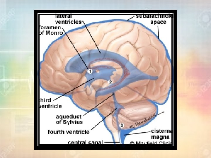
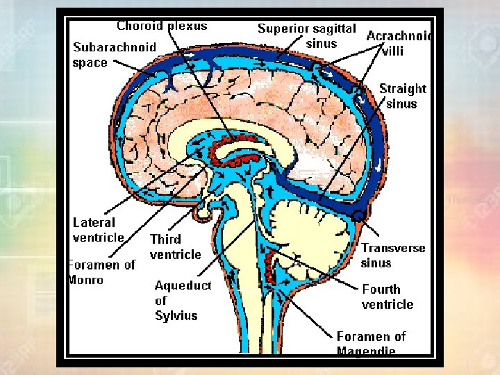
Ø The brain ventricles are four cavities located within the brain that contain cerebral spinal fluid (CSF). two lateral ventricles , the third ventricle and the fourth ventricle Ø It protects the brain by allowing it to “float” in a fluid bath and provides a shock absorber against head trauma. The CSF itself also helps to provide nutrients to the brain and to keep the brain in chemical balance. CSF • The normal volume of CSF is 140 ml. • CSF is produced by the choroid plexus in the lateral ventricles at a rate of 0. 4 ml per minute (or about 500 ml in 24 hours).

NORMAL CSF CIRCULATION

CSF circulation • The CSF flows from the lateral ventricles through the foramen of Monro into the 3 rd ventricle, via the aqueduct of Sylvius into the 4 th ventricle and then through the foramina of Magendie and Luschka into the subarachnoid space and basal cisterns. • The CSF circulates throughout the spinal subarachnoid space and the basal cisterns up through the tentorial hiatus. It flows over the cerebral hemispheres and is largely absorbed by the arachnoid villi of the dural sinuses.

• Hydrocephalus is an abnormal enlargement of the ventricles due to an excessive accumulation of CSF resulting from a disturbance of its flow, absorption or, uncommonly, secretion.

Classification of hydrocephalus • Obstructive hydrocephalus—when there is an obstruction to the flow of CSF through the ventricular system. • Communicating hydrocephalus—when there is no obstruction to the flow of CSF within the ventricular system but the hydrocephalus is due either to obstruction to CSF flow outside the ventricular system or to failure of absorption of CSF by the arachnoid villi.
 lateral ventricle obstruction by tumours.")
Causes of hydrocephalus • 1 Obstructive hydrocephalus: • (a) lateral ventricle obstruction by tumours. • (b) 3 rd ventricular obstruction, due to colloid cyst of the 3 rd ventricle or glioma of the 3 rd ventricle. • (c) occlusion of the aqueduct of Sylvius (either primary stenosis or secondary to a tumour). • (d) 4 th ventricular obstruction due to posterior fossa tumour.
 obstruction to flow of CSF through the")
• 2 Communicating hydrocephalus: • (a) obstruction to flow of CSF through the basal cisterns. • (b) failure of absorption of CSF through the arachnoid granulations over the cerebral hemispheres. • The most common causes of communicating hydrocephalus are infection (especially bacterial and tuberculous) and subarachnoid haemorrhage (either spontaneous, traumatic or postoperative).

• The most common congenital cause is stenosis of the aqueduct of Sylvius. This is a major cause of hydrocephalus in children with spina bifida and myelomeningocele. • The acquired forms of hydrocephalus occur most frequently after intracranial bleeding, in meningitis and because of tumours.

Aqueductal stenosis • The normal aqueduct measures about 1 mm in diameter, and is about 11 mm in length.
. – Aqueduct")
Aquductal stenosis • Is the most common cause of congenital hydrocephalus (43%). – Aqueduct develops about the 6 th week of gestation. – M: F = 2: 1. – Other congenital anomalies (16%): thumb deformities. – Prognosis: 11 -30% mortality.

Etiology of Aquductal stenosis • Intrinsic Pathology of the Aqueduct • Gliosis of the Aqueduct: Aqueduct Usually of infectious origin showing a marked gliofibrillary response. The lumen is clear of ependyma. ( proliferation + • Stenosis of the Aqueduct: Aqueduct Narrowed aqueduct without evidence of gliosis. This may have hereditary basis. • Forking of the Aqueduct: Typically, there are two channels seen in midsagittal plane unable to handle CSF volume. Most often seen with spina bifida. • Septum or Membrane Formation: Formation A thin membrane of neuroglia may occlude the aqueduct. There may be a primary developmental defect or it may follow granular ependymitis from intrauterine infections. This is the rarest of the types of narrowing.

Etiology of Aquductal stenosis • Extrinsic Pathology of the Aqueduct: • Infectious: Abscesses. • Neoplastic: Pineal tumors, brainstem gliomas, medulloblastoma, ependymoma. • Vascular: AVM, aneurysm. • Developmental: Arachnoid cysts.

Clinical features of aqueductal stenosis • Obstructive hydrocephalus: presents with macrocephaly and/or intracranial hypertension. • Parinaud's syndrome: Inability to elevate eyes. • Collier's sign: Retraction of the eyelids.

Imaging of aqueductal stenosis • Ultrasonography can detect aqueductal stenosis in utero.

Imaging of aqueductal stenosis • CT and MRI is essential if third ventriculostomy is to be considered.

Treatment of aqueductal stenosis • Remove underlying cause of obstruction if possible. • Third ventriculostomy as initial treatment of choice. • VP shunt if technical reasons do not allow third ventriculostomy or if the child fails after ventriculostomy. • Aqueductal stent can be placed if technically feasible. Usually rarely done due to risk of upper brain stem injury.

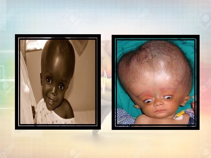
Presenting features Hydrocephalus in infants • failure to thrive. • increased skull circumference (compared with normal growth curves). • tense anterior fontanelle. • ‘cracked pot’ sound on skull percussion. ( hyper-resonance) • transillumination of cranial cavity with strong light. • severe, impaired conscious level and Vomiting. • ‘setting sun’ appearance due to lid retraction and impaired upward gaze from 3 rd ventricular pressure on the midbrain tectum. • thin scalp with dilated veins.

Presenting features Hydrocephalus in Adult • acute onset and deterioration or • gradual onset and slowly progressive deterioration.

Acute-onset adult hydrocephalus • This type of presentation occurs particularly in patients with tumours causing obstructive hydrocephalus, hydrocephalus although it may occur with any of the causes of hydrocephalus and an acute rapid neurological deterioration may occur in patients who have had longstanding chronic hydrocephalus. The major presenting features are due to the signs and symptoms of raised intracranial pressure : • headache • vomiting • papilledema • deterioration of consciousness. • Upgaze will often be impaired due to pressure of the dilated 3 rd ventricle on the superior colliculus of the tectum.

Gradual onset adult hydrocephalus • this type of onset occurs less frequently than the previous type. The symptoms of raised intracranial pressure are only very gradually progressive and late diagnosis is common. • Early features in the adolescent involve deteriorating school performance as a result of headaches, failing mental function, memory loss and behavioural disturbances. • Endocrine abnormalities such as infantilism and precocious puberty can occur in association with chronic hydrocephalus in older children and adolescents due to disturbance of the hypothalamus and possible compression of the pituitary gland. • If the condition is unrecognized progressive visual failure will occur, secondary to papilloedema and optic atrophy. acute decompensation may occur and the patient may suddenly develop a rapid deterioration of conscious state.

Radiological investigation • The most important investigation is either a CT scan or MRI of the brain which will show which ventricles are dilated. • If the lateral ventricles and 3 rd ventricle are all very dilated, and the 4 th ventricle is small, it is likely that the obstruction is at the level of the aqueduct of Sylvius. • In a communicating hydrocephalus all the ventricles are dilated.

CT MRI

• Ultrasonography through the open anterior fontanelle is useful in assessing ventricular size in infants and may obviate the need for repeated CT scans. • Records of the head circumference and its comparison with body weight and length centile charts are an integral part of postnatal follow-up of any child, and post op.

Treatment

Treatment • Management of hydrocephalus will depend on the underlying cause. • 123 - Options include : removing a causative mass lesion. ventricular shunting. third ventriculostomy.

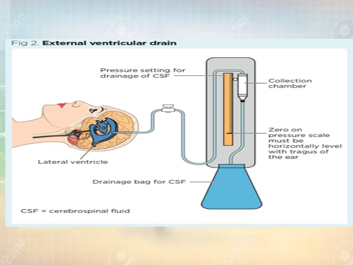
Removing a causative mass lesion • In some circumstances it may be appropriate to treat the hydrocephalus by tumour removal and decompression of the CSF pathways, perhaps with the insertion of an external ventricular drain (EVD) to cover the early postoperative period.

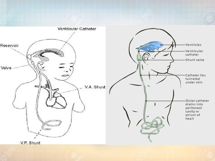
Ventriculoperitoneal shunt • involves the insertion of a catheter into the lateral ventricle. The catheter is then connected to a shunt valve under the scalp and finally to a distal catheter, which is tunneled subcutaneously down to the abdomen and inserted into the peritoneal cavity. • If the CSF pressure exceeds the shunt valve pressure, then CSF will flow out of the distal catheter and be absorbed by the peritoneal lining.

Ventriculoperitoneal shunt

Ventriculoperitoneal shunt
 (ventriculo-atrial shunt): from")
• Other options for distal catheter placement include: Ø 1) (ventriculo-atrial shunt): from the right atrium via the deep facial and jugular vein , it’s necessary when there has been marked intraperitoneal sepsis or multiple abdominal operations. Ø 2) (lumboperitoneal shunts) : The lumboperitoneal shunt involves drainage of the CSF from the lumbar theca rather than the ventricle. (pseudotumor cerebri)

LP SHUNT
 infection : • Shunt infection affects between 1% and 7%")
Complications of shunt 1) infection : • Shunt infection affects between 1% and 7% of shunt insertions and is usually caused by skin commensals, such as Staphylococcus epidermidis. Neonates are susceptible to Escherichia coli and haemolytic streptococcal infections. • Most infections become apparent clinically by 6 weeks and over 90% are apparent within 6 months. • o o Risk factors for infection include : very young children. open myelomeningocele. longer operative time. excessive staff movement into and out of theatre.

Complications of shunt • Treatment is by : removal of the shunt, external CSF drainage and treatment of infection prior to reinsertion of the shunt at a different site. • The introduction of antibiotic-impregnated catheters has resulted in a reduction in shunt infection rates.
 shunt blockage : • Shunt blockage may affect the ventricular")
Complications of shunt 2) shunt blockage : • Shunt blockage may affect the ventricular catheter, shunt , valve or distal catheter. • More than one-half of cases of shunt blockage are subsequently shown to be infected. • Causes of blockage : choroid plexus adhesion, blood, cellular debris or misplacement of the distal catheter in the preperitoneal space.
 intracranial hemorrhage : • Shunt systems may over drain leading")
Complications of shunt 3) intracranial hemorrhage : • Shunt systems may over drain leading to subdural haemorrhage. • Other complications are common to intracranial surgery and include seizures (5%), CSF leak, stroke and (< 1%) intracerebral haemorrhage.
 or the")
External drains • External drains can be placed within the ventricle (EVD) or the lumbar thecal sac (lumbar drain). • These are useful for temporary CSF drainage and can be used to administer intrathecal antibiotics to treat CSF infection. , take sample and decrease ICP

Endoscopic third ventriculostomy • involves the insertion of a neuroendoscope into the frontal horn of the lateral ventricle and then into the third ventricle through the foramen of Munro. A stoma can be created in the floor of the third ventricle in between the mamillary bodies and infundibular (pituitary) recess. • CSF can then communicate freely between the ventricular system and interpeduncular subarachnoid space. • The technique is particularly useful when there is obstruction of the CSF pathways below the third ventricle such as with aqueduct stenosis or posterior fossa mass lesions.

• It has an advantage over shunting in that no tubing is left in the patient and therefore infection rates are lower. • ETVs may block off, however, with about onehalf of these patients ending up with a shunt. Rare, but serious, complications include basilar artery rupture or memory impairment from injury to the fornix. • The procedure is less useful for communicating types of hydrocephalus or in infants of less than 6 months of age, but has a success rate of over 70% for accepted indications.

Normal-pressure hydrocephalus • Normal-pressure hydrocephalus was first discussed by describing a group of patients with triad of symptoms; symptoms dementia, ataxia and incontinence, where the radiological studies showed hydrocephalus but the lumbar CSF pressure was normal.

Etiology • in a large percentage the communicating hydrocephalus may have resulted from obliteration of the subarachnoid pathways in the basal cisterns following an episode of meningitis or subarachnoid haemorrhage, from either rupture of an aneurysm, arteriovenous malformation or following trauma. • Although lumbar puncture pressure is within the normal range, continuous monitoring of the intracranial pressure in these patients will frequently reveal abnormal wave formation, especially at night.

Investigation • The CT scan or MRI will show dilated ventricles without significant cortical atrophy. The difficulty arises that normal-pressure hydrocephalus may occur in patients with a scan appearance of cortical atrophy, but in these patients the degree of ventricular dilation should be more than would be expected just to compensate for the degree of atrophic change. • Other investigations, including isotope cisternography, neuropsychological assessment and CSF infusion studies, studies have been used but they have a high failure rate in predicting which patients will benefit from a CSF shunt.

Treatment • The following criteria can be used to assess the patients with the greatest chance of improvement following a shunt: • Aclinical presentation of the classic triad, particularly if the features of gait disturbance predominate. • The CT scan or MRI showing marked hydrocephalus with minimal cortical atrophy. • Aclearly defined cause for the hydrocephalus, such as a past episode of subarachnoid haemorrhage, trauma or meningitis. • Abnormal pressure waves on continuous intracranial pressure monitoring. • Naturally, a patient who has all these positive criteria deserves a shunt and should make a good recovery following the operation.

Hydrocephalus ex-vacuo • occurs when there is damage to the brain caused by stroke or injury, and there may be an actual shrinkage of brain substance. Although there is more CSF than usual, the CSF pressure itself is normal

External hydrocephalus • A condition that occurs in infancy and early Childhood. • characterised by: enlargement of the subarachnoid space, typically in the frontal areas and interhemispheric fissure with raised intracranial pressure without significantly enlarged ventricles. • It is attributed to an absorption deficiency and typically resolves within a year.

Benign External Hydrocephalus

Normal pressure hydroceph benign intracranial hypertension Pressure Normal , no S & S of increased ICP Very high Volume Very high Normal or low Epidemiology Old age males Young obese females Presentation ( 3 W`s triad ) : wacky ( dementia ) Wubbly ( ataxia ) Wet ( incontenence ) S & S of increased ICP : headache , blurred vision , papilloedema Imaging Dilated ventricles Normal size or small ventricles Treatment VP shunt -conservative : 1 -weight loss (sucsess rate 20%) 2 -dimox (acetazolamide ) 30% respond -surgery : theco-peritoneal shunt
- Slides: 52