HYDROCEPHALUS Kaan Yaltrk MD Hydrocephalus From Greek hydrokephalos


,")


 hydrocephalus are a result of developmental malformations. Other")
 Aqueduct develops about the")


 Type 2 malformation of brain seen most exclusively with")


")
- Slides: 36
HYDROCEPHALUS Kaan Yaltırık, MD
Hydrocephalus • From Greek hydrokephalos, from hydr- + kephal. E head • Definition: An abnormal increase in the amount of cerebrospinal fluid within the cranial cavity that is accompanied by expansion of the cerebral ventricles, enlargement of the skull and especially the forehead, and atrophy of the brain
A syndrome, or sign, resulting from disturbances in the dynamics of cerebrospinal fluid (CSF), which may be caused by several diseases.
Incidence Occurs in 3 -4 of every 1000 births. Cause may be congenital or acquired. Congenital- may be due to maldevelopment or intrauterine infection Acquired- may be due to infection, neoplasm or hemorrhage.
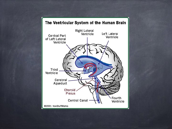
Pathophysiology CSF is formed by two mechanisms: Secretion by the choroid plexus, Lymphatic-like drainage by the extracellular fluid in brain. CSF circulates thru ventricular system and is absorbed within subarachnoid spaces by unknown mechanism.
Overview of CSF production The CSF volume of an average adult ranges from 80 to 160 ml The ventricular system holds approximately 20 to 50 ml of CSF is produced in the choroid plexuses at a daily rate of 14 -36 ml/hr
Overview of CSF circulation The CSF flows from the lateral ventricles downward to the foramina of Magendie and Luschka, to the perimedullary and perispinal subarachnoid spaces, and then upward to the basal cistern and finally to the superior and lateral surfaces of the cerebral
Overview of CSF production The choroid plexuses are the source of approximately 80% of the CSF The blood vessels in the subependymal regions, and pia also contribute to the formation of CSF
The pressure gradient is highest in the lateral ventricles and diminishes successively along the subarachnoid space CSF circulation + Arterial pulsations in the choroid plexuses help drive the fluid from the ventricular system Normally, the periventricular tissues offer little resistance to the flow of CSF -
The function of the CSF The CSF acts as a “water jacket” for the brain and spinal cord The 1300 g adult brain weighs approximately 45 g when suspended in CSF
The CSF acts like a “sink”, effectively flushing waste products as new fluid is secreted reabsorbed The function of the CSF A constant CSF electrolyte composition helps maintain a stable medium for excitable cells (neurons)
Mechanisms of Fluid Imbalance Hydrocephalus results from: 1. Impaired absorption of CSF within the subarachnoid space (communicating hydrocephalus), or 2. Obstruction to the flow of CSF through the ventricular system (non-communicating hydrocephalus)
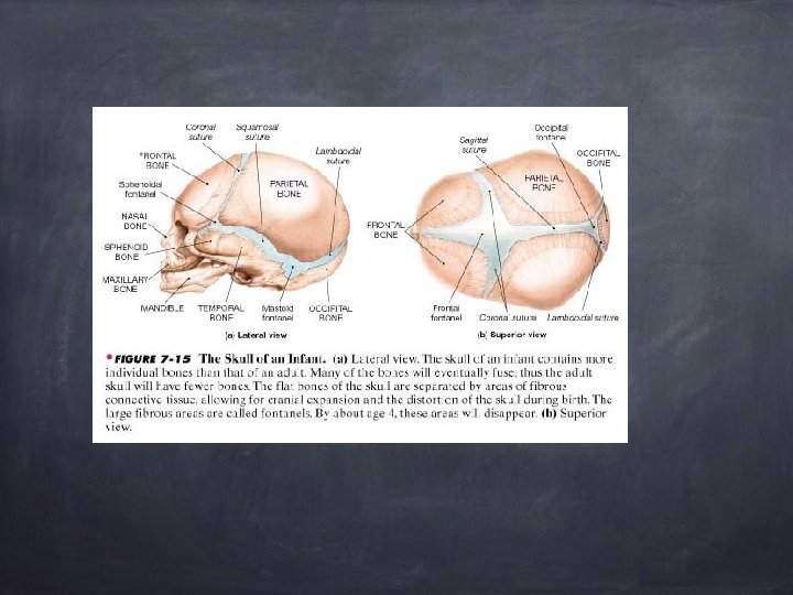
Mechanisms of fluid imbalance Both lead to increase accumulation of CSF in the ventricles! Ventricles become dilated and compress the brain. When this happens before cranial sutures are closed, skull enlarges. In children <10 -12, previously closed sutures may open.
Hydrocephalus Most cases of non-communicating (obstructive) hydrocephalus are a result of developmental malformations. Other causes: neoplasms, intrauterine infections, trauma. Developmental defects account for most causes of hydrocephalus from birth to 2 years of age. (Table 11 -3, page 497 sites and types of hydrocephalus)
Aqueductal stenosis Is the most common cause of congenital hydrocephalus(43%) Aqueduct develops about the 6 th week of gestation M: F = 2: 1 Other congenital anomalies (16%): thumb deformities Prognosis: 11 -30% mortality
Etiology of aqueductal stenosis Intrinsic Pathology of the Aqueduct • Septum or Membrane Formation: A thin membrane of neuroglia may occlude the aqueduct. It commonly occurs caudally. There may be a primary developmental defect or it may follow granular ependymitis from intrauterine infections. This is the rarest of the types of narrowing. • Forking of the Aqueduct: Typically, there are two channels seen in midsagittal plane unable to handle CSF volume. Most often seen with spina bifida. • Gliosis of the Aqueduct: Usually of infectious origin showing a marked gliofibrillary response. The lumen is devoid of ependyma. • Stenosis of the Aqueduct: Narrowed aqueduct without evidence of gliosis. This may have hereditary basis.
Clinical features of aqueductal stenosis Obstructive hydrocephalus: presents with macrocephaly and/or intracranial hypertension. Parinaud's syndrome. Inability to elevate eyes Collier's sign. Retraction of the eyelids
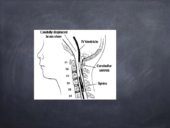
Common Defects Arnold-Chiari Malformation (ACM) Type 2 malformation of brain seen most exclusively with myelomeningocele, is characterized by herniation of a small cerebellum, medulla, pons, and fourth ventricle into the cervical spinal canal through an enlarged foramen magnum.
Causes of communicating hydrocephalus Overproduction of CSF Blockage of CSF circulation Blockage of CSF resorption Hydrocephalus exvacuo Normal pressure
Clinical manifestations Clinical picture depends on acuity of onset and presence of preexisting structural lesions.
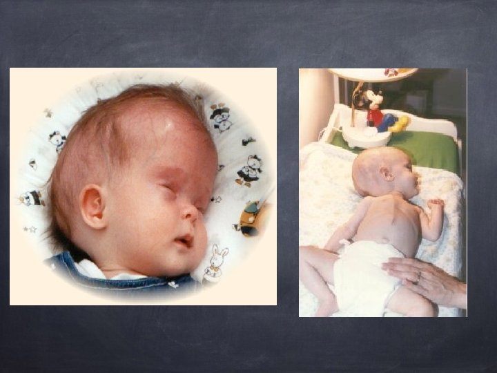
Infancy Head grows at alarming rate with hydrocephalus. First signs- bulging of fontanels without head enlargement. Tense, bulging, non-pulsatile anterior fontanel Dilated scalp veins, esp. when crying Thin skull bones with separated sutures (cracked pot sounds on percussion)
Infancy Protruding forehead or bossing. Depressed eyes or setting-sun eyes (eyes rotating or downward with sclera visible above pupil) Pupils sluggish with unequal response to light Irritability, lethargy, feeds poorly, changes in LOC, arching of back (opisthotonos), lower extremity spasticity. May cry when picked up or rocked; quiets when allowed to lay still.
Infancy Swallowing difficulties, stridor, apnea, aspiration, respiratory difficulties and arm weakness may indicate brain stem compression. If hydrocephalus progresses, difficulty sucking and feeding, and a high-pitched shrill cry results. (lower brain stem dysfunction)
Infancy Emesis, somnolence, seizures, and cardiopulmonary distress ensues and hydrocephalus progresses. Severely affected infants may not survive neonatal period.
Childhood Signs and symptoms caused by increased ICP. Manifestations caused by posterior neoplasms and aqueduct stenosis, manifestations associated with space-occupying lesions.
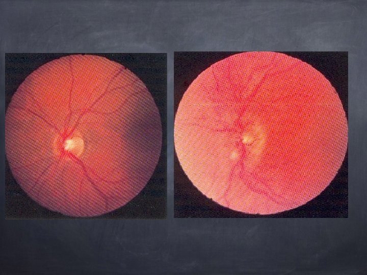
Childhood Headache on awakening with improvement following emesis or sitting up. Papilledema (swelling of optic disc DT obstruction), strabismus, and extrapyramidal tract signs such as ataxia Irritability, lethargy, apathy, confusion, and often incoherent
Childhood Dandy-Walker syndrome- congenital defect-late onset. Obstruction of foramen of Lushka and Magendie Bulging occiput, nystagmus, ataxia, cranial nerve palsies Female predominance (3: 1) Absence or occlusion of ventricles
Diagnostic Evaluation Antenatal- fetal ultrasound as early as 14 weeks Infancy- based on head circumference crosses one or more grid lines on the infant growth chart within a 4 week period and there are progressive neuro signs. CT and MRI to localize site of obstruction; reveal large ventricles
Therapeutic management Goals: Relieve hydrocephaly Treat complications Manage problem resulting from effects of disorder on psychomotor development USUALLY SURGICAL!
Surgical Treatment Therapy of choice! Direct removal of source of obstruction (neoplasm, cyst, or hematoma) Most require shunt procedure to drain CSF from ventricles to extracranial area; usually peritoneum(VP shunt), or right atrium (VA shunt) for absorption.
Treatment of hydrocephalus The two most commonly used shunt systems are the ventriculoatrial (VA) and ventriculoperitoneal (VP) shunts. The VP shunt is most commonly used as it is simpler to place, extra tubing may be placed in the peritoneum and the consequences of infection are less.