Human Visual System Image formation Cornea Lens Exposure


 Vitreous Humor Eyelens Fovea")


")


 This example shows best")

 4")


- Slides: 75
Human Visual System Image formation • Cornea • Lens Exposure Control Detection Processing • Iris/pupil • Photoreceptor sensitivity • Retina • Rods • Cones • Brain
Human Visual System Image formation • Cornea • Lens Exposure Control Detection Processing • Iris/pupil • Photoreceptor sensitivity • Retina • Rods • Cones • Brain
Human Eye Ciliary Muscle Sclera Iris Pupil Ear side (Temporal) Vitreous Humor Eyelens Fovea Retina Optic Nerve Cornea Nose side (Nasal) Aqueous Humor Suspensory ligament Choroid ¬ Human eye is a complete imaging system.
Image Formation Object ¬ The curved surfaces of the eye focus the image onto the back surface of the eye. Image
Cornea Sclera Cornea ¬ The outer wall of the eye is formed by the hard, white sclera. ¬ Cornea is the clear portion of the sclera. ¬ 2/3 of the refraction takes place at the cornea.
Iris and Pupil Iris ¬ Colored iris controls the size of the opening (pupil) where the light enters. ¬ Pupil determines the amount of light, like the aperture of a camera. Pupil Iris open Dilated pupil Iris closed Constricted pupil
Lens Ciliary muscle Lens Suspensory Ligament Transparent Fibers Cross section of the eye lens ¬ Eye lens is made of transparent fibers in a clear membrane. ¬ Suspended by suspensory ligament. ¬ Used as a fine focusing mechanism by the eye; provides 1/3 of eye’s total refracting power. ¬ Non-uniform index of refraction.
Accommodation Distant object Near object Relaxed muscle Taut ligaments ¬ The suspensory ligaments attach the lens to the ciliary muscle. ¬ When the muscle contracts, the lens bulges out in the back, decreasing its focal length. ¬ The process by which the lens changes shape to focus is called accommodation. Contracted muscle Slack ligaments
Aqueous Humor and Vitreous Humor Aqueous Humor ¬ Transparent gelatinous liquid filling the eye. ¬ Provides nutrients to the cornea and eye lens. ¬ Also helps maintain the eyeball shape with its pressure.
Retina Fovea Optic Nerve ¬ Retina is the photosensitive “detector” for the eye. ¬ Two types of receptors in the retina: rods for low light level, and cones for color. ¬ Located at the center of the retina, fovea contains a greater concentration of cones. ¬ Signals from the receptors leave through the optic nerve to the brain.
Plexiform Layer ¬ The retina is made of three layers: Fovea Light Plexiform Layer Optic Nerve – Plexiform layer is a network of nerves which carry the signals from the photo receptors. Photo receptors – Photo receptors. – Choroid provides nourishment to the receptors, as well as absorb any light that didn’t get absorbed by the photo receptors, like a antihalation backing in film. Choroid
Eye Defects Object at infinity ¬ Image focuses on the retina for a normal eye. Normal ¬ Distant objects look blurry for a myopic (near sighted) eye. Myopic Hyperopic Eyes at relax state. ¬ Near objects look blurry for a hyperopic (far sighted) eye.
Myopia - Near sightedness Far object Near object Far object ¬ Distant objects look blurry because the eye cannot Myopic eye relaxed relax any farther so that the Blurry image is focused before the retina. Myopic eye relaxed ¬ Near object in focus In focus without accommodation. ¬ Corrected with a negative lens. Myopia corrected with a negative lens The virtual image from the diverging lens appears to be closer.
Hyperopia - Far sightedness Far object Near object ¬ Near objects look blurry because the eye cannot Hyperopic eye Partially accommodated accommodate enough for In focus near objects. ¬ Far object in focus. Hyperopic eye Fully accommodated ¬ Corrected with a positive Blurry lens. Hyperopia corrected with a positive lens Light from the converging lens looks as though it is coming from the distance.
Contact Lens Contact lens Cornea Fluid ¬ Contact lens is an alternative to corrective lenses. ¬ Changes the curvature of the cornea by adhering to the surface with some fluid.
Astigmatism Cornea Object ¬ The cornea is not spherical; Focal length different from one plane to a perpendicular plane. F’ horizontal Direction of blur F’ Vertical Image at F’ Horizontal Image at F’ Vertical
Astigmatism Cylindrical lens Rays in the horizontal plane are focused ¬ Correction of astigmatism is done through the use of a cylindrical lens. ¬ Cylindrical lens converge rays in one plane but not the perpendicular plane. Rays in the vertical plane are undeviated
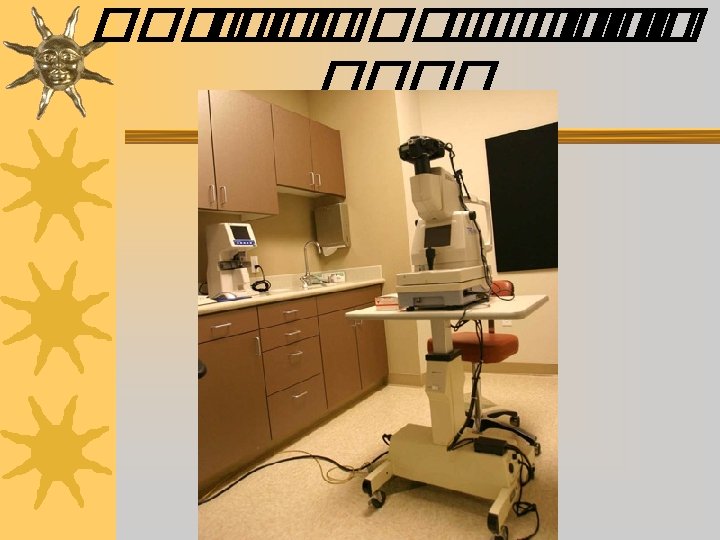
Eye unit
Slit lamp
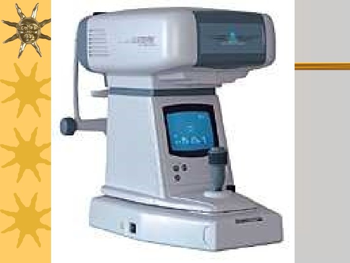
Retinal Camera
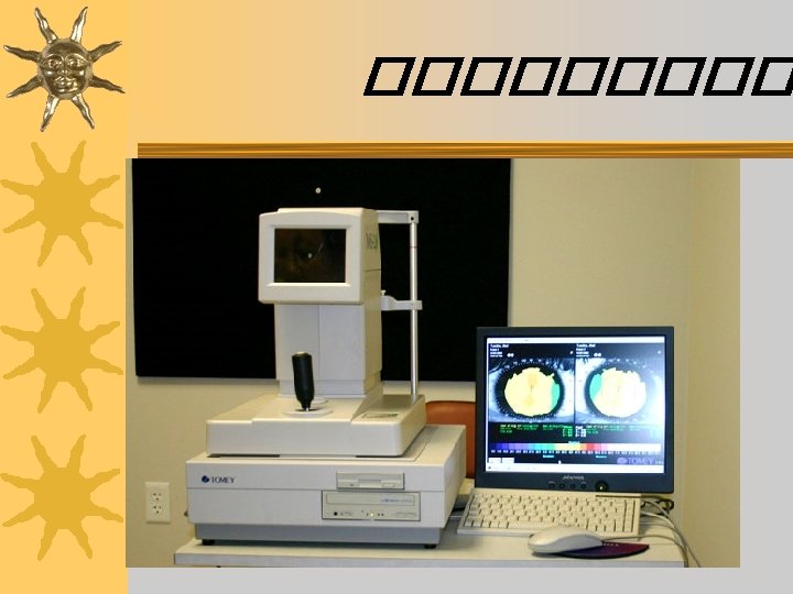
Scan & Pachymeter
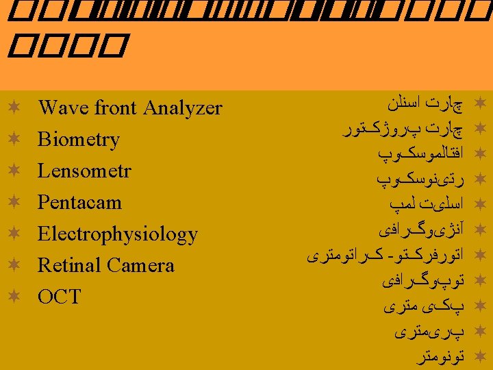
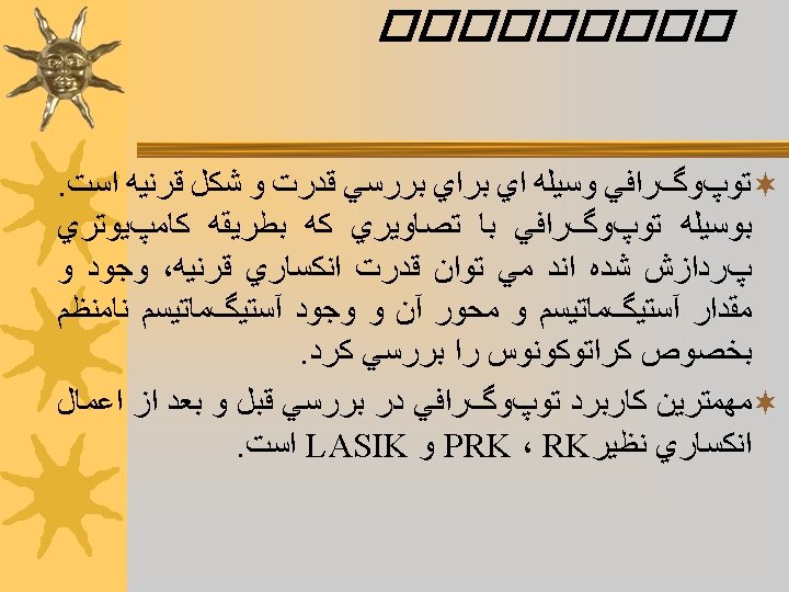
WAVEFRONT ANALYZER
Hartmann Shack Messung CCD Sensor deformed perfect wave front as sensor image Lenslet array perfect wave deformed front wave front
Easy fokussing and x/y adjustment … X / Y grid pattern Illumination spots help to find correct focus of the system … before accquisition
Datentransfer ORK-Software und Schwind Database 200 Hz Pulsfrequenz 0. 8 mm Gaußprofil 330 Hz Eyetracker
Simulation of visual accuity Simulation of uncorrected visual accuity (UCVA) This example shows best corrected visual accuity (BCVA), because ony sphere and cylinder are selected Selection of coefficients simulate the corrected visual accuity based individual aberrations on the snellen “E”
Principle of Hartmann Shack Aberrometrie SLD CCDsensor Lenslet array wavefront Permeable mirror Retinal image
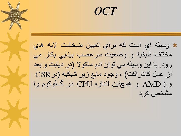
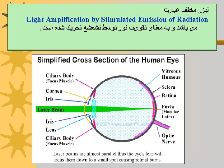
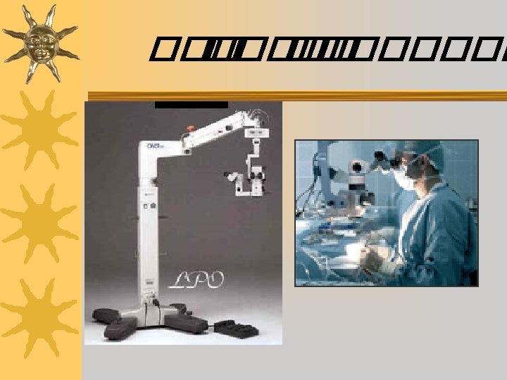
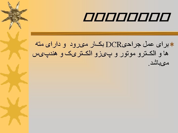
ﻋﻮﺍﻣﻞ ﻣﻮﺛﺮ ﺩﺭ ﻟﻴﺰﺭ avelength Exposure time 3 - Spot diameter (mm) 4 - power (m. W) density (W/cm 2)
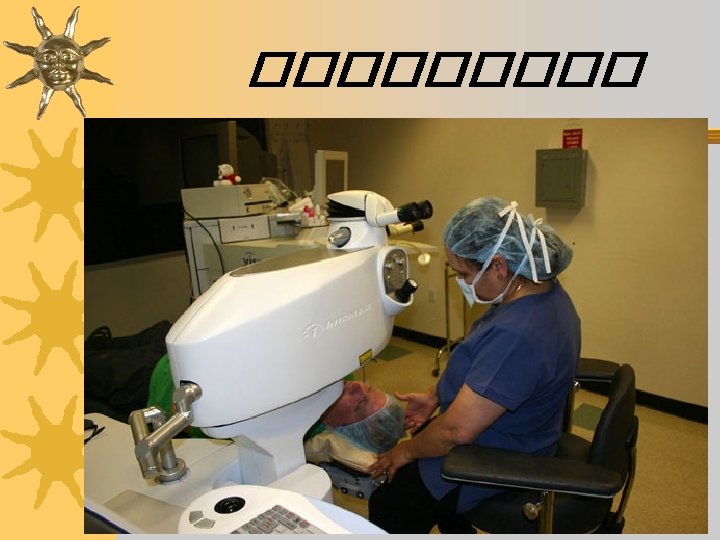
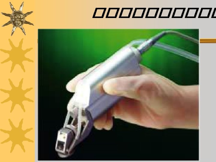
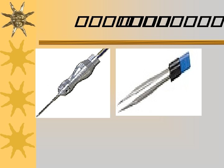
Handpice
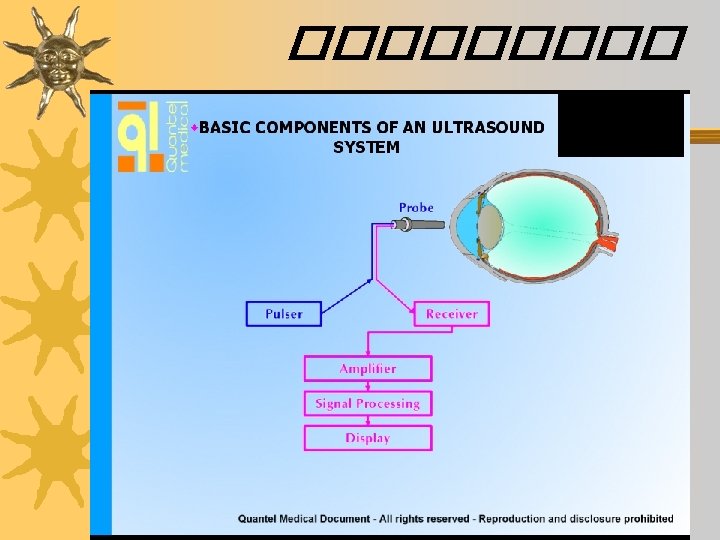
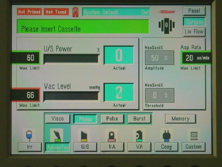
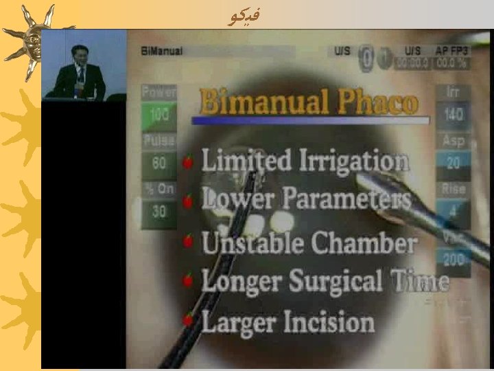
Traditional Pulse Ultrasound ¬ Power is applied in bursts, with fixed 50/50 “burst/rest” periods comprising each “pulse” ¬ Energy buildup occurs during long “burst” periods; “rest” periods may be too brief for cooling to occur