Human Physiology Synaptic Transmission by Talib F Abbas

Human Physiology Synaptic Transmission by Talib F. Abbas

Synaptic transmission and Reflexes � Impulses are transmitted from one nerve cell to another cell at synapses. �presynaptic cell. �postsynaptic cell. �chemical or electrical synapse. �At chemical synapses, a synaptic cleft. �In electrical synapses, there are gap junctions form between the cells.
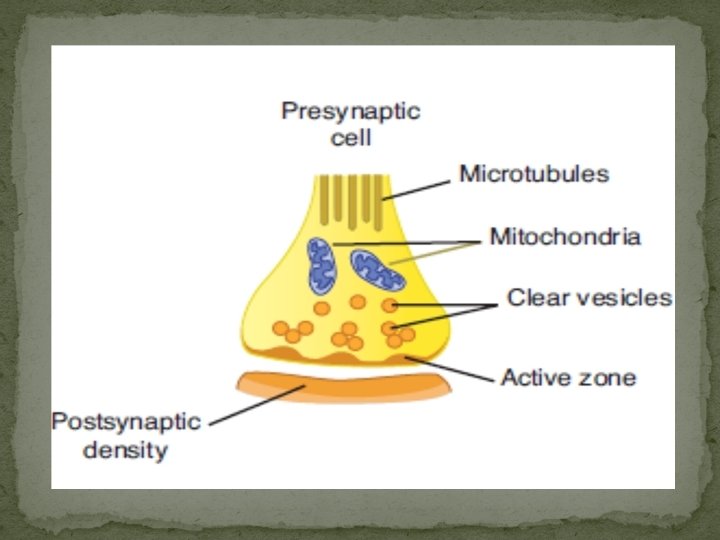

synaptic vesicles � small, clear synaptic vesicles that contain acetylcholine, glycine, GABA, or glutamate; small vesicles with a dense core that contain catecholamines; and large vesicles with a dense core that contain neuropeptides. � These vesicles fuse with the cell membrane and release transmitters through exocytosis and are then recovered by endocytosis. � vesicle discharges its contents through a small hole (kiss-and-run discharge). � The Ca 2+ that triggers exocytosis of transmitters enters the presynaptic neurons, and transmitter release starts within 200μs � neurexins
 by measuring")
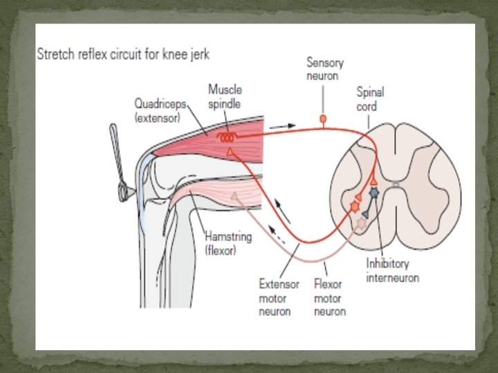
Reflexes �Reflex pathway is monosynaptic or polysynaptic (contains more than one synapse) by measuring the delay in transmission from the dorsal to the ventral root across the spinal cord. �A single stimulus applied to the sensory nerves characteristically does not lead to the formation of a propagated action potential in the postsynaptic neuron. �transient partial depolarization or a transient hyper polarization. �excitatory postsynaptic potential (EPSP) The EPSP is produced by depolarization of the postsynaptic cell membrane immediately under the presynaptic ending.

Depolerization of Synapses �The initial depolarizing response produced by a single stimulus to the proper input begins about 0. 5 ms after the afferent impulse enters the spinal cord. It reaches its peak 11. 5 ms later and then declines exponentially. �excitatory postsynaptic potential (EPSP). �The EPSP due to activity in one synaptic knob is small, but the depolarizations produced by each of the active knobs summate. �inhibitory postsynaptic potential (IPSP), localized increase in Cl– transport. �(reversal potential).

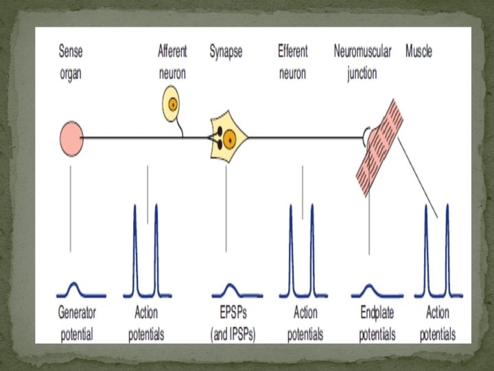
Reflex arc �This arc consists of a sense organ, an afferent neuron, one or more synapses within a central integrating station, an efferent neuron, and an effector. �the connection between afferent and efferent somatic neurons is generally in the brain or spinal cord. �Bell–Magendie law. �This generates all-or none action potentials in the afferent nerve, the number of action potentials being proportional to the size of the generator potential. �The simplest reflex arc is monosynaptic. �more interneuron is interposed is polysynaptic.
 a group of specialized intrafusal muscle fibers with")
STRUCTURE OF MUSCLE SPINDLES � (1) a group of specialized intrafusal muscle fibers with contractile polar ends and a noncontractile center � (2) large diameter myelinated afferent nerves (types Ia and II) originating in the central portion of the intrafusal fibers � (3) small diameter myelinated efferent nerves supplying the polar contractile regions of the intrafusal fibers. � Changes in muscle length are associated with changes in joint angle; thus muscle spindles provide information on position (ie, proprioception). � The intrafusal fibers are positioned in parallel to the extrafusal fibers (the regular contractile units of the muscle). Imanpour
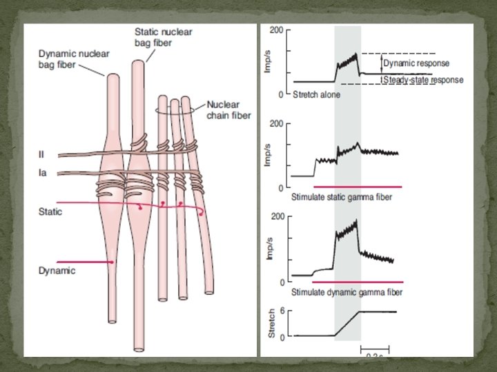

Intrafusal Fiber �Intrafusal fibers do not contribute to the overall contractile force of the muscle, but rather serve a pure sensory function. �With many nuclei in a dilated central area , is called a nuclear bag fiber (dynamic and static). �The second intrafusal fiber type, the nuclear chain fiber. �There are two kinds of sensory endings in each spindle, a single primary (group Ia) ending and up to eight secondary (group II) endings.

CORTICAL PLASTICITY �phantom limb pain: remarkable changes in cortical and thalamic organization that occur in response to limb amputation. �Amputated digit > somatosensory map > surrounding cortex. �Extensive, long-term deafferentation of limbs leads to even more dramatic shifts in somatosensory representation in the cortex, with, for example, the limb cortical area responding to touching the face. �Plasticity of this type occurs not only with input from cutaneous receptors but also with input in other sensory systems.

CORTICAL PLASTICITY �PET scanning in humans also documents plastic changes, sometimes from one sensory modality to another. Thus, for example, tactile and auditory stimuli increase metabolic activity in the visual cortex in blind individuals. Conversely, deaf individuals respond faster and more accurately than normal individuals to moving stimuli in the visual periphery. Plasticity also occurs in the motor cortex. These findings illustrate the malleability of the brain and its ability to adapt.

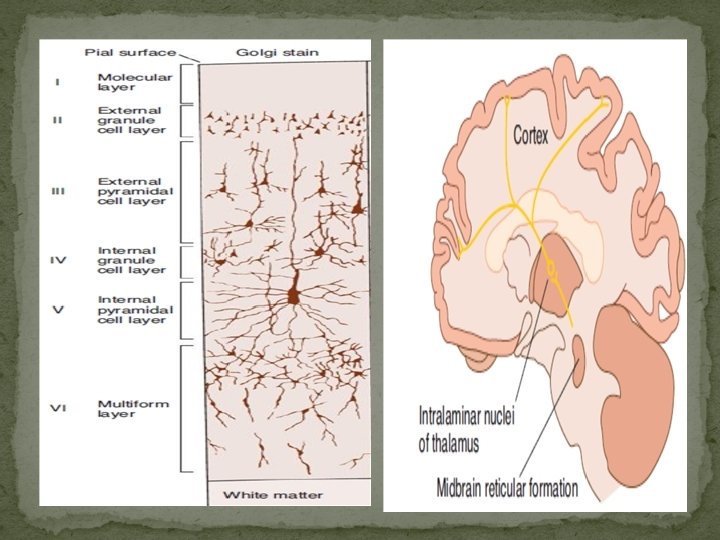
Higher function of nervous system �Thalamus: The thalamus is a large collection of neuronal groups within the diencephalons; it participates in sensory, motor, and limbic functions, “gateway” to the cerebral cortex. �Neocortex: The neocortex is generally arranged in six layers. The most common neuronal type is the pyramidal cell with an extensive vertical dendritic tree that may reach to the cortical surface. � Pyramidal neurons are the only projection neurons of the cortex, and they are excitatory neurons that release glutamate at their terminals. �The other Interneurons (basket cells and chandelier cells which is inhibitory release GABA.
 �The reticular formation, the phylogenetically old reticular core of the")
Reticular activating system (RAS) �The reticular formation, the phylogenetically old reticular core of the brain, occupies the midventral portion of the medulla and midbrain. It is primarily an anatomic area made up of various neural clusters and fibers with discrete functions. IMN �it contains the cell bodies and fibers of many of the serotonergic, noradrenergic, and cholinergic systems. It also contains many of the areas concerned with regulation of heart rate, blood pressure, and respiration.

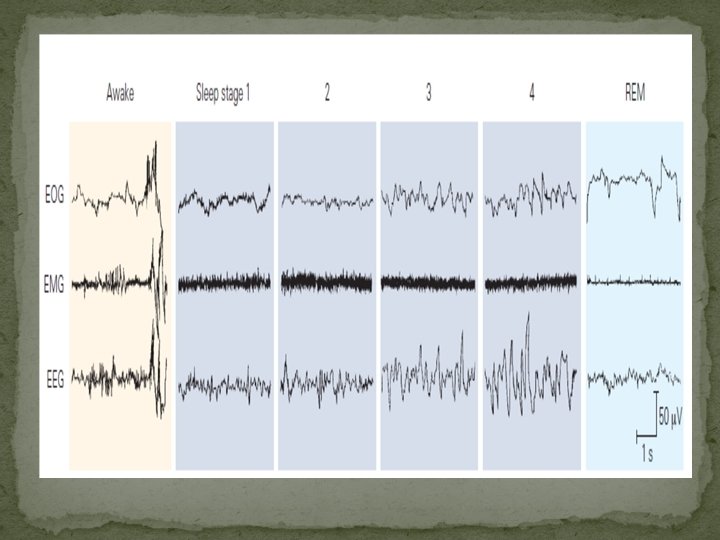
Electroencephalogram �firstly used by German Hans Berger, to denote the record of the variations in brain potential. Its either by one electrode (unipolar) or two electrode dipolar ( study the fluctuations in cortex potentials). The cerebellar cortex and the hippocampus are two other parts of the central nervous system (CNS) where many complex, parallel dendritic processes are located subpially over a layer of cells. In both areas, characteristic rhythmic fluctuations occur in surface potential similar to that observed in the cortical EEG. It has been widely used in study the seizure, Parkinson, and Alzheimer diseases.

EEG and Sleep –wake recordings of waves �EEG is a fairly regular pattern of waves at a frequency of 8– 13 Hz and amplitude of 50– 100μV called Alpha rhythem. �When attention is focused on something, the alpha rhythm is replaced by an irregular 13– 30 Hz lowvoltage activity, the beta rhythm. This phenomenon is called alpha block. �Alerting response, EEG activity seen in the alert state is also synchronized, but at a higher rate. Therefore, the term desynchronization is misleading. Gamma oscillations at 30– 80 Hz are often seen when an individual is aroused and focuses attention on something.

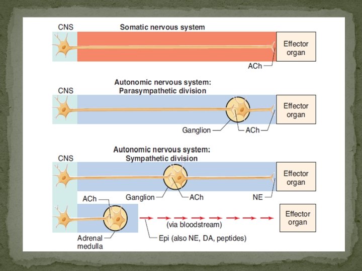
The autonomic nervous system ANS �Although survival is possible without an ANS, the ability to adapt to environmental stressors and other challenges is severely compromised. The ANS has two major divisions: the sympathetic and parasympathetic nervous systems. �the ANS includes the enteric nervous system within the gastrointestinal tract. The classic definition of the ANS is the preganglionic and postganglionic neurons within the sympathetic and parasympathetic divisions.

Thank you
- Slides: 23