Human parasitology tapeworm common features of Class Cestoda
")


")
 1 -The adult worm lives in the intestine of humans.")
 4 -8 m , 1000 -2000 segments Scolex Neck Immature")

 Note : Bilobed ovary (Carmine stained)")
 Hermaphroditic testes, sperm duct ovary, uterus genital pore vitellaria (yolk gland)")


 show evaginated sscolex (right) The scolex is similar")
")
 Taenia solium The uterus")


")
 1 The adult worm lives in the intestine of humans.")


")


 a small central lobe Note: Trilobed ovary")
 India ink")
.")


 Taenia solium (pork tapeworm) is the main cause")


")


 It is soybean-like in shape, has an small scolex invaginated into")


- Slides: 71
Human parasitology (tapeworm)
common features of Class Cestoda 1. Adult worm is flattened ribbon-like, without body cavity. 2. The body is composed of a head, neck and segmented strobilus The head has suckers, rostellum and hooklets or sucking grooves. The neck is the budding zone from which segments are formed. The strobilus consists of immature, mature and pregnant proglottides. 3. They are hermaphroditic. There is a set of female and male reproductive organs in every mature proglottid. 4. Digestive tract is absent. Nutrition is absorbed by villi of body surface. 5. They are biohelminths. Intermediate hosts are indispensable.
6. All adult worms parasitize digestive tracts of mammals. 7. The developing stages in intermediate hosts are called metacestode, such as cysticercus, hydatid cyst, cysticercoid, procercoid, plerocercoid. 8. Tapeworms are classified into two orders: Cyclophyllidea : The head is spherical with suckers, hooklets. The uterus has no opening. One intermediate host is required. The eggs contain an oncosphere. They are medically important, such as Taenia solium, Taenia saginata and Echinococcus granulosus. Pseudophyllidea : The head is spear-like with sucking grooves. The uterus has an opening. Two or more intermediate hosts are required. The eggs contain a coracidium and have to get into water to develop. Human being occasionally get infection. This worms include Spirometra mansoni and Diphyllobothrium latum.
Taenia saginata (Beef tapeworm)
Taenia saginata (Beef tapeworm) 1 -The adult worm lives in the intestine of humans. 2 - The larvae ( Cysticercus bovis) localize in beef mainly.
Taenia saginata (beef tapeworm) 4 -8 m , 1000 -2000 segments Scolex Neck Immature segment Mature segment Gravid segment
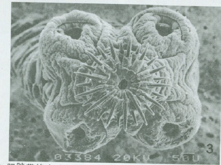
Scolex and Neck 1. 5 - 2 mm in diameter Without rostellum and hooks Four suckers ( unarmed tapeworm)
Mature segment (ovary with 2 lobes) Note : Bilobed ovary (Carmine stained)
Mature segments (proglottids) Hermaphroditic testes, sperm duct ovary, uterus genital pore vitellaria (yolk gland) excretory canal
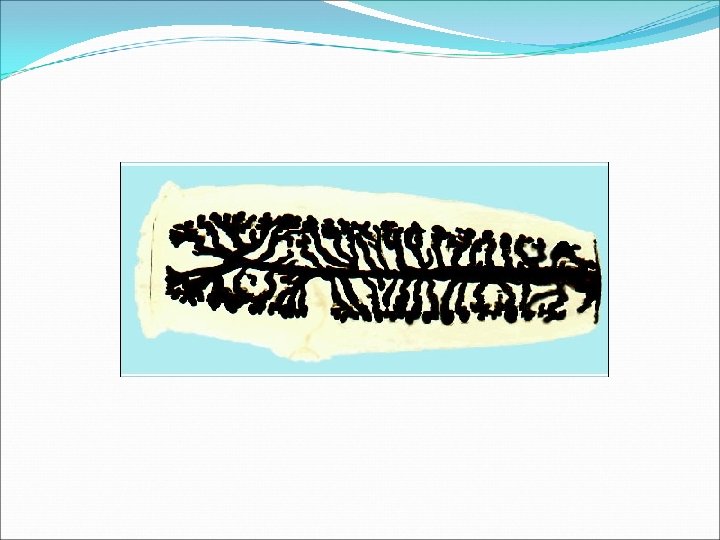
Gravid segment India ink technique Note : More than 15 lateral uterine branches (one side).
Taenia eggs The eggs of Taenia saginata and T. solium are indistinguishable morphologically.
Taenia egg spherical 31 to 43 µm a thick embryophore an oncosphere inside an egg shell outside (usually break away from the eggs in the feces)
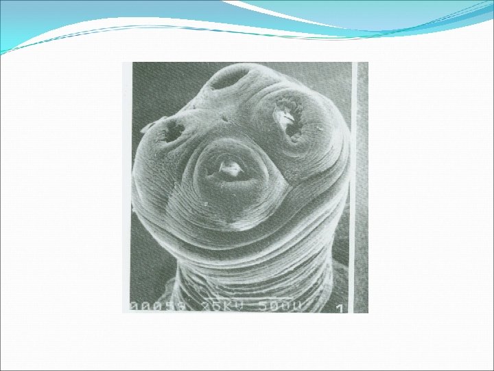
Cysticercius bovis Show invaginated scolex (left) show evaginated sscolex (right) The scolex is similar to that of adult worm in morphology Cysticercius bovis
Cysticercus bovis showing the bladder and the scolex (measly beef)
Gravid segment ( the primary lateral branches of the uterus) Taenia solium The uterus has 7 to 13 lateral branches on each side Taenia saginata The uterus has 15 to 30 lateral branches on each side
Life cycle of Taenia saginata The life cycle of Taenia saginata is similar to that of Taenia solium, but the intermediate host is cattle. The custicercus bovis (the beef bladder worm ) nearly does not parasite in human body. Humans serve as the final hosts only. (beef worm infection)
Life cycle
A cysticercus can survive for several years in the animal. Humans become infected by ingesting raw or undercooked infected meat. In the human intestine, the cysticercus develops over 2 months into an adult tapeworm, which can survive for years. The adult tapeworms attach to the small intestine by their scolex and reside in the small intestine The adults produce gravid proglottids which detach from the tapeworm, and migrate to the anus or are passed in the stool.
Geographic Distribution Both species are worldwide in distribution. Taenia saginata is more prevalent in communities where humans live in close contact with Cattle and eat undercooked beef.
Clinical Features Taenia saginata taeniasis produces mild abdominal symptoms and may cause malnutrition. (Epigastric pain, vomiting, diarrhea) The most striking feature consists of the passage (active and passive) of proglottids. The patients may find gravid proglottids themselves and take the segments to see doctors. Occasionally, appendicitis or cholangitiscan result from migrating proglottids.
Laboratory Diagnosis Microscopic identification of eggs and proglottids in feces is diagnostic for taeniasis. Repeated examination and concentration techniques will increase the likelihood of detecting light infections. Species determination of Taenia is impossible if solely based on microscopic examination of eggs, because all Taenia species produce eggs that are morphologically identical.
Eggs of Taenia sp. are also indistinguishable from those produced by cestodes of the genus Echinococcus. Microscopic identification of gravid proglottids (or, more rarely, examination of the scolex) allows species determination.
Diagnostic findings Microscopy: 1 egg * The eggs can not be used as species identification.
Taenia solium (pork tapeworm)
Taenia solium (pork tapeworm) 1 The adult worm lives in the intestine of humans. 2 The larvae ( Cysticercus cellulosae , bladder worm) localize in pigs mainly. 3 Cysticercus cellulosae can also infect humans causing cysticercosis.
Morphology Adult : 2 -4 m long, 700 -1000 segments: Scolex Neck Immature segment Mature segment Gravid segment
scolex
Immature segment note that the reproductive organs are just beginning to differentiate. (Carmine stained) Developing reproductive organs
Mature segment
Mature proglottid
Mature segment ( Ovary with 3 lobes) a small central lobe Note: Trilobed ovary ( Carmine stained )
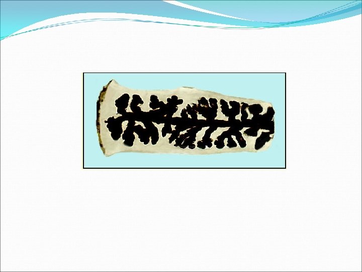
Gravid segment Note : Less than 14 lateral uterine branches (one side) India ink stained
India Ink Technique Note : less than 13 lateral uterine branches (one side).
t
Cysticercus cellulosae cyst Scolex The cyst is filled with fluid
Cysticercus cellulosae It is soybean-like in shape, has an small scolex invaginated into the translucent cyst. (left) The scolex evaginated from the cyst (right) Cysticercius cellulosae
Under stimulation of bile The scolex invaginates in the bladder The scolex evaginates
Cystcercus cellulosae in muscles of pigs
Life cycle of Taenia solium Humans serve as final hosts as well as intermediate hosts in the life cycle of Taenia solium. ( both adult and bladder worm parasite in humans) 1 The adult worm lives in the small intestine of humans ( pork tapeworm infection) 2 The larvae (Cysticercus cellulosea, bladder worm ) localize and develop in pigs (intermediate host) 3 The cysticercus cellulosae may also infect humans causing cystcercosis (humans as intermediate hosts)
Life cycle
Humans are the only definitive hosts for Taenia saginata and Taenia solium. Eggs or gravid proglottids are passed with feces The eggs can survive for days to months in the environment. Cattle (T. saginata) and pigs (T. solium) become infected by ingesting eggs or gravid proglottids In the animal's intestine, the oncospheres hatch , invade the intestinal wall, and migrate to tissues, where they develop into cysticerci.
Cysticercosis Causal Agent: The cestode (tapeworm) Taenia solium (pork tapeworm) is the main cause of human cysticercosis.
Life Cycle
Cysticercosis is an infection of both humans and pigs with the larval stages of Taenia solium. This infection is caused by ingestion of eggs of a human tapeworm carrier. Pigs and humans become infected by ingesting eggs or gravid proglottids Humans are infected either by ingestion of food contaminated with feces, or by autoinfection.
In autoinfection, a human infected with adult T. solium can ingest eggs either through fecal contamination or, possibly, from proglottids carried into the stomach by reverse peristalsis.
Three Modes of infection : 1 internal autoinfection 2 external autoinfection. 3 heteroinfection
Once eggs are ingested, oncospheres hatch in the intestine , invade the intestinal wall, and migrate to striated muscles, as well as the brain, liver, and other tissues, where they develop into cysticerci. In humans, cysts can cause serious results if they localize in the brain, resulting in neurocysticercosis
Clinical Features The symptoms of cysticercosis are caused by the development of cysticerci in various sites. Of great comcern is cerebral cysticercosis (or neurocysticercosis), which can cause manifestations including seizures, mental disturbances, focal neurologic deficits, and signs of space-occupying intracerebral lesions. Death can occur suddenly
Cerebral cysticercosis
Extracerebral cysticercosis can cause ocular, cardiac, or spinal lesions with associated symptoms Asymptomatic subcutaneous nodules and calcified intramuscular nodules can be encountered.
Cysticercosis Cysticercus cellulosae in heart ( Cardiac cysticercosis )
Note this cysticercus in the tongue
Ocular type: The cysticercus is usually found in the vitreous body or subretina. Visual disturbance often occurs. The died body of worm may provokes local inflammation causing blindness.
Ocular cysticercosis Patient may complain of blurred vision even blindness. serious symptoms usually occur after the death of the cysticercus。
Laboratory Diagnosis The definitive diagnosis consists of demonstrating the cysticercus in the tissue involved. Persons who are found to have eggs or proglottids in their feces should be evaluated serologically since autoinfection, resulting in cysticercosis, can occur.
Diagnostic findings Antibody detection provides a useful adjunct in specific diagnosis Improved imaging techniques such as CT and MR can be very useful in detecting cysticerci in various organs.
Antibody detection: Sera from patients with cysticercosis react with at least one of the specific proteins (left) whereas sera from patients with echinococcosis do not react with any of the seven diagnostic proteins. (right)
Treatment Infections are generally treated with antiparasitic drugs in combination with anti-inflammatory drugs. Surgery is sometimes necessary to treat infection in the eyes, cases that are not responsive to drug treatment, or to reduce brain edema. The use of albendazole and praziquantel is controversial.
Cysticercus (bladder worm) It is soybean-like in shape, has an small scolex invaginated into the translucent cyst. Cysticercius cellulosae The cyst is filled with fluid. The scolex is exactly similar to that of adult worm in morphology Cysticercius bovis
Summary Organism Taenia saginata Taenia solium Transmis sion Symptoms Diagnosis Treatment Measly beef Epigastric pain, vomiting, diarrhea Proglottids or eggs in stool Praziquant or perianal el area Measly pork Epigastric pain, vomiting, diarrhea Proglottids or eggs in stool Praziquant or perianal el area Muscle pain and weakness, ocular and neural CT , MR T. solium Oral. Cysticercos fecal is antibody detection Praziquant el or Albendazo
Antibody detection may prove useful especially in the early invasive stages, when the eggs and proglottids are not apparent in the stools.
Geographic Distribution Both species are worldwide in distribution. Taenia solium is more prevalent in poorer communities where humans live in close contact with pigs and eat undercooked pork, and in very rare in Muslim countries.
Taenia solium taeniasis is less symptomatic than Taenia saginata taeniasis. The main symptom is often the passage (passive) of proglottids. The most important feature of Taenia solium taeniasis is the risk of development of cysticercosis.
Treatment: Treatment is simple and very effective. Praziquantel is the drug of choice. This drug is approved by the FDA, but considered investigational for this purpose.
TAKE EXTREME CARE IN PROCESSING THE SAMPLES! INGESTION OF EGGS CAN RESULT IN CYSTICERCOSIS! The prognosis for intestinal taeniasis is good, but the infection should be terminated to reduce the risk of cysticercosis!
Summary on differentiations between the two species of tapeworms Feature T. solium T. saginata Body length 2 -4 m 4 -8 m No. of segments 700 -1000, thinner, transparent 1000 -2000, thicker, untransparent Scolex globular (1 mm), with a rostellum and hooks pyriform (1. 5 - 2 mm), without rostellum and hooks Mature segment trilobed ovary ( a small central lobe) bilobed ovary Gravid segment The uterus has 7 -13 lateral braches on each side 15 -30 lateral branches on each side
Differential Morphology of the Diagnostic stages of the two species: gravid segment Species (Gravid segment) Taenia solium Size 12 mm in length x 5 -7 mm wide Appearance of Uterus Other Central "stem" with 7 -13 main lateral branches on each side. Usually on surface of fecal material. May be in short chains of 2 -3 proglottids. Taenia saginata 16 -20 mm long Central "stem" × 5 -7 mm wide. with 15 -30 main lateral branches on each side. Usually on surface of fecal material. May be single detached proglottids