
Human Health and Disease Lecture 2 Cell Signaling
- Slides: 32
Human Health and Disease Lecture 2
Cell Signaling �Cells can not live in an isolated environment. Ø Prokaryotes communicate with each other, other organisms and surrounding environment. Ø Eukaryotes e. g yeasts, slime molds, and protozoans mate, differentiate and respond to the environment by secreting pheromones �Cells are able to receive and process �Individual cells receive many signals. simultaneously, and they then integrate the information they receive into a unified action plan. �They also send out messages to other cells both near and far.
What kind of signals do cells receive? �Most cell signals are chemical in nature. �Prokaryotic organisms have sensors that detect nutrients and help them navigate toward food sources. �In multicellular organisms, growth factors, hormones, neurotransmitters, and extracellular matrix components are some of the many types of chemical signals cells use. �These substances can exert their effects locally, or they might travel over long distances. �Some cells also respond to mechanical stimuli. For example sensory cells in skin
Signaling in plants and animals �In plants and animals, extra cellular signaling molecules control �Metabolism �Growth and differentiation of tissues �Synthesis and secretion of proteins �Composition of intracellular and extracellular fluids
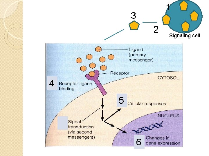
General principle signaling 1. 2. 3. 4. 5. 6. Synthesis of signaling molecules by the signaling cells Release of signaling molecules Transport of the signal to the target cell Detection of a signal by a specific receptor protein present on the target cell A change in cellular metabolism, function or development triggered by the receptor-signal complex Removal of the signal, which often terminate the cellular response
1 3 2 4 5 6 Signaling cell
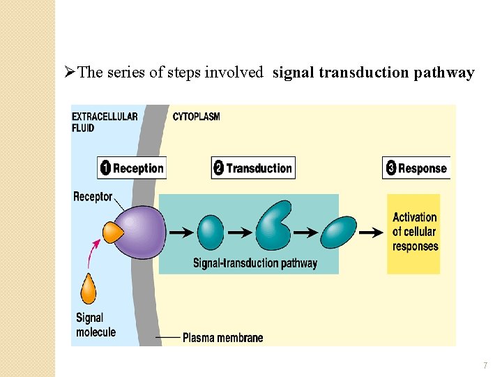
ØThe series of steps involved signal transduction pathway 7
Cellular responses due to cell signaling �Changes in the activity or function of specific enzymes and other proteins present in the cells �Changes in the amount of protein produced by a cell e. g. modification of transcription factors that stimulate or repress gene expression
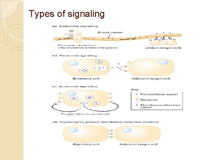
Types of signaling
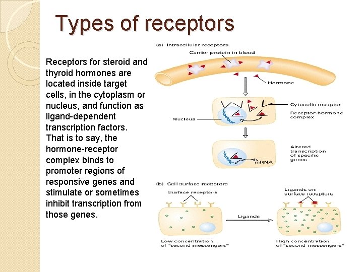
Types of receptors Receptors for steroid and thyroid hormones are located inside target cells, in the cytoplasm or nucleus, and function as ligand-dependent transcription factors. That is to say, the hormone-receptor complex binds to promoter regions of responsive genes and stimulate or sometimes inhibit transcription from those genes.
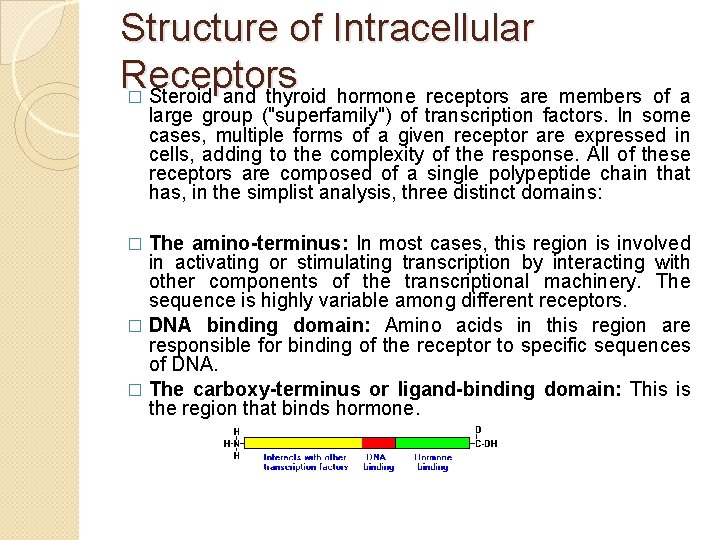
Structure of Intracellular Receptors � Steroid and thyroid hormone receptors are members of a large group ("superfamily") of transcription factors. In some cases, multiple forms of a given receptor are expressed in cells, adding to the complexity of the response. All of these receptors are composed of a single polypeptide chain that has, in the simplist analysis, three distinct domains: The amino-terminus: In most cases, this region is involved in activating or stimulating transcription by interacting with other components of the transcriptional machinery. The sequence is highly variable among different receptors. � DNA binding domain: Amino acids in this region are responsible for binding of the receptor to specific sequences of DNA. � The carboxy-terminus or ligand-binding domain: This is the region that binds hormone. �
NLS in Intracellular Receptors �In addition to three core domains, two other important regions of the receptor protein are a nuclear localization sequence, which targets the protein to nucleus, and a dimerization domain, which is responsible for latching two receptors together in a form capable of binding DNA.
Hormone-Receptor Binding and Interactions with DNA � Being lipids, steroid hormones enter the cell by simple diffusion across the plasma membrane. Thyroid hormones enter the cell by facilitated diffusion. The receptors exist either in the cytoplasm or nucleus, which is where they meet the hormone. When hormone binds to receptor, a characteristic series of events occurs: Receptor activation is the term used to describe conformational changes in the receptor induced by binding hormone. The major consequence of activation is that the receptor becomes competent to bind DNA. � Activated receptors bind to "hormone response elements", which are short specific sequences of DNA which are located in promoters of hormone-responsive genes. In most cases, hormone-receptor complexes bind DNA in pairs. � Transcription from those genes to which the receptor is bound is affected. Most commonly, receptor binding stimulates transcription. The hormone-receptor complex thus functions as a transcription factor. �
Classification of hormones �Lipophillic Hormones with intracellular receptors e. g steroid, thyroxine, retinoic acid �Hydrophillic with cell-surface receptors e. g peptide hormones (insulin growth factor and glucagon), small charge molecules (epinephrine and histamine) �Lipophillic with cell surface receptor e. g. prostaglandins
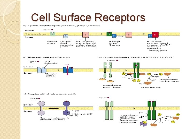
Cell Surface Receptors
Receptor protein exhibit ligand binding effect �Receptor present on Plasma or nuclear membrane has ligand binding sites �Signaling molecules (hormones, pheromones or neurotransmitters) act as ligands �Confirmational change occurs in the receptor that initiate a sequence of chemical reactions �Receptor proteins are specific for each horomone
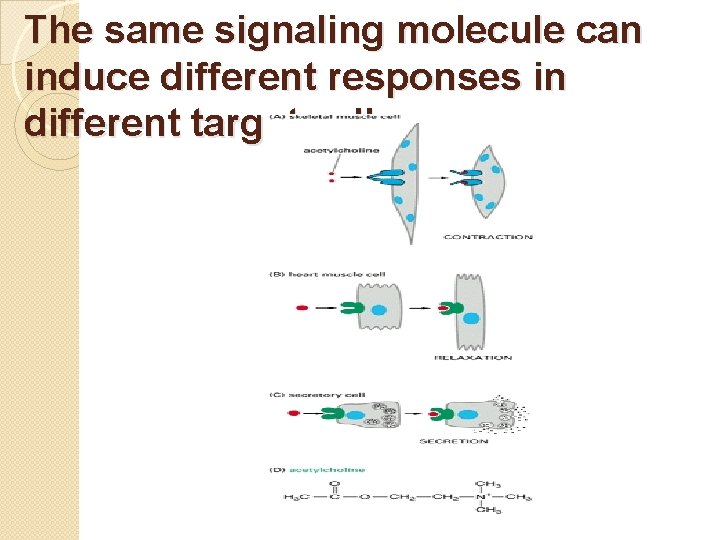
�Different cells have different sets of receptor for the same ligand each of which induces a different response �Different cells respond in a variety of way to the same ligand (e. g. acetylcholine) �Different ligands can induce the same cellular response in some cells (glucagon/epinephrine) �In most receptor-ligand system, the ligand do not have any function except to bind to receptor �Upon binding it changes the properties of receptor which then produce signals to the cell that a specific product is present �Target cells often degrade or modify the ligand to terminate or modify their response
The same signaling molecule can induce different responses in different target cells
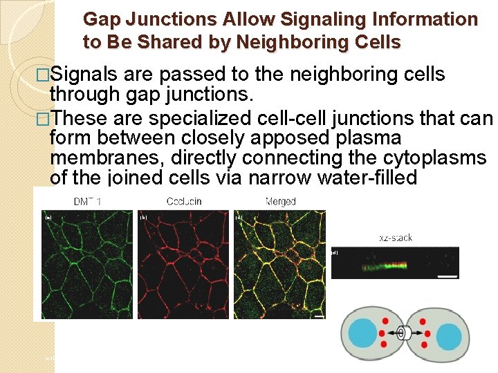
Gap Junctions Allow Signaling Information to Be Shared by Neighboring Cells �Signals are passed to the neighboring cells through gap junctions. �These are specialized cell-cell junctions that can form between closely apposed plasma membranes, directly connecting the cytoplasms of the joined cells via narrow water-filled channels. (a) (b) (c) (d) (c 1) (c 2) (c 3) (c 4) (c 5)
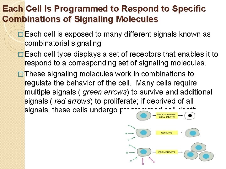
Each Cell Is Programmed to Respond to Specific Combinations of Signaling Molecules � Each cell is exposed to many different signals known as combinatorial signaling. � Each cell type displays a set of receptors that enables it to respond to a corresponding set of signaling molecules. � These signaling molecules work in combinations to regulate the behavior of the cell. Many cells require multiple signals ( green arrows) to survive and additional signals ( red arrows) to proliferate; if deprived of all signals, these cells undergo programmed cell death.
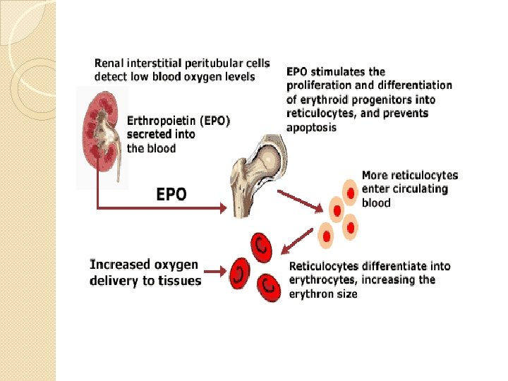
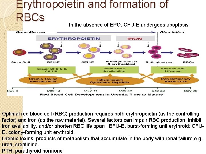
Erythropoietin and formation of RBCs In the absence of EPO, CFU-E undergoes apoptosis Optimal red blood cell (RBC) production requires both erythropoietin (as the controlling factor) and iron (as the raw material). Several factors can impair RBC production; inhibit iron availability, and/or shorten RBC life span. BFU-E, burst-forming unit erythroid; CFUE, colony-forming unit erythroid. Uremic toxins: products of metabolism that accumulate in the body with renal failure e. g. urea, creatinine PTH: parathyroid hormone
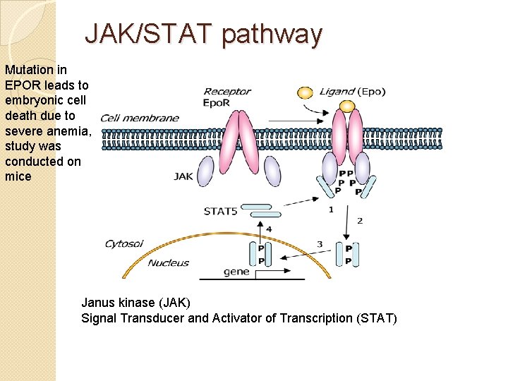
JAK/STAT pathway Mutation in EPOR leads to embryonic cell death due to severe anemia, study was conducted on mice Janus kinase (JAK) Signal Transducer and Activator of Transcription (STAT)
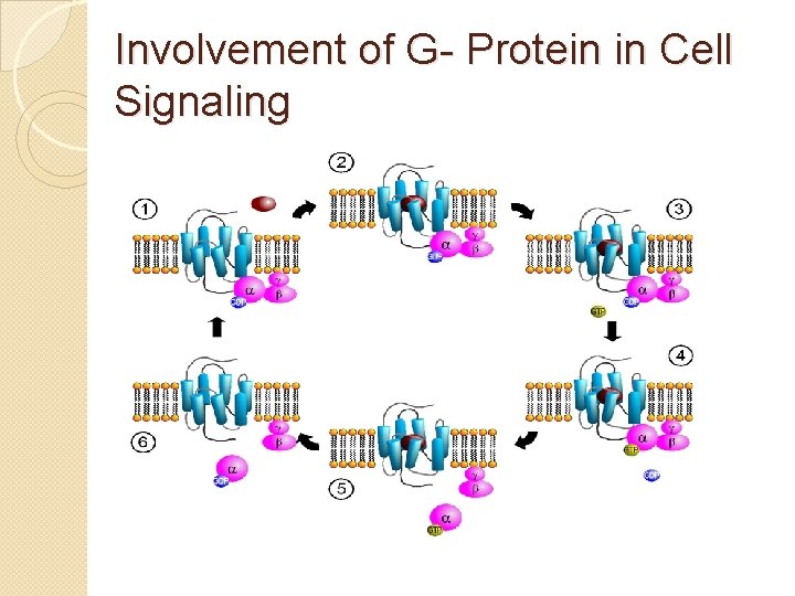
Involvement of G- Protein in Cell Signaling
General elements of GPCRs �Most abundant class of receptors �Found in organisms from yeast to man 1. A receptor with 7 membrane-spanning domains 2. A coupled trimeric G protein 3. A membrane bound effector protein 4. Feedback regulation and desensitization of the signalling pathway 5. A 2 nd messenger present in many
�Second messengers are molecules that relay signals from receptors on the cell surface to target molecules inside the cell, in the cytoplasm or nucleus. �These components of GPCRs can be mixed and matched to achieve an astonishing number of different pathways �GPCR pathways usually have short term effects in the cells �Allow the cells to respond rapidly to a variety of signals like environmental stimuli (light) or hormonal stimuli (epinephrine)
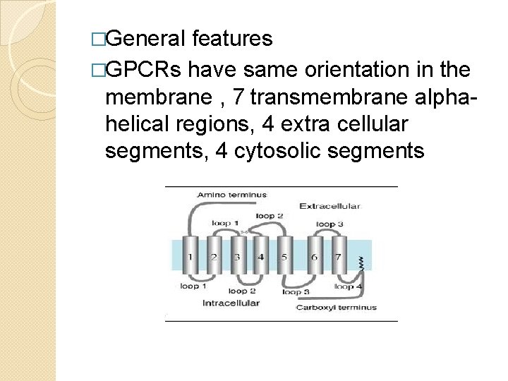
�General features �GPCRs have same orientation in the membrane , 7 transmembrane alphahelical regions, 4 extra cellular segments, 4 cytosolic segments
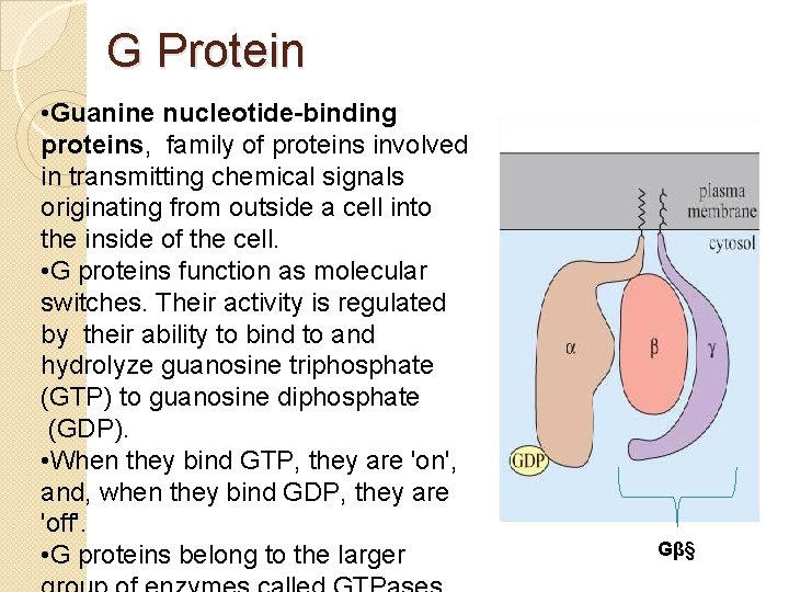
G Protein • Guanine nucleotide-binding proteins, family of proteins involved in transmitting chemical signals originating from outside a cell into the inside of the cell. • G proteins function as molecular switches. Their activity is regulated by their ability to bind to and hydrolyze guanosine triphosphate (GTP) to guanosine diphosphate (GDP). • When they bind GTP, they are 'on', and, when they bind GDP, they are 'off'. • G proteins belong to the larger Gβ§
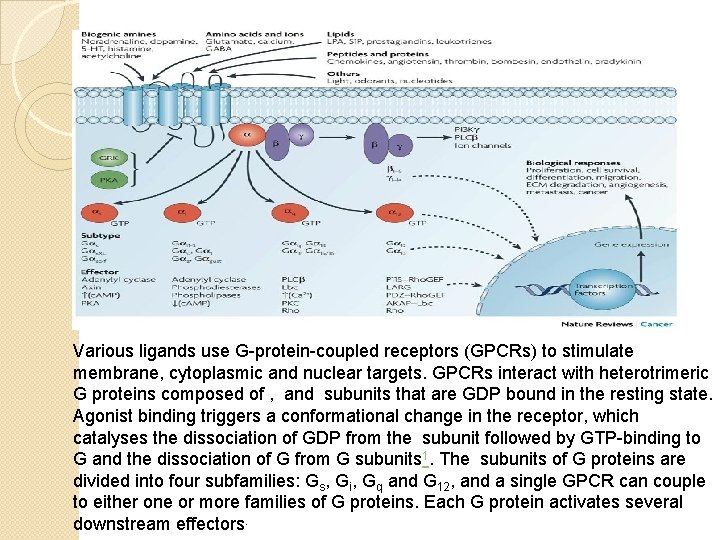
Various ligands use G-protein-coupled receptors (GPCRs) to stimulate membrane, cytoplasmic and nuclear targets. GPCRs interact with heterotrimeric G proteins composed of , and subunits that are GDP bound in the resting state. Agonist binding triggers a conformational change in the receptor, which catalyses the dissociation of GDP from the subunit followed by GTP-binding to G and the dissociation of G from G subunits 1. The subunits of G proteins are divided into four subfamilies: Gs, Gi, Gq and G 12, and a single GPCR can couple to either one or more families of G proteins. Each G protein activates several downstream effectors.
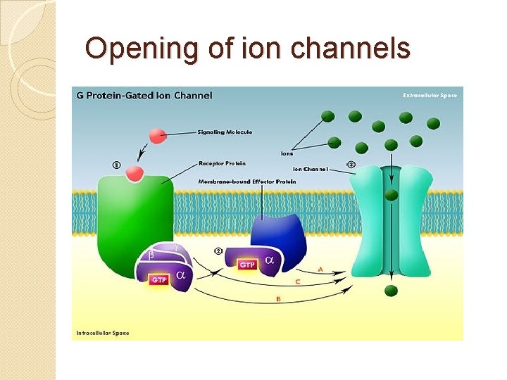
Opening of ion channels
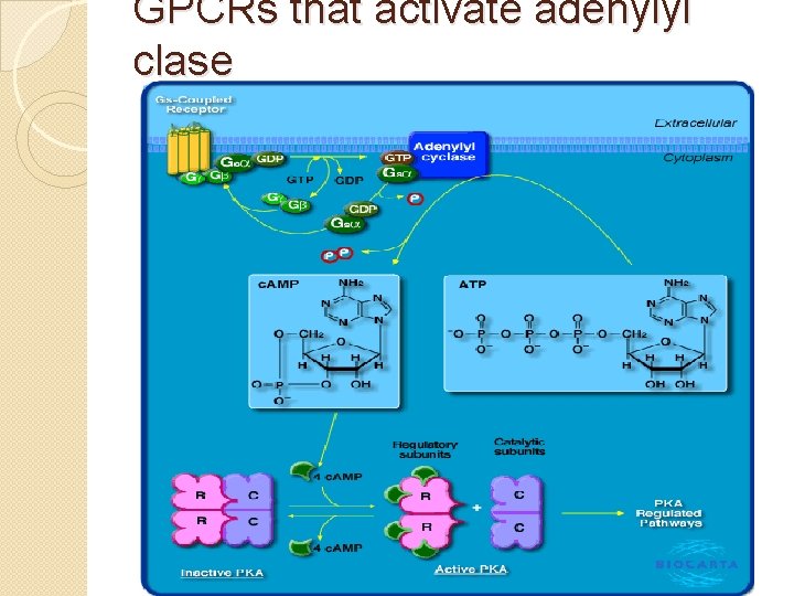
GPCRs that activate adenylyl clase
Lecture prepared from �The Cell: A Molecular Approach. 2 nd edition http: //www. ncbi. nlm. nih. gov/books/NBK 9898/ �Molecular Cell Biology, Lodish and co 5 Edition, Chapter 15