Human Dentition Introduction Dental Anatomy Includes 1 Nomenclature


 Upper First premolar & Second premolar Lower")


")


 The")
 “FDI” Federation Dentaire International First Digit = quadrant")

 “FDI” Federation Dentaire First. International Digit = quadrant")


 1. LABIAL is facial")


- Slides: 62
Human Dentition Introduction Dental Anatomy Includes: 1 - Nomenclature & terminology. 2 - The external morphology and internal composition of individual teeth. 3 -How tooth form serves its function. 4 - The relationship of teeth to each other and to the jaw bones.
Dental Anatomy Nomenclature The Jaws & Dental Arches
The Jaws and Dental Arches l A. The maxilla is two bones forming the upper jaw; they are rigidly attached to the skull. l B. The mandible is a horse-shoe shaped bone which articulates with the skull by way of the temporo-mandibular joint the TMJ.
Right Quadrants: right & left quadrants Left Maxillary right and left. Mandibular right and left. 4 Quadrants Right Left
Functions of Teeth 1 - Mastication: teeth are designed to perform this function. Incisors Canine Premolars Chisel like Wedge like At least two projections Cutting or Cutting (cusps). incising and Tearing and tearing grinding Molars Multiple projections (cusps) Grinding
2 - Appearance: - Well arranged clean teeth with proper alignment give nice appearance to the face. – Teeth give support to the facial expressions. 3 - Speech: for clear pronunciation and production of sound. 4 - Growth of jaws: The teeth play a role in the growth of the jaws in some periods of life.
Types of Teeth
Types of Teeth Anterior Teeth for cutting & tearing food Posterior Teeth
Anterior Teeth - Upper Canine lateral Central Incisors
Anterior Teeth - Lower Canine lateral - Central Incisors
Premolars: (in permanent only) Upper First premolar & Second premolar Lower
Molars: Upper First & second Lower Wisdom tooth = Third permanent molar
Types of Dentitions: Primary & Permanent Dentition
Types of Dentitions: I-Primary Dentition Deciduous , baby, milk teeth: a. Twenty ( 20) primary teeth. b. 10 in each arch c. 5 in each quadrant In function: 2 years 12 years
I-Primary Dentition
Mixed Dentition Period 6 years 12 years Eruption of first permanent molar Shedding of last primary molar
6 years eruption of first permanent molar
12 years Shedding of last primary molar
II-Permanent Dentition In function: 12 years through out life
Permanent Teeth
Tooth Identification Systems Numbering or Coding Systems 1. Palmer Notation System 2. Universal System 3. International FDI System 4. (two digit system)
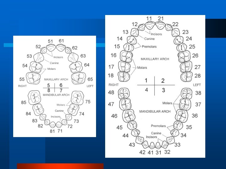
1 -Palmer Notation System for Permanent Teeth Right Left 8 -1 4 5 1 1 2 3 3 2 1 -8 4 5 6 6 7 7 8 8 8 7 6 8 -1 1 2 3 5 4 1 -8
It represents the four quadrants of the dentition as if you are facing the patient. In upper right In upper left In lower right In lower left Horizontal and vertical lines = symbol for the quadrant The permanent teeth are numbered from 1 -8 on each side from the midline. Upper right Upper left 8 7 6 5 4 3 2 1 1 2 3 4 5 6 7 8 Lower right Lower left
Palmer Notation System for Primary Teeth E-A A B C D A-E E E-A A-E
The deciduous teeth are lettered from A-E on each side from the midline Upper right Upper left E D C B A A B C D E Lower right Lower left
Palmer Notation System
2 -The International Numbering System “FDI” Federation Dentaire International (the two digit system) The teeth are designated by using two-digits: a. The first digit of the code is located at the left side of the number and indicates the quadrant: In permanent dentition In deciduous dentition U. R. 1 2 U. L. U. R. 5 6 L. R. 4 3 L. L. R. 8 7 U. L L. L.
2 -International System (Two Digit System) “FDI” Federation Dentaire International First Digit = quadrant 1 2 4 3 For permanent Teeth 11 21 22 12 13 23 14 24 15 25 16 26 17 27 28 18 48 38 37 47 36 46 35 45 44 34 43 42 41 31 3233
Second Digit = Tooth number in the quadrant b- The second digit is located at the right side of the number and indicates the number of the tooth in the quadrant. The two digits should be pronounced separately. Permanent teeth 18 17 16 15 14 13 12 11 21 22 23 24 25 26 27 28 48 47 46 45 44 43 42 41 31 32 33 34 35 36 37 38
2 -International System (Two Digit System) “FDI” Federation Dentaire First. International Digit = quadrant For Primary Teeth 52 8 62 53 63 54 64 55 First Digit = quadrant 5 51 61 6 85 7 84 5 6 8 7 65 75 74 83 82 81 71 72 73
For Primary Teeth 5 6 First Digit = quadrant 8 7 Second Digit = Tooth number in the quadrant Primary Teeth 55 54 53 52 51 61 62 63 64 65 85 84 83 82 81 71 72 73 74 75
Universal system for Permanent Teeth
Universal System for Primary Teeth
Macro & Micro-anatomy of Teeth crown neck root
Surrounding Bone: Crypt developing tooth Socket erupted tooth root
Anatomical Crown & Clinical Crown
Single-rooted Multi-rooted
Micro-anatomy of Teeth
Pulp Cavity 1 -Coronal pulp: • Pulp chamber • Pulp horns 2 -Radicular pulp: • Root canal • Apical foramen
Pulp Cavity in Root 2 -Radicular pulp: • Root canal • Apical foramen
Surfaces of Teeth
Surfaces of teeth are identified by the relationship to surrounding orofacial structures Each tooth has Five surfaces: 1. Facial 2. Lingual, 3. Mesial, and 4. Distal (proximal) 5. Functioning surf. ) Incisal) occlusal
FACIAL SURFACE any surface on the outside (towards the face) 1. LABIAL is facial surface of the anterior teeth )green , ( 2. BUCCAL is the facial surface of the posterior teeth (blue).
Inter-proximal surfaces: Surfaces in between two teeth: 1. Facing towards the midline: MESIAL. 2. Facing away from the midline: DISTAL
LINGUAL The surfaces facing the tongue PALATAL on the maxilla. palatal
Functioning Surfaces: chewing/biting surfaces of teeth: OCCLUSAL: chewing surfaces of posterior teeth INCISAL: the anteriors’ biting surface
Line and Point Angles Division into Thirds
Line and Point Angles Line angle: It is formed by the junction of two surfaces and its name is derived from both surfaces Point angle: It is formed by the junction of three surfaces and its name is derived from these surfaces.
Line Angles of Anterior Teeth
Line Angles of Posterior Teeth
Point Angles Meeting of 3 surfaces
Division into Thirds
Anatomical Landmarks of the Crown Developmental data: Lobe: • It is one of the primary centers of calcification and growth formed during the crown development. • Each tooth begins to develop from four lobes or more. • The pulp chamber has pulp horns corresponding to these lobes.
Anterior teeth premolars
Maxillary molars Mandibular molars
A- Crown Elevations: 1 -Mamelons They are three small rounded projections of enamel present in the incisal third of newly erupted incisors 2 -Cingulum It is the bulge on the cervical third of lingual surface of the crown in anterior teeth 3 -Cusps They are pyramidal projections on the incisal portion of the canine and on the occlusal surfaces of the premolars and molars
4 -Tubercle It is a small elevation produced by excessive formation of enamel. Tubercle is noticed at : • the palatal surface of E & 6 • sometimes at the lingual surface of incisors over the cingulum. N. B. Tubercle differs from cusp , it is formed of enamel only while cusp is formed of pulp horn covered by dentin and enamel.
Marginal 5 -Ridges Triangular Ridge or elevation of enamel forming the margin of the surface of a tooth; • on mesial /distal margins of lingual surfaces anterior teeth. • mesial & distal margins of occlusal surfaces posterior teeth Transverse Ridge Oblique Ridge Linear • Linear ridges: Elevated portions of tooth run in a line named for their location • Cervical • Incisal • Labial • Buccal • Lingual
B-Crown Depressions Linear Depressions Irregular Depressions 1 -Developmental grooves: 1 -Fossa Depression or concavity Shallow grooves that separate cusps or primary parts of teeth 2 - Supplemental grooves: • Lingual • Small , irregularly placed auxiliary grooves. fossa • Branches from developmental grooves. • They do not denote union of primary lobes. • The third molars followed by second permanent molars are characterized by high number of supplemental grooves. • triangular fossae • Central fossa
Linear Depressions 3 - Fissures • Found in the bottom of developmental grooves. • Result from incomplete union of the primary lobes. • Represent a fault in enamel. Irregular Depressions 2 - PITS: • b- FAULTY PIT: • Develop as a result • of incomplete formation of enamel. • It is located at the end of the buccal developmental grooves of the lower molars or palatal developmental grooves of the upper molars.