
HUMAN CHROMOSOMAL ABERRATIONS ITS IMPLICATIONS BY Prof S
HUMAN CHROMOSOMAL ABERRATIONS & ITS IMPLICATIONS BY Prof. S. R. RAMESH Department of Studies in Zoology UNIVERSITY OF MYSORE Manasagangotri, MYSORE – 570 006
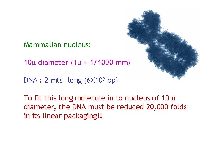
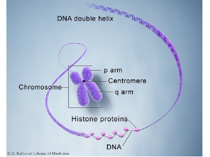
Mammalian nucleus: 10 diameter (1 = 1/1000 mm) DNA : 2 mts. long (6 X 10⁹ bp) To fit this long molecule in to nucleus of 10 diameter, the DNA must be reduced 20, 000 folds in its linear packaging!!
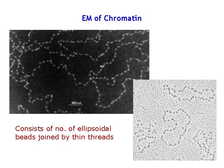
EM of Chromatin Consists of no. of ellipsoidal beads joined by thin threads
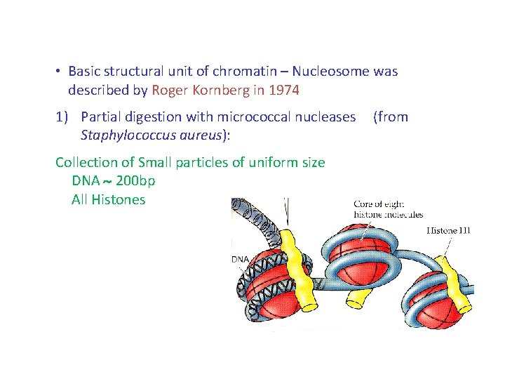
• Basic structural unit of chromatin – Nucleosome was described by Roger Kornberg in 1974 1) Partial digestion with micrococcal nucleases Staphylococcus aureus): Collection of Small particles of uniform size DNA 200 bp All Histones (from
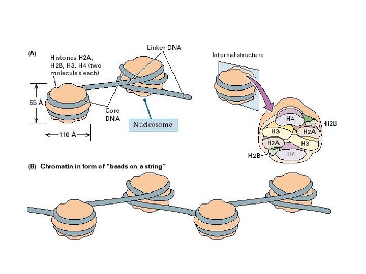
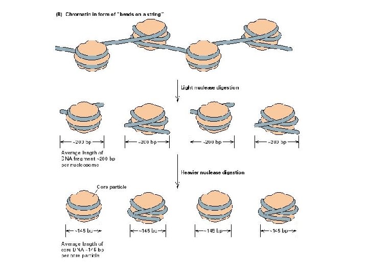
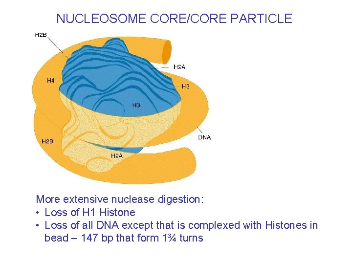
NUCLEOSOME CORE/CORE PARTICLE More extensive nuclease digestion: • Loss of H 1 Histone • Loss of all DNA except that is complexed with Histones in bead – 147 bp that form 1¾ turns
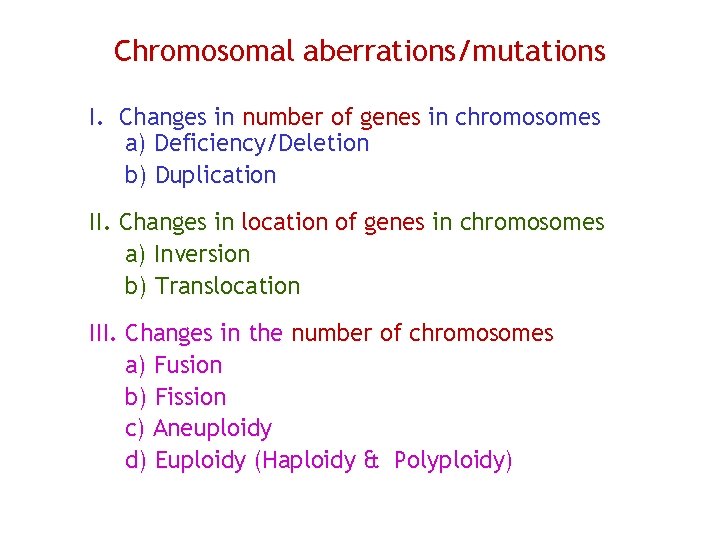
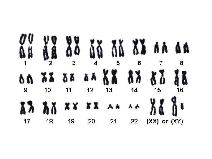
Chromosomal aberrations/mutations I. Changes in number of genes in chromosomes a) Deficiency/Deletion b) Duplication II. Changes in location of genes in chromosomes a) Inversion b) Translocation III. Changes in the number of chromosomes a) Fusion b) Fission c) Aneuploidy d) Euploidy (Haploidy & Polyploidy)
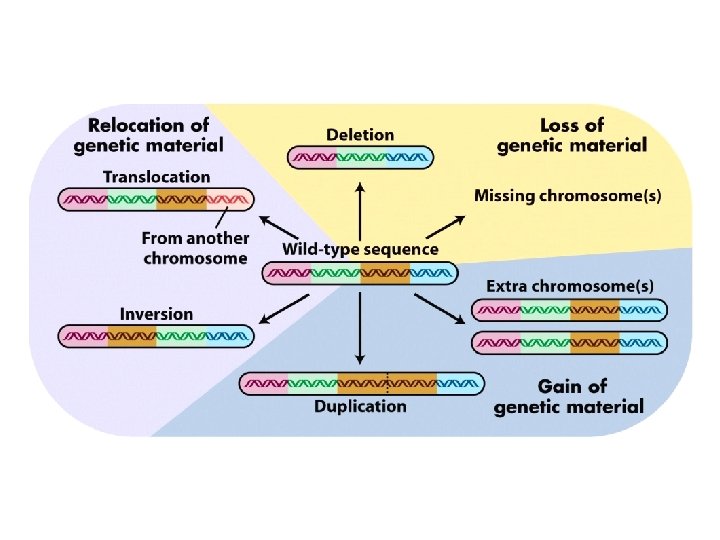
May occur spontaneously under natural conditions ……Rare events Could be induced by Physical agents/ Chemicals/Ionizing Radiations May produce changes in • Phenotype • Expected genetic ratios • Linkage relationships of certain genes
Deletions/Deficiencies • Chromosome/Chromatin breaks • Types: a) Size: Small: Intragenic • Inefficient product • Truncated peptide - Nonfunctional • Inactivate gene Large: Multigenic (Usually lethal) b) Position: Terminal (Single break) Interstitial (Two breaks – followed by ‘Healing’)
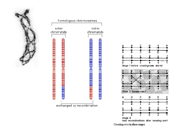
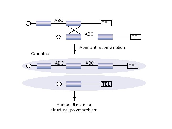
GENETIC CONSEQUENCES OF DELETIONS • Failure of the chromosome/individual to survive as a homozygote (also occurs in lethal mutation) • Chromosomes/individuals with deletions can never revert to normal (can be detected/ realized only when a specific phenotype is associated with deletion)
• Deletion of a region (segment) on one homologue results in the expression of recessive alleles present on the other homologue – leading to unexpected phenotype in the progeny • This apparent dominance of a recessive gene is called PSEUDODOMINANCE • This is one way to recognize deletions Expression of Waltzing in mice Eg.
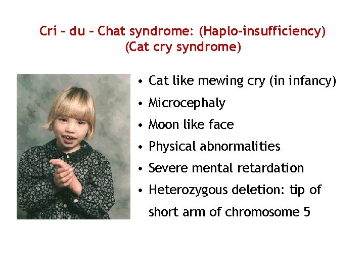
Cri – du – Chat syndrome: (Haplo-insufficiency) (Cat cry syndrome) • Cat like mewing cry (in infancy) • Microcephaly • Moon like face • Physical abnormalities • Severe mental retardation • Heterozygous deletion: tip of short arm of chromosome 5
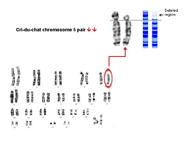
Cri-du-chat chromosome 5 pair
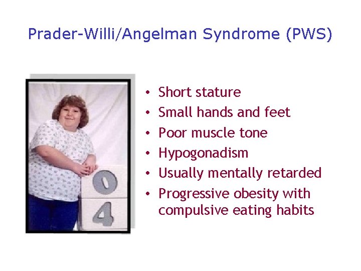
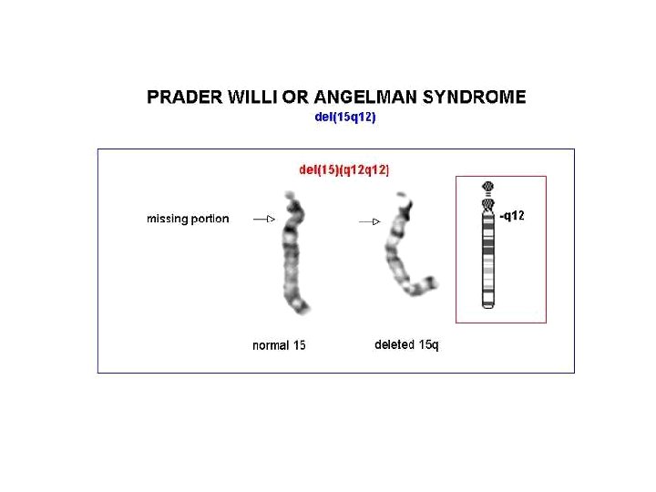
Prader-Willi/Angelman Syndrome (PWS) • • • Short stature Small hands and feet Poor muscle tone Hypogonadism Usually mentally retarded Progressive obesity with compulsive eating habits

Wolf-Hirschhorn syndrome/Pitt-Rogers-Dank syndrome) • Low birth weight - 2 kg • Microcephaly • High forehead • Broad nose in prolongation of the eyebrows line • Ocular malformations, hare-lip / cleft palate • Long, slender, manicured fingers Malformations: heart (50%) • IQ = 20; seizures; often bedridden Karyotype: deletion of band 4 p 16 gives full phenotype; critical segment narrowed to 200 kb.
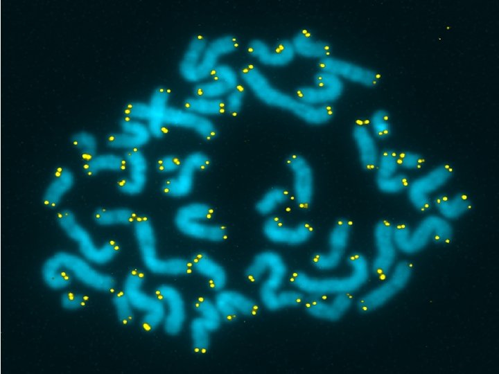
Microdeletion syndromes: • Deletion is missing a part of chromosome delay in developmental milestones in children mental retardation/dysmorphic physical features or both • Very very small deletions viz. , ‘microdeletions’ cannot be seen by routine analysis chromosomes appear normal • Can be the cause of specific microdeletion syndromes • Can be detected by FISH - Smith Magenis syndrome (SMS)
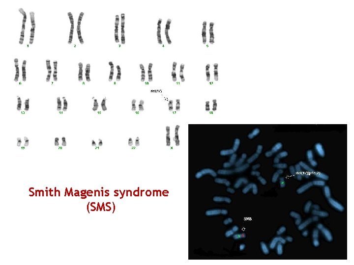
Smith Magenis syndrome (SMS)
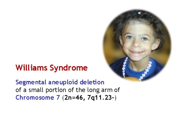
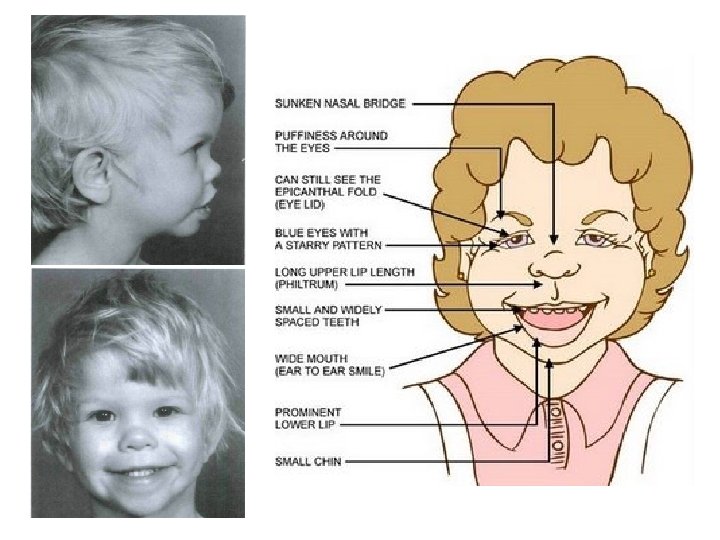
Williams Syndrome Segmental aneuploid deletion of a small portion of the long arm of Chromosome 7 (2 n=46, 7 q 11. 23 -)
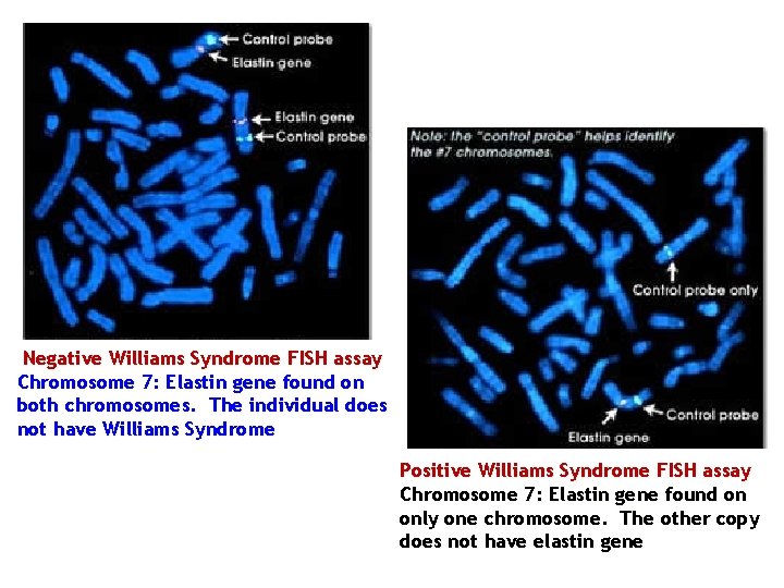
Negative Williams Syndrome FISH assay Chromosome 7: Elastin gene found on both chromosomes. The individual does not have Williams Syndrome Positive Williams Syndrome FISH assay Chromosome 7: Elastin gene found on only one chromosome. The other copy does not have elastin gene
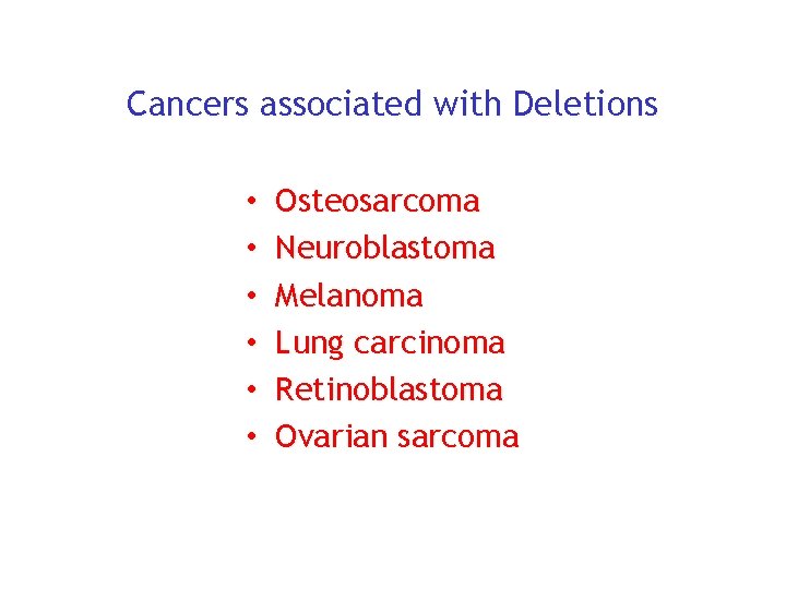
Cancers associated with Deletions • • • Osteosarcoma Neuroblastoma Melanoma Lung carcinoma Retinoblastoma Ovarian sarcoma
• Large deletions: Often lethal/seriously deleterious even in heterozygotes presumably due to developmental imbalance • Deletions of small chromosomal regions might not be lethal in homozygotes!! Whenever deleted genes had been previously duplicated ……. . and their function could still be carried out by the remaining genes
GENETIC CONSEQUENCES OF DUPLICATIONS
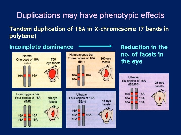
Duplications may have phenotypic effects Tandem duplication of 16 A in X-chromosome (7 bands in polytene) Incomplete dominance Reduction in the no. of facets in the eye
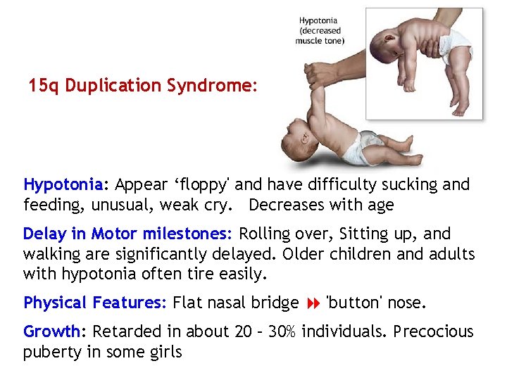
15 q Duplication Syndrome: Hypotonia: Appear ‘floppy' and have difficulty sucking and feeding, unusual, weak cry. Decreases with age Delay in Motor milestones: Rolling over, Sitting up, and walking are significantly delayed. Older children and adults with hypotonia often tire easily. Physical Features: Flat nasal bridge 'button' nose. Growth: Retarded in about 20 – 30% individuals. Precocious puberty in some girls
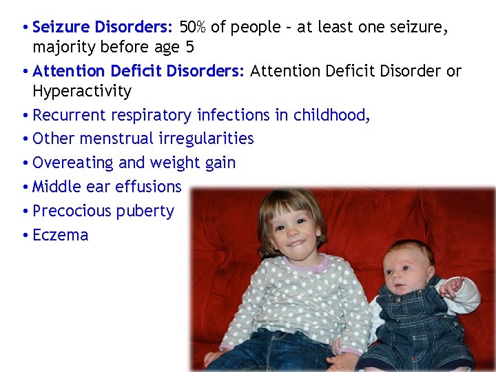
• Seizure Disorders: 50% of people – at least one seizure, majority before age 5 • Attention Deficit Disorders: Attention Deficit Disorder or Hyperactivity • Recurrent respiratory infections in childhood, • Other menstrual irregularities • Overeating and weight gain • Middle ear effusions • Precocious puberty • Eczema
22 q 11. 2 duplication syndrome (2. 09 – 3. 06 Mb): Deletion in same region Di. George syndrome Most frequent reported symptoms: • Mental retardation/learning disabilility (97%) • Delayed psychomotor development (67%) • Growth retardation (63%) • Muscular hypotonia (43%) • Congenital heart malformation • Visual and hearing impairment • Seizures, microcephaly, ptosis, & urogenital abnormalities
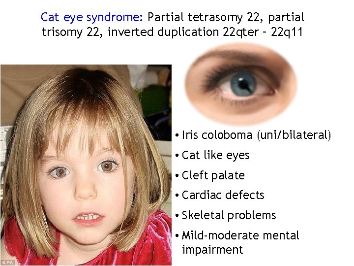
Cat eye syndrome: Partial tetrasomy 22, partial trisomy 22, inverted duplication 22 qter – 22 q 11 • Iris coloboma (uni/bilateral) • Cat like eyes • Cleft palate • Cardiac defects • Skeletal problems • Mild-moderate mental impairment
Multiple copies of r. RNA genes E. coli : 7 r. RNA genes Human: 400 r. RNA genes/genome (on 5 chromosomes) Xenopus: 1200 r. RNA genes/genome (Single cluster)
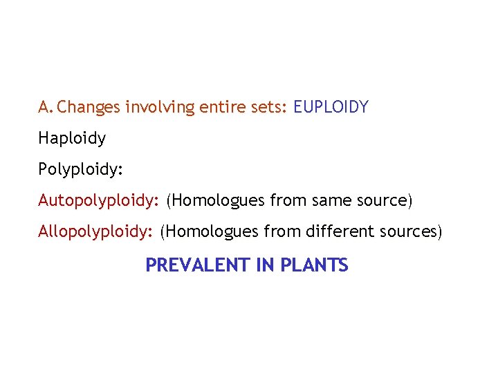
A. Changes involving entire sets: EUPLOIDY Haploidy Polyploidy: Autopolyploidy: (Homologues from same source) Allopolyploidy: (Homologues from different sources) PREVALENT IN PLANTS
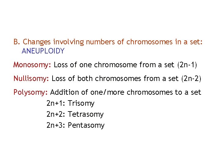
B. Changes involving numbers of chromosomes in a set: ANEUPLOIDY Monosomy: Loss of one chromosome from a set (2 n-1) Nullisomy: Loss of both chromosomes from a set (2 n-2) Polysomy: Addition of one/more chromosomes to a set 2 n+1: Trisomy 2 n+2: Tetrasomy 2 n+3: Pentasomy
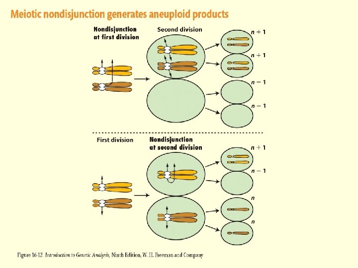
Figure 16 -12
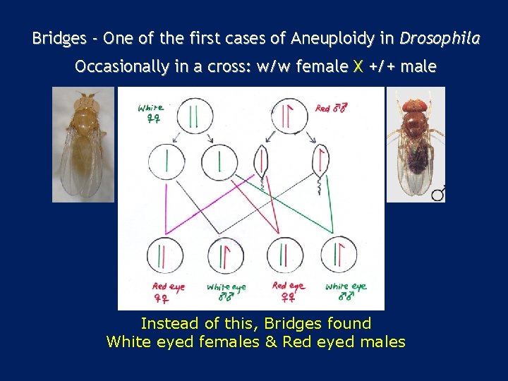
Bridges - One of the first cases of Aneuploidy in Drosophila Occasionally in a cross: w/w female X +/+ male Instead of this, Bridges found White eyed females & Red eyed males
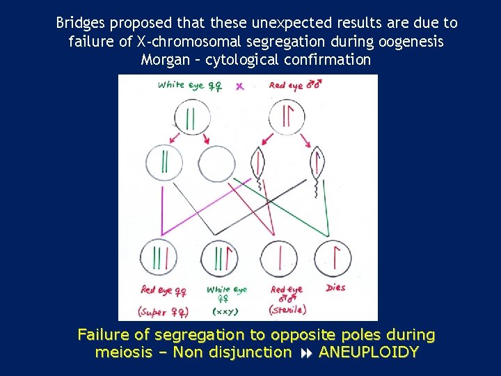
Bridges proposed that these unexpected results are due to failure of X-chromosomal segregation during oogenesis Morgan – cytological confirmation Failure of segregation to opposite poles during meiosis – Non disjunction ANEUPLOIDY
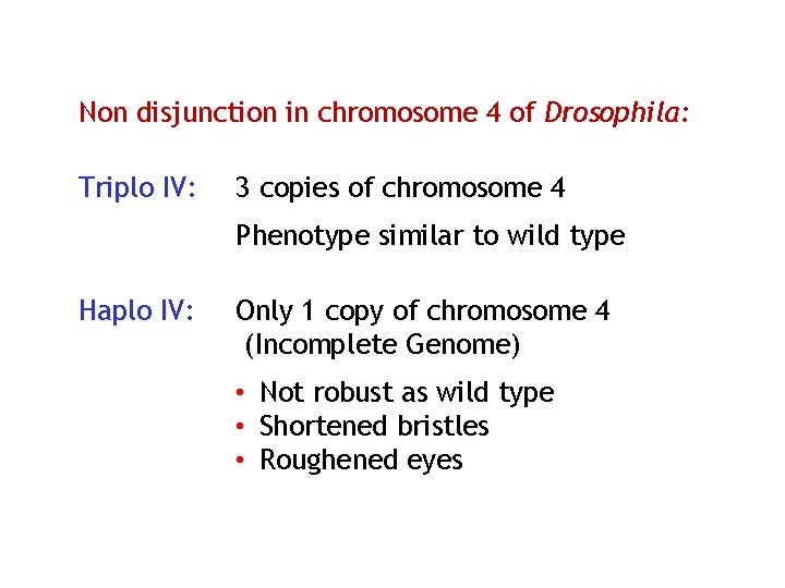
Non disjunction in chromosome 4 of Drosophila: Triplo IV: 3 copies of chromosome 4 Phenotype similar to wild type Haplo IV: Only 1 copy of chromosome 4 (Incomplete Genome) • • • Not robust as wild type Shortened bristles Roughened eyes
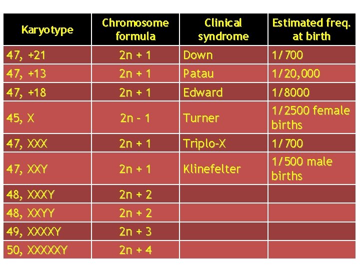
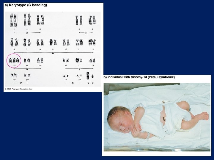
Karyotype Chromosome formula Clinical syndrome Estimated freq. at birth 47, +21 2 n + 1 Down 1/700 47, +13 2 n + 1 Patau 1/20, 000 47, +18 2 n + 1 Edward 45, X 2 n – 1 Turner 47, XXX 2 n + 1 Triplo–X 1/8000 1/2500 female births 1/700 47, XXY 2 n + 1 48, XXXY 2 n + 2 48, XXYY 2 n + 2 49, XXXXY 2 n + 3 50, XXXXXY 2 n + 4 Klinefelter 1/500 male births
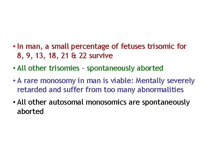
• In man, a small percentage of fetuses trisomic for 8, 9, 13, 18, 21 & 22 survive • All other trisomies – spontaneously aborted • A rare monosomy in man is viable: Mentally severely retarded and suffer from too many abnormalities • All other autosomal monosomics are spontaneously aborted
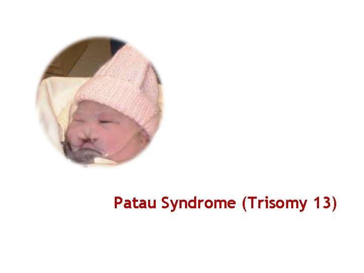
Patau Syndrome (Trisomy 13)
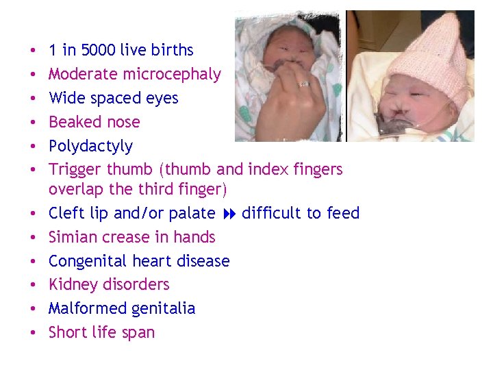
• • • 1 in 5000 live births Moderate microcephaly Wide spaced eyes Beaked nose Polydactyly Trigger thumb (thumb and index fingers overlap the third finger) Cleft lip and/or palate difficult to feed Simian crease in hands Congenital heart disease Kidney disorders Malformed genitalia Short life span
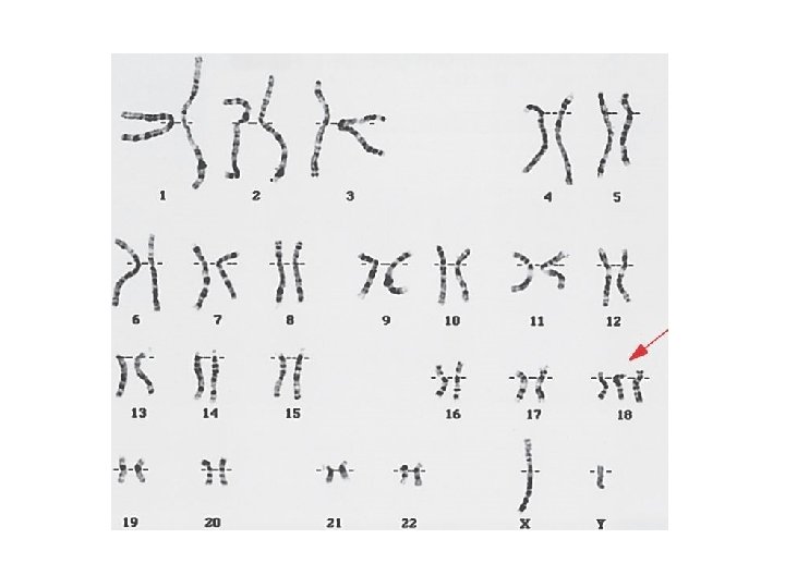
Edward’s syndrome (Trisomy 18)
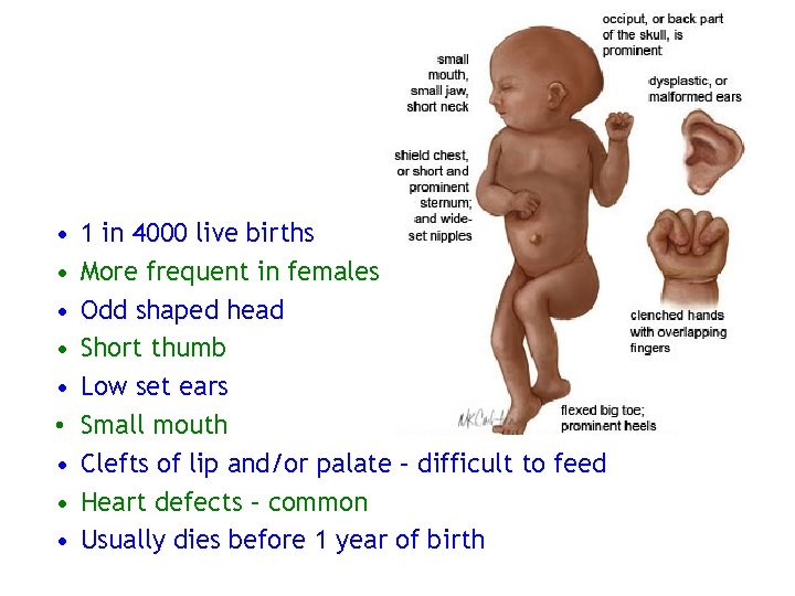
• • • 1 in 4000 live births More frequent in females Odd shaped head Short thumb Low set ears Small mouth Clefts of lip and/or palate – difficult to feed Heart defects – common Usually dies before 1 year of birth
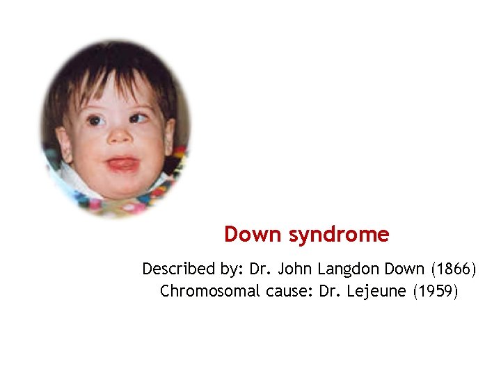
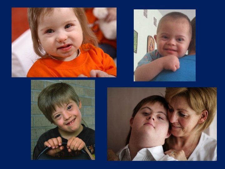
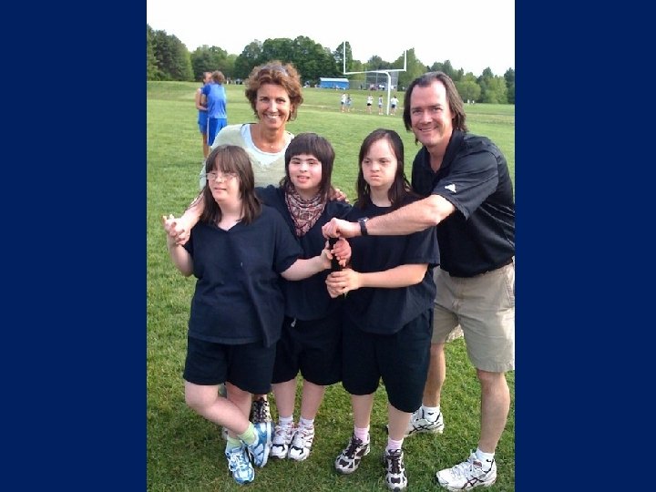
Down syndrome Described by: Dr. John Langdon Down (1866) Chromosomal cause: Dr. Lejeune (1959)
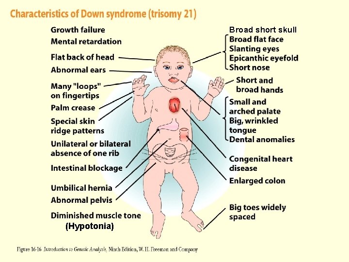
Figure 16 -16 (Hypotonia) Broad short skull
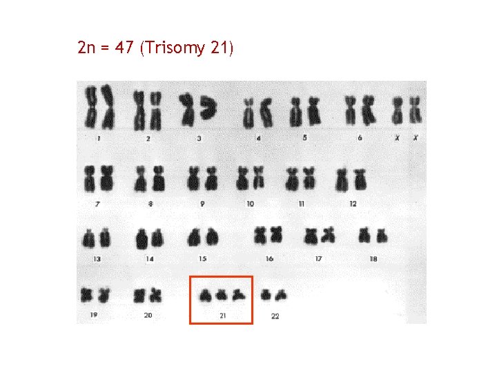
2 n = 47 (Trisomy 21)
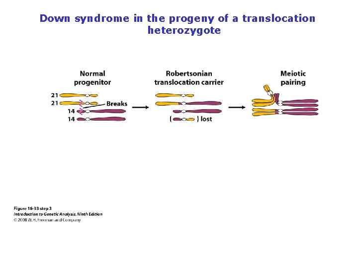
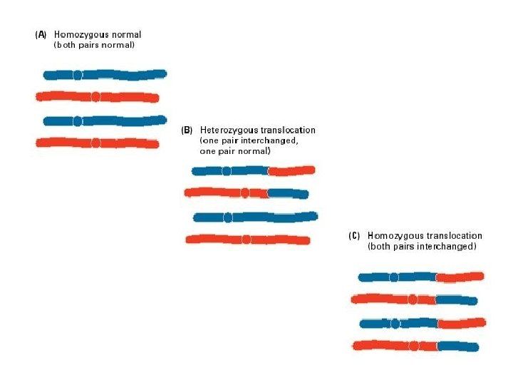
Down syndrome in the progeny of a translocation heterozygote
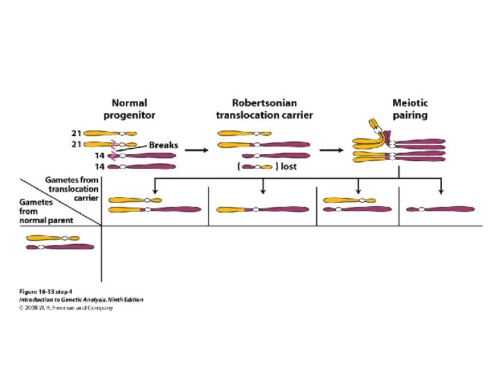
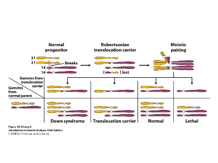
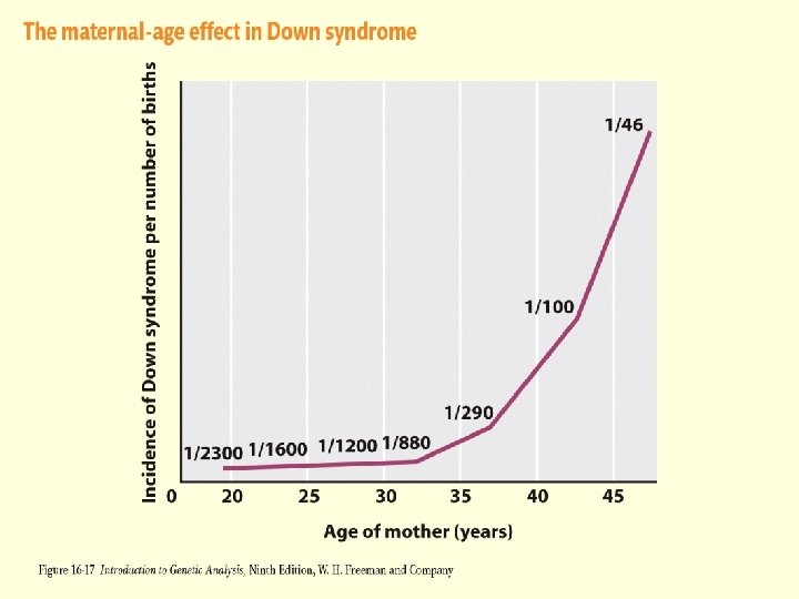
Figure 16 -17
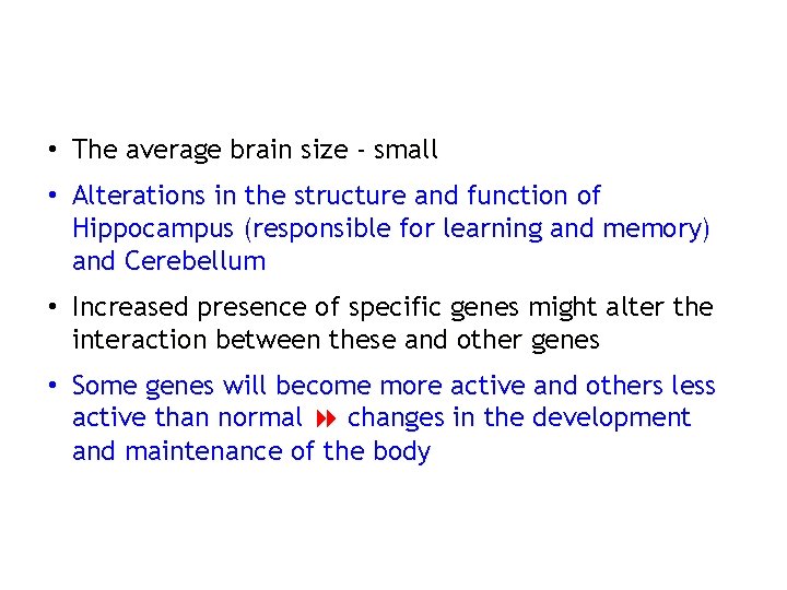
• The average brain size - small • Alterations in the structure and function of Hippocampus (responsible for learning and memory) and Cerebellum • Increased presence of specific genes might alter the interaction between these and other genes • Some genes will become more active and others less active than normal changes in the development and maintenance of the body
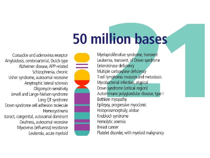
• Chromosome 21 – 60, 000 kb DNA • Consensus region for Downs – Distal half of long arm close to SOD-1 locus • Among some gene loci mapped to long arm of chromosome 21, 3 are present on chromosome 16 of mouse! this region is evolutionarily conserved between mouse and man!? ! • Is there a Downs region in this common segment? ?
Downs mouse (Trisomy 16): • Cardiovascular malformation • Reduced brain weight • Relatively short lived Chimps & Orangutan (Trisomy 22, homologous to human chromosome 21): • Phenotype resemble that of Human Down syndrome
Turner syndrome Dr. Turner (1938) – described as incompletely developed females
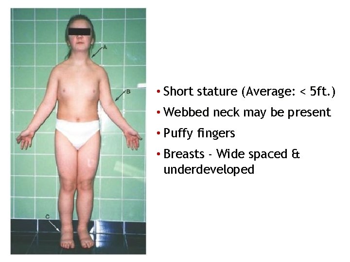
• Short stature (Average: < 5 ft. ) • Webbed neck may be present • Puffy fingers • Breasts - Wide spaced & underdeveloped
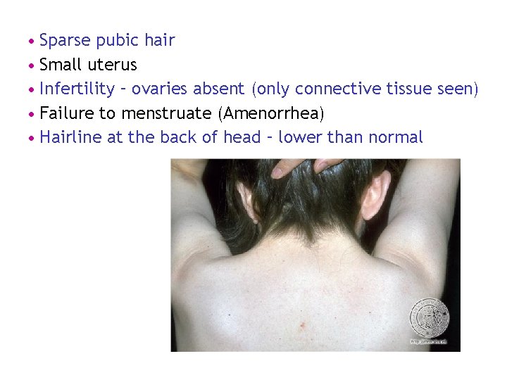
• Sparse pubic hair • Small uterus • Infertility – ovaries absent (only connective tissue seen) • Failure to menstruate (Amenorrhea) • Hairline at the back of head – lower than normal
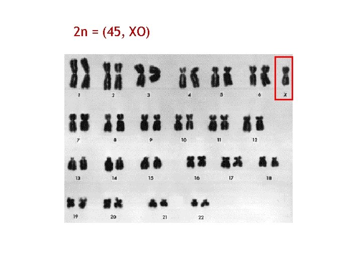
2 n = (45, XO)
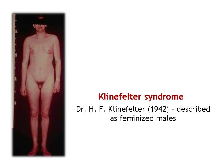
Klinefelter syndrome Dr. H. F. Klinefelter (1942) – described as feminized males
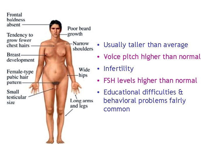
• Usually taller than average • Voice pitch higher than normal • Infertility • FSH levels higher than normal • Educational difficulties & behavioral problems fairly common
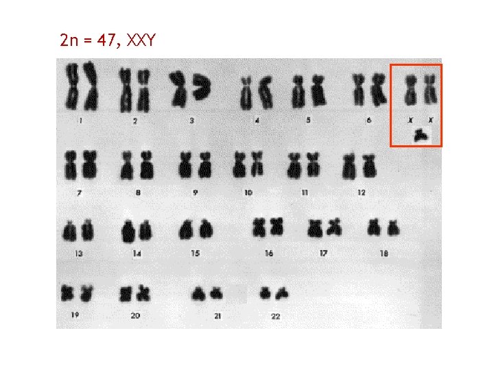
2 n = 47, XXY
Several kinds of cancers such as: 1. Chronic lymphocytic leukemia 2. Small lymphocytic lymphoma 3. Acute monocytic leukemia 4. Acute myelomonocytic leukemia 5. Acute nonlymphocytic leukemia are associated with occurrence of inversions
Transfer of chromosomal segments between Non -homologous chromosomes 1. Reciprocal (Symmetrical) – Exchange 2. Non- Reciprocal (Non-Symmetrical) - One way transfer
Patients with Salivary gland tumors Ovarian carcinoma Burkitt’s lymphoma Acute lymphocytic leukemia HAVE TRANSLOCATIONS !!
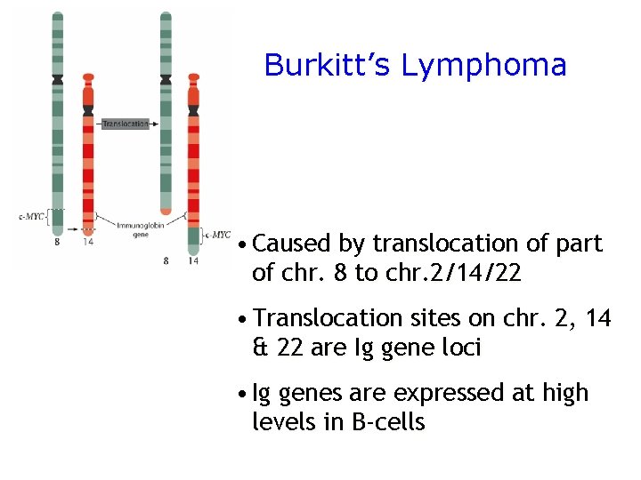
Burkitt’s Lymphoma • Caused by translocation of part of chr. 8 to chr. 2/14/22 • Translocation sites on chr. 2, 14 & 22 are Ig gene loci • Ig genes are expressed at high levels in B-cells
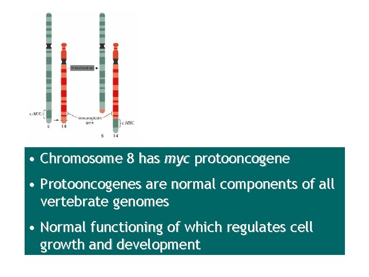
• Chromosome 8 has myc protooncogene • Protooncogenes are normal components of all vertebrate genomes • Normal functioning of which regulates cell growth and development
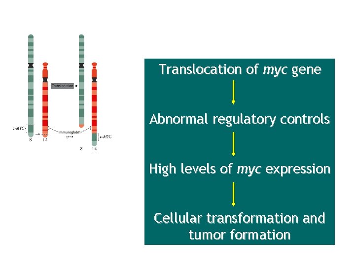
Translocation of myc gene Abnormal regulatory controls High levels of myc expression Cellular transformation and tumor formation
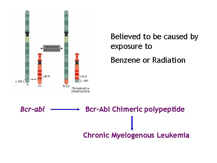
Believed to be caused by exposure to Benzene or Radiation Bcr-abl Bcr-Abl Chimeric polypeptide Chronic Myelogenous Leukemia
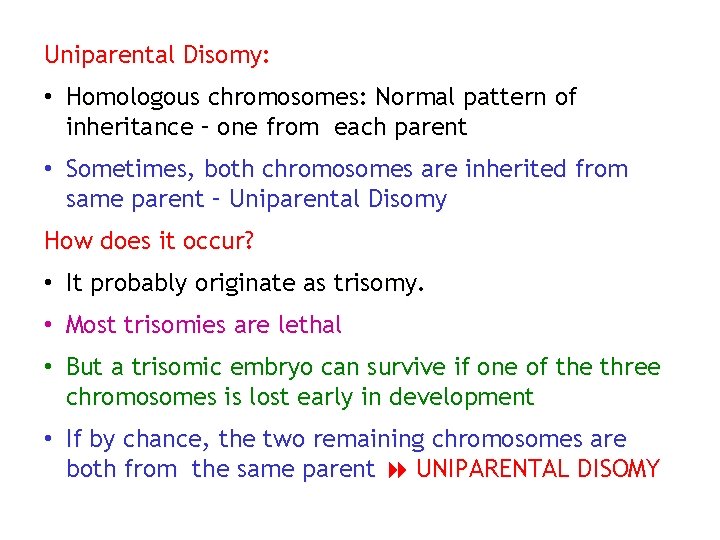
Uniparental Disomy: • Homologous chromosomes: Normal pattern of inheritance – one from each parent • Sometimes, both chromosomes are inherited from same parent – Uniparental Disomy How does it occur? • It probably originate as trisomy. • Most trisomies are lethal • But a trisomic embryo can survive if one of the three chromosomes is lost early in development • If by chance, the two remaining chromosomes are both from the same parent UNIPARENTAL DISOMY
- Slides: 82