
HUMAN ANATOMY PHYSIOLOGY BY DR STELLA SELENA M

HUMAN ANATOMY & PHYSIOLOGY BY: DR. STELLA SELENA M. B. B. S. , DGO. ACADEMY DIRECTOR MOTHER TERESA INSTITUTE OF PHARMACEUTICAL EDUCATION AND RESEARCH
Two Branches Of Science Anatomy : Is the science of the body structures and their relationships among them Ana : up tomy cutting It was first studied by dissection Physiology: Physio = Nature, logy = study of Is the science of body functions from body parts - work

Sub Disciplines Of Anatomy q. Gross anatomy Structures/ body parts can be examined without using microscope q. Surface anatomy Anatomical land marks on surface of body by visualization / palpation q. Systemic anatomy Structure of specific systems of the body ex: Nervous system, Respiratory system q. Regional anatomy head or chest. Specific region of the body such as

Radio-graphic anatomy: Radio-ray Graphic- To write Body structures that are visualized with X – Rays Histology: Hist – Tissue Microscopic structure of tissues Embryology: Embryo Structure that emerge from the time of the fertilized egg through the eight week into utero Pathological anatomy: disease Structural changes associated with
Sub disciplines of physiology: Neurophysiology Neuro-nerve Functional properties of new cells Endocrinology Endo: with Crine: Secretion Hormones(chemical regulations in the blood) how they control body functions Cardiovascular Physiology Cardi : Heart Vascular : Blood vessels Functions of heart and blood vessels
Immunology: Immuno : not suseptable How body depends itself against disease causing agents Respiratory Physiology: Respira : To breathe Functions of air passages and lungs Renal Physiology : Ren: kidney Functions of kidney Patho physiology: Patho : Disease Functional changes associated with disease and aging
Basic Terminology : Anatomical Terminology To communicate clearly scientists and health care people professionals use common language of special terms when referring the body structure and their functions. Body positions: Descriptions of any origin/part of human body in a specific stance called – anatomical position. In anatomical position: 1. 2. 3. 4. 5. Body upright Stands erect facing observer with the head level and the eyes facing directly forward Feet are flat on the floor and Arms at sides Palms turned forward
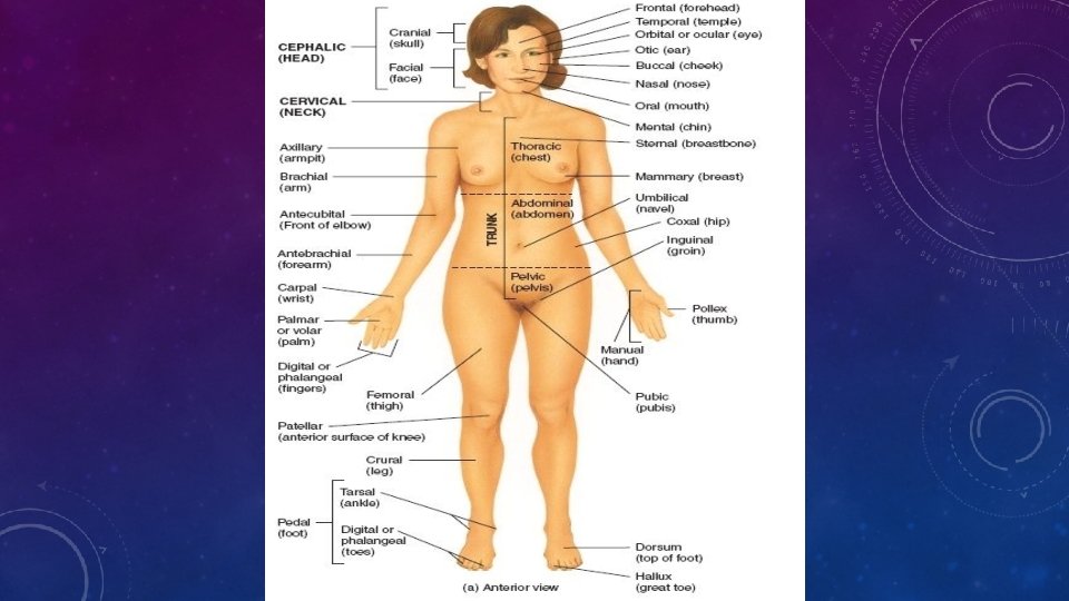
Two terms described a reclining body Prone position: If a body is lying face down Superior position: If body is lying face up Regional names: Human body is divided into several major regions Head – consists of skull and face (is front position of head includes eyes, nose, mouth, cheeks, forehead and chin) Neck – Supports head and attaches it to trunk Trunk – Consists of chest, abdomen and pelvis

Upper limbs – Each upper limb attaches to trunk and consists shoulders Lower limbs – Attaches to the trunk and consists of Buttock, thighs, leg, ankle and foot & arm pit, arm, fore arm, wrist and hand Groin: Area on the front surface of the body marked by a crease on each side when trunk attaches to the thighs. Directional terms: To locate various body structures specific directional lens and in use to describe the position of one body part to other. They are in pairs that have opposite meanings like - Anterior(front) : Posterior (back)
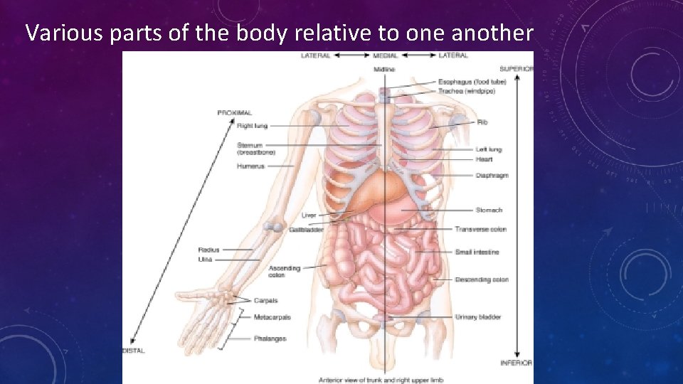
Various parts of the body relative to one another

Directional Terms Definitions Examples Superior (cranial) Towards head Heart superior to liver Inferior (cardial) lower part away from head Stomach inferior to lungs Anterior( ventral ) Front of the body Sterinum (breast bone)anterior to head Posterior (Dorsal) Back of the body Oesophagus posterior to trachea Medial Nearer to midline Ulna is medial to radius Lateral Further from midline Lungs lateral to heart Intermediate Between two structures Transverse colon is intermediate to ascending and descending colons Ipsilateral on the same side of the body Gall bladder and ascending colon are ipsilateral

Contraletaral on opposite side of the body Proximal nearest to attachment of limb to the trunk nearer to origination of structure failure from attachment failure from origination of structure. towards or on the body surface Away from the surface of the body Distal Superficial Deep Ascending and d desecending color are contraletive Hermecus to proximal to radius Phalanges are distal to the carpil Ribs are superficial to the lungs Ribs are deep to the skin of the chest and back.
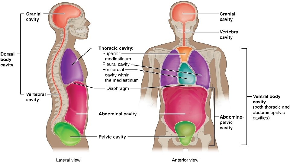
Body Cavities: Spaces within the body that help to protect, separate and support internal organs. Cavities: Cranial cavity formed by cranial bones contains brain Vertebral cavity formed by vertebral column and contains spinal cord and beginnings of spinal nerves
Thoracic Cavity Chest cavity; contains pleural and pericardial cavities and mediastinum Pleural Cavity Each surounds a lung; the serous membrane of the pleural cavities is the pleura. Pericardial Cavity Surrounds the heart; the serous membrane of the pericardial cavity is the pericardium. Mediastinum Central portion of thoracic cavity between the lungs; extends from sternum to vertebral column and from neck to diaphragm; contains heart, thymus, esophagus, trachea, and several large blood vessels.
Abdominopelvic cavity and pelvic cavities - Subdivided into abdominal Abdominal Cavity Contains stomach, spleen, liver, gallbladder, small intestine, and most of large intestine; the serous membrane of the abdominal cavity is the peritoneum. Pelvic cavity Contains urinary bladder, portions of large intestine and internal organs of reproduction.
- Slides: 18