HUMAN ANATOMY LECTURE TWENTY URINARY SYSTEM FUNCTIONS Excretion


 on the posterior")


- Slides: 17
HUMAN ANATOMY LECTURE TWENTY URINARY SYSTEM
FUNCTIONS • Excretion - removing waste products from the blood • Blood volume control - regulating urine production (H 2 O loss) • Ion concentration regulation - Na+, Cl-, K+, Ca+2, HPO 4 -2 • p. H regulation - p. H of extracellular fluid by controlling H+ and HCO 3 loss in urine • Nutrient conservation - preventing excretion of required substances • Red blood cell concentration • Vitamin D synthesis
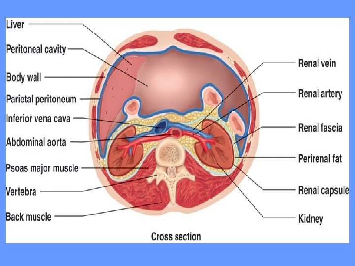
KIDNEY ANATOMY LOCATION • Bean-shaped organs lying behind the peritoneum (retroperitoneal) on the posterior abdominal wall on either side of the vertebral column • Protected by lumbar vertebrae and rib cage • Right kidney is slightly lower than left EXTERNAL ANATOMY • Superior surface capped by adrenal gland • Surrounded by RENAL CAPSULE fibrous connective tissue layer
• RENAL FAT PAD around renal capsule for protection • RENAL FASCIA - dense fibrous connective tissue outer layer anchors kidney to surrounding structures • HILIUM - on medial side where nerves and blood vessels enter and exit - opens into RENAL SINUS (cavity filled with adipose tissue and loose C. T. ) INTERNAL ANATOMY • Outer RENAL CORTEX • Inner RENAL MEDULLA surrounding renal sinus • RENAL PYRAMIDS - triangle shaped structures at boundary of cortex and medulla with tips projecting toward hilium
• Apex of renal pyramid is RENAL PAPILLA - projects into renal sinus • RENAL COLUMNS separate the renal pyramids • RENAL LOBES consist of renal pyramid, area of renal cortex surrounding renal columns • Urine drains from renal papilla into MINOR CALYX, 4 -5 then drain into a MAJOR CALYX, 2 -3 drain into the RENAL PELVIS • URETER drains the renal pelvis, exiting through the hilium to the URINARY BLADDER
NEPHRON • Microscopic functional unit of the kidney • Production of urine • Bowman’s capsule, proximal and distal tubules - within renal cortex • Loop of Henle, collecting ducts, and papillary ducts - within renal medulla
RENAL CORPUSCLE Made up of Bowman’s capsule and glomerulus BOWMAN’S CAPSULE • Outer parietal layer - simple squamous epithelium becomes cuboidal where Bowman’s capsule ends and proximal tubule begins • Inner visceral layer - specialized podocytes (large cells with “feet”) that wrap around the glomerulus GLOMERULUS • Network of capillaries that form a ball surrounded by Bowman’s C.
GLOMERULAR CIRCULATION • AFFERENT ARTERIOLE - supplies blood to glomerulus from renal artery • EFFERENT ARTERIOLE - drains glomerulus and branches into … • PERITUBULAR CAPILLARIES - form a plexus surrounding the proximal and distal tubules and upper area of loop of Henle • VASA RECTA - specialized areas of the peritubular capillaries that extend into medulla and surround loop of Henle
JUXTAGLOMERULAR APPARATUS • Specialized structure where distal tubule projects between afferent and efferent arterioles and Bowman’s capsule • Endocrine structure that secretes erythropoietin (hormone) and renin (enzyme) • Macula densa - dense connective tissue with tall cells and clustered nuclei associated with smooth muscle fibers (juxtaglomerular cells) in wall of afferent arteriole
FILTRATION MEMBRANE • FENESTRAE - window-like openings in the endothelial cells of the glomerular capillaries • FILTRATION SLITS - gaps between the cell processes of podocytes - basement membrane sandwiched between endothelial cells of glomerular capillaries and podocytes • FILTRATION MEMBRANE - glomerular capillary endothelium, basement membrane, podocytes - first stage of urine formation occurs here when fluid from blood in capillaries moves across filtration membrane into the Bowman’s capsule
PROXIMAL TUBULE • Connected to Bowman’s capsule • Lining is simple cuboidal epithelium with microvilli for absorption of nutrients, ions, water, plasma proteins from the blood LOOP OF HENLE • Descending limb - first portion similar in structure to proximal tubule - latter portion simple squamous epithelium and thinner (permeable to water) • Ascending limb - first portion simple squamous epithelium and thin - later portion thicker and simple cuboidal
DISTAL TUBULE • Initial segment passes between the afferent and efferent arterioles (near juxtaglomerular apparatus) • Smaller diameter and simple cuboidal cells with few microvilli • Site of secretion of ions, drugs, acids, toxins, selective reabsorption of ions, and water COLLECTING DUCT • Many distal tubules drain into • Larger in diameter with simple cuboidal epithelium • Several collecting ducts drain into larger PAPILLARY DUCT that empties into a minor calyx
URINE FORMATION
URETERS • Muscular tubes extending from kidneys to urinary bladder • Begins with funnel-shaped renal pelvis within kidney • Lined with transitional epithelium (specialized to stretch) inside walls of smooth muscle
BLADDER • Hollow triangle-shaped muscular container lying in pelvic cavity just posterior to pubic symphysis • Held in place by umbilical ligaments • Stores urine - lined with transitional epithelium and rugae to stretch • Trigone - interior triangular area between ureter opening and exit of urethra (acts as a funnel) • Internal urethral sphincter at neck of bladder - in males keeps semen from entering bladder
URETHRA • Extends from urinary bladder to outside body • In males – divided into prostatic urethra (through center of prostate gland), membranous urethra (penetrates floor of pelvic cavity), spongy urethra (through penis) • Passes through urogenital diaphragm (circular band of skeletal muscle) that form the external urethral sphincter - under voluntary control • Lining is stratified columnar and then stratified squamous epithelium with elastic connective tissue