HUMAN ANATOMY LECTURE SEVENTEEN LYMPHATIC SYSTEM LYMPHATIC SYSTEM

 enters lymphatic")


- Slides: 10
HUMAN ANATOMY LECTURE SEVENTEEN LYMPHATIC SYSTEM
LYMPHATIC SYSTEM FUNCTIONS • Fluid balance - extra interstitial fluid (from capillaries) enters lymphatic vessels - now called lymph • Fat absorption - absorbs fat from digestive tract (villi) into lacteals • Defense - microorganisms and other foreign substances are filtered from the lymph by lymph nodes Lymphatic system consists of: • lymph - fluid component • lacteals - remove fat from digestive tract • lymphatic vessels - carry lymph • lymphoid tissues and organs - tonsils, lymph nodes, spleen, thymus gland
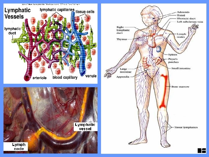
LYMPHATIC VESSELS • Carry lymph in one direction - from tissues to circulatory system • Fluid moves from extra cellular space into lymphatic capillaries - tiny, closed ended vessels with very thin walls of simple squamous epithelium - overlapping squamous cells act as valves to prevent backflow - found in all body tissues except CNS, bone marrow and tissues without blood vessels (epidermis, cartilage) • Lymphatic capillaries join to form lymphatic vessels - similar to small veins with one-way valves - superficial lymphatics and deep lymphatics collect lymph from capillaries • Lymph drains into the cisterna chyli (saclike chamber) of the thoracic duct and into the left subclavian vein - from abdomen, pelvis, lower limbs • Lymph from head, neck, chest, upper limbs drains into the right lymphatic duct and empties into the right subclavian vein
LYMPH ORGANS • Made up of lymphatic tissue consisting of many lymphocytes held by fine reticular fibers • Lymphocytes are formed in the red bone marrow and act as part of the immune system by increasing in number when exposed to microorganisms
TONSILS • Large lymphatic nodules on walls of pharynx • 3 kinds: phayngeal - near internal opening of nasal cavity palatine - on each side of posterior opening of oral cavity “tonsils” lingual - posterior surface of tongue • Form protective ring around opening of oral and nasal cavities
LYMPH NODES • Small, round nodules distributed along lymphatic vessels - lymph passes through before entering blood • Three aggregations of nodes: - inguinal nodes in the groin - axillary nodes in the armpits - cervical nodes in the neck • Surrounded by a dense connective tissue capsule and divided by trabeculae into compartments of lymphatic tissue (lymphocytes) and lymph sinuses (spaces containing macrophages within fibrous network) • As lymph travels through the node (i) immune system may be activated to produce more lymphocytes (ii) removal of microorganisms by macrophages
SPLEEN • • Contains the largest amount of lymphatic tissue in the body Located in the left, superior corner of abdominal cavity Surrounded by a capsule of collagen and elastic fibers Contain two kinds of lymphatic tissue: White pulp - surrounds arteries within the spleen, resemble lymph nodes Red pulp - associated with the veins, contains large number of macrophages
• Cells detect and respond to foreign substances in the blood and destroy worn out RBC’s as blood passes through the red pulp • Stores iron as RBC’s broken down • Initiates immune response in response to antigens in the blood • Acts as a resevoir for blood - can let blood out in an emergency (ie/ hemorrgage)
THYMUS • • Located in superior mediastium just posterior to the sternum Capsule divides it into two thymic lobes separated by the septa Each lobule consists of a dense cortex and a central medulla Lymphocytes (T cells) divide in the cortex and move to the medulla – where they mature and eventually move into the blood