Human Anatomy Introduction n Anatomical position a common


 plane - Lies vertically and divides body into")


 n n n Tightly-joined closely-packed cells One side of epithelium exposed")
")
 n n Main function: binding and support other tissues Large amount")
 n Composed muscle fibers n Contraction movement")
 n n Senses stimuli and transmits signals called nerve impulses from")

- Slides: 27
Human Anatomy Introduction
n Anatomical position – a common visual reference point ¨ Person stands erect with feet together and eyes forward ¨ Palms face anteriorly with the thumbs pointed away from the body
n Regional terms – names of specific body areas Axial region – the main axis of the body ¨ Appendicular region – the limbs ¨ n Directional terminology Refers to the body in anatomical position ¨ Standardized terms of directions are paired terms ¨
Orientation and Directional Terms
Orientation and Directional Terms
Orientation and Directional Terms
Regional Terms
Regional Terms
Body Planes and Sections
Body Planes and Sections Coronal (frontal) plane - Lies vertically and divides body into anterior (front) and posterior (back) parts n Sagittal plane – lies vertically and divides the body into left and right sides. n ¨ Median (midsagittal) plane - Specific sagittal plane that lies vertically in the midline n Transverse plane - runs horizontally and divides body into superior (up) and inferior (down) parts
Body Planes and Sections n Oblique section through the trunk Figure 1. 6
Body Cavities and Membranes n n Dorsal body cavity Cavity subdivided into the cranial cavity and the vertebral cavity. ¨ Cranial cavity houses the brain. ¨ Vertebral cavity runs through the vertebral column and encloses the spinal cord
Body Cavities and Membranes n Ventral body cavity – subdivided into: ¨Thoracic cavity – divided into three parts Two lateral parts each containing a lung surrounded by a pleural cavity n Mediastinum – contains the heart surrounded by the pericardial sac n
Body Cavities and Membranes n Ventral body cavity ¨Abdominopelvic cavity – divided into two parts Abdominal cavity – contains the liver, stomach, kidneys, and other organs n Pelvic cavity – contains the bladder, some reproductive organs, and rectum n
Body Cavities and Membranes n Serous cavities – a slit-like space lined by a serous membrane ¨ Pleura, n n pericardium, and peritoneum Parietal serosa – outer wall of the cavity Visceral serosa covers the visceral organs
Body Cavities and Membranes
Other Body Cavities n n n Oral cavity Nasal cavity Orbital cavities Middle ear cavities Synovial cavities
Abdominal Regions and Quadrants n Abdominal regions divide the abdomen into nine regions
Abdominal Quadrants n Abdominal quadrants divide the abdomen into four quadrants Right upper and left upper quadrants ¨ Right lower and left lower quadrants ¨
FOUR TYPES OF ANIMAL TISSUES
EPITHELIAL TISSUE (COVERING) n n n Tightly-joined closely-packed cells One side of epithelium exposed to air or internal fluid, other side attached to a basement membrane, a dense mat of extracellular matrix (connective tissue) Covers the outside of the body and lines the internal organs and cavities Barrier against mechanical injury, invasive microorganisms, and fluid loss Provides surface for absorption, excretion and transport of molecules
TYPES OF EPITHELIAL TISSUE n n n Cell shape ¨ Squamous (flat & thin) ¨ Cuboidal (box or square) ¨ Columnar (rectangular) Number of cell layers ¨ Simple (one) ¨ Stratified (two or more) ¨ Pseudostratified (one but appears to be two) RELATE STRUCTURE TO FUNCTION!
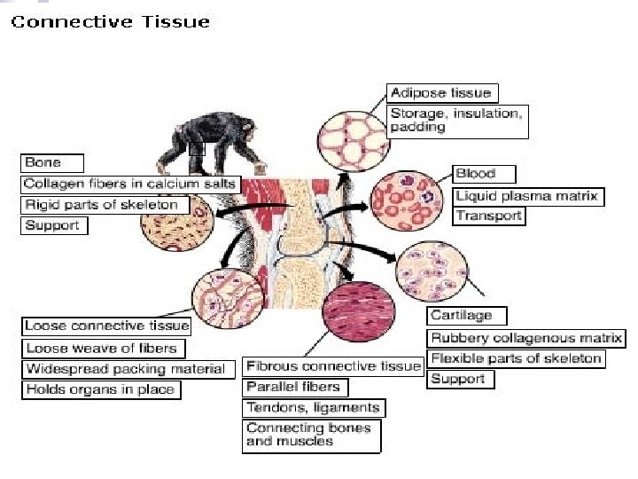
CONNECTIVE TISSUE (FRAMEWORK) n n Main function: binding and support other tissues Large amount of extra -cellular matrix with fewer cells Connective tissue cells secrete the extra -cellular matrix Extracellular matrix consists of network of fibers in liquid, jellylike or solid matrix
MUSCLE TISSUE of long cells called (MOVEMENT) n Composed muscle fibers n Contraction movement
NERVOUS TISSUE (CONTROL) n n Senses stimuli and transmits signals called nerve impulses from one part of an animal to another Consists of a cell body and long extensions called dendrites (towards cell body) and axons (towards another cell or an effector) Axon Dendrite Cell body
Tissue Type Epithelial Connective Muscle Nerve Cell Shape Flattened, cuboidal, columnar Irregular or round Elongated Cell appendages branched Cell Arrangement Single multilayered Scattered in matrix In sheets or bundles Isolated or networked Location Body covering or lining organs or cavities Supports other organs Lining internal organs, make skeletal muscles Concentrated in brain and spinal cord + all over the body Surface Feature of Cells Cilia, microvilli - - - Matrix Type Basement membrane Varied – protein fibers + liquid, gelatinous, firm to calcified - - Matrix Amount Minimal Extensive Absent Unique Feature No direct blood supply, except for glands Cartilage has no blood supply Can generate electrical signals, force and movement Can generate electrical signal