
Human Anatomy Chapter 3 Table of Contents n
Human Anatomy Chapter 3
Table of Contents n Introduction n Terms and Concepts Worth Knowing – Anatomical Position – Directional Terms – Planes of the Body n The Musculoskeletal System – Bones – Joints – Muscles Kinesiology Books Publisher 2
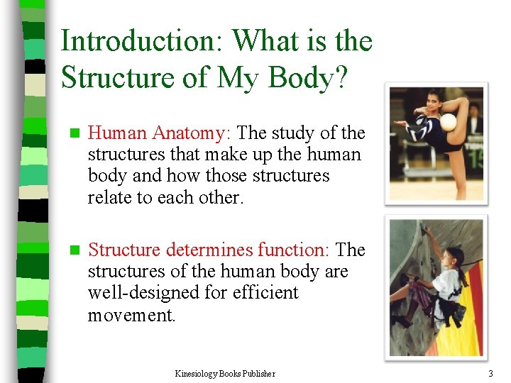
Introduction: What is the Structure of My Body? n Human Anatomy: The study of the structures that make up the human body and how those structures relate to each other. n Structure determines function: The structures of the human body are well-designed for efficient movement. Kinesiology Books Publisher 3

Terms and Concepts Worth Knowing

Anatomical Position Kinesiology Books Publisher 5
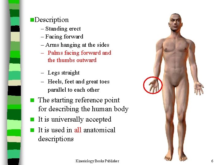
n. Description – Standing erect – Facing forward – Arms hanging at the sides – Palms facing forward and the thumbs outward – Legs straight – Heels, feet and great toes parallel to each other The starting reference point for describing the human body n It is universally accepted n It is used in all anatomical descriptions n Kinesiology Books Publisher 6
Directional Terms Kinesiology Books Publisher 7
Reminder: All directional terms are based on the assumption that the body is in the anatomical position. Kinesiology Books Publisher 8
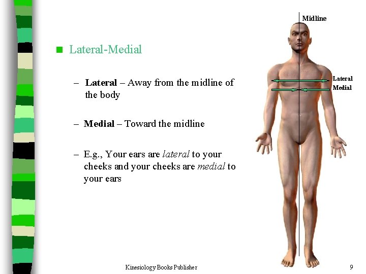
Midline n Lateral-Medial – Lateral – Away from the midline of the body Lateral Medial – Toward the midline – E. g. , Your ears are lateral to your cheeks and your cheeks are medial to your ears Kinesiology Books Publisher 9
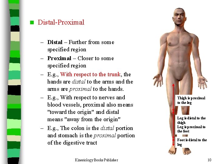
n Distal-Proximal – Distal – Further from some specified region – Proximal – Closer to some specified region – E. g. , With respect to the trunk, the hands are distal to the arms and the arms are proximal to the hands. – E. g. , With respect to nerves and blood vessels, proximal also means "toward the origin" and distal means "away from the origin" – E. g. , The colon is the distal portion and stomach is the proximal portion of the digestive tract Kinesiology Books Publisher Thigh is proximal to the leg Leg is distal to the thigh Leg is proximal to the foot Foot is distal to the leg 10
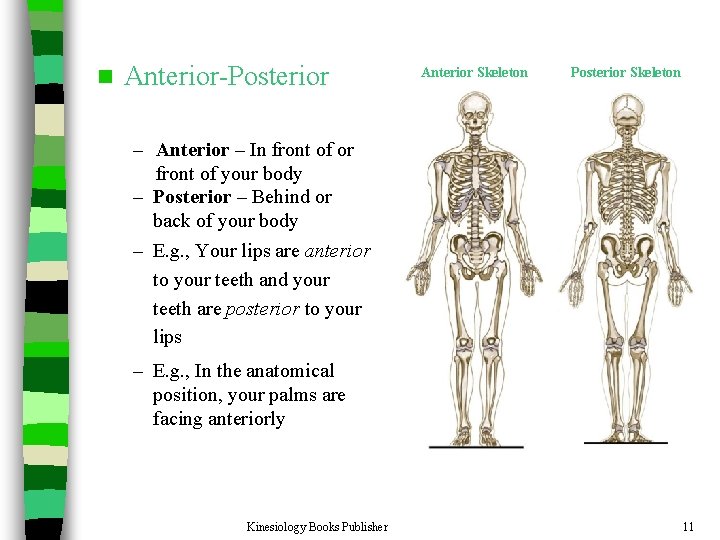
n Anterior-Posterior Anterior Skeleton Posterior Skeleton – Anterior – In front of or front of your body – Posterior – Behind or back of your body – E. g. , Your lips are anterior to your teeth and your teeth are posterior to your lips – E. g. , In the anatomical position, your palms are facing anteriorly Kinesiology Books Publisher 11
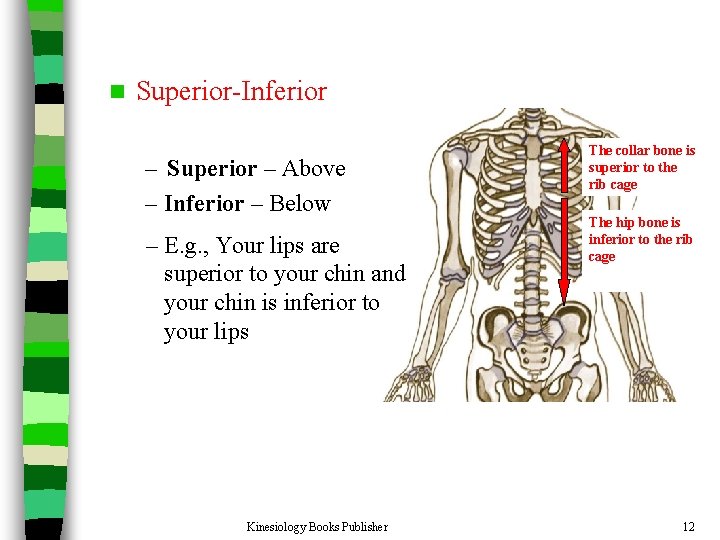
n Superior-Inferior – Superior – Above – Inferior – Below – E. g. , Your lips are superior to your chin and your chin is inferior to your lips Kinesiology Books Publisher The collar bone is superior to the rib cage The hip bone is inferior to the rib cage 12
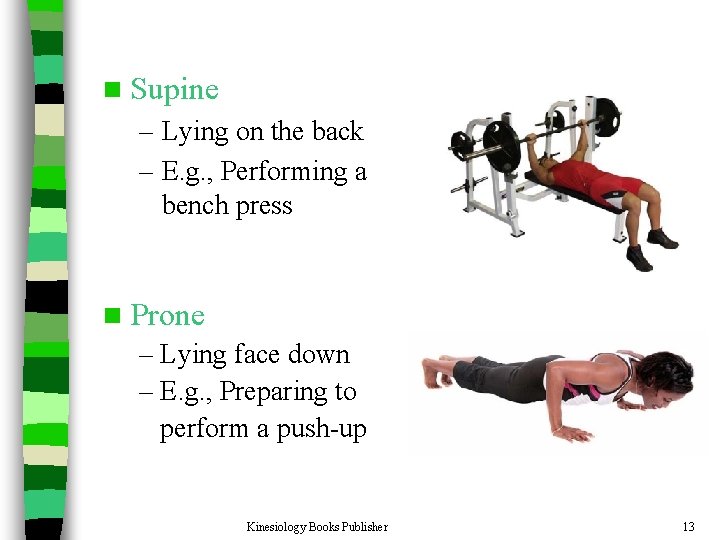
n Supine – Lying on the back – E. g. , Performing a bench press n Prone – Lying face down – E. g. , Preparing to perform a push-up Kinesiology Books Publisher 13
Planes of the Body Kinesiology Books Publisher 14
Planes n Imaginary flat surfaces that divide the human body n They are used to: – divide the body for further identification of particular areas – describe different movements or actions n Always refer to the body in the anatomical position Kinesiology Books Publisher 15
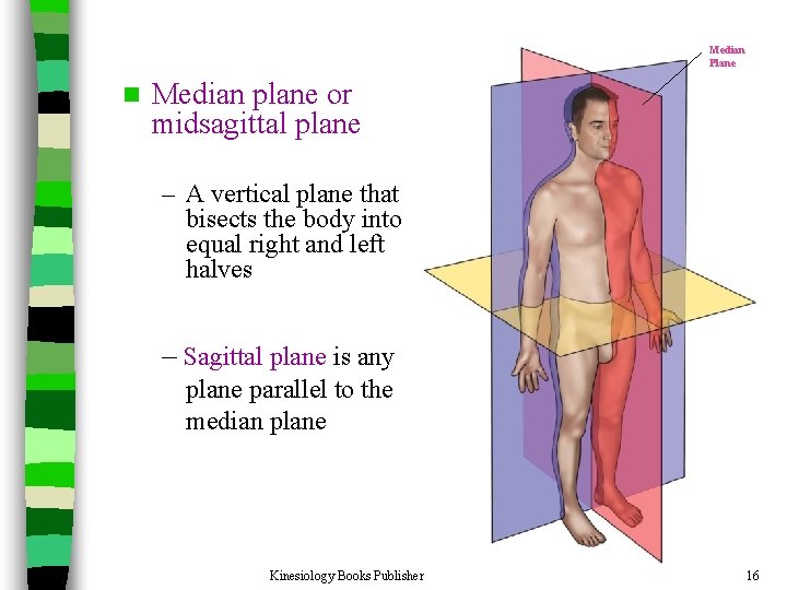
Median Plane n Median plane or midsagittal plane – A vertical plane that bisects the body into equal right and left halves – Sagittal plane is any plane parallel to the median plane Kinesiology Books Publisher 16
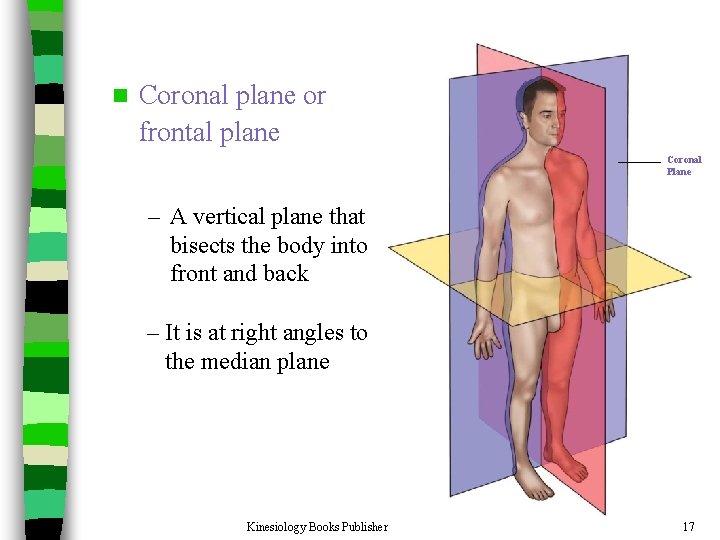
n Coronal plane or frontal plane Coronal Plane – A vertical plane that bisects the body into front and back – It is at right angles to the median plane Kinesiology Books Publisher 17
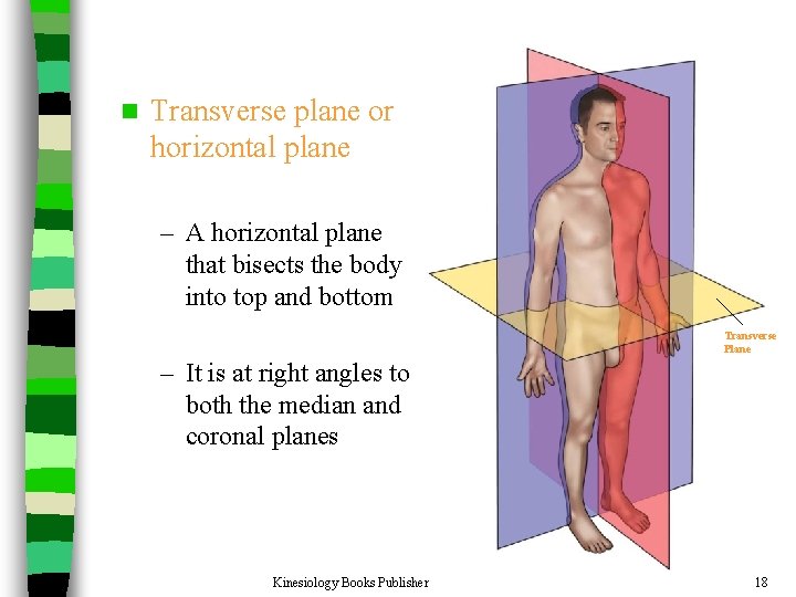
n Transverse plane or horizontal plane – A horizontal plane that bisects the body into top and bottom Transverse Plane – It is at right angles to both the median and coronal planes Kinesiology Books Publisher 18
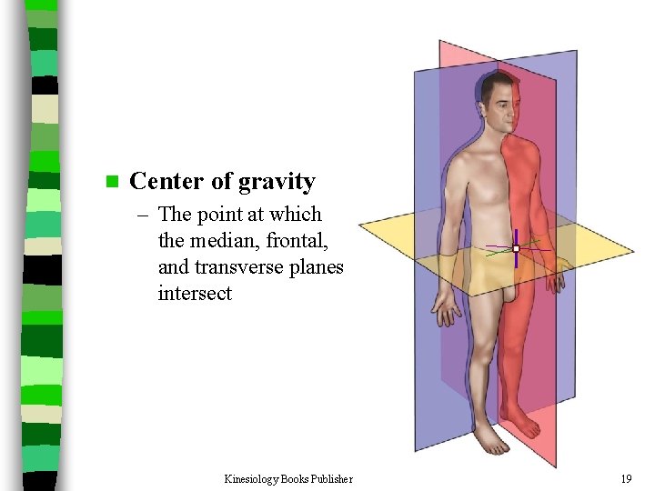
n Center of gravity – The point at which the median, frontal, and transverse planes intersect Kinesiology Books Publisher 19
Movements Kinesiology Books Publisher 20
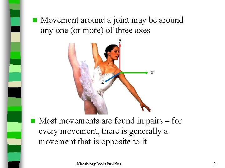
n Movement around a joint may be around any one (or more) of three axes Y X Z n Most movements are found in pairs – for every movement, there is generally a movement that is opposite to it Kinesiology Books Publisher 21
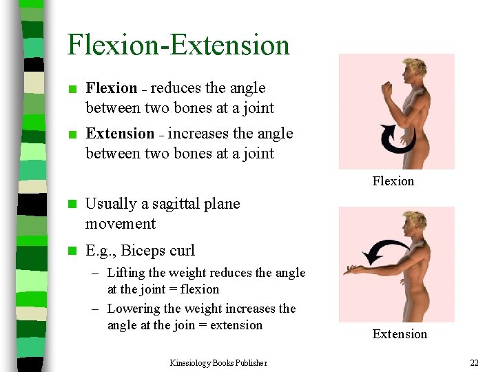
Flexion-Extension Flexion – reduces the angle between two bones at a joint Extension – increases the angle between two bones at a joint Flexion n Usually a sagittal plane movement n E. g. , Biceps curl – Lifting the weight reduces the angle at the joint = flexion – Lowering the weight increases the angle at the join = extension Kinesiology Books Publisher Extension 22
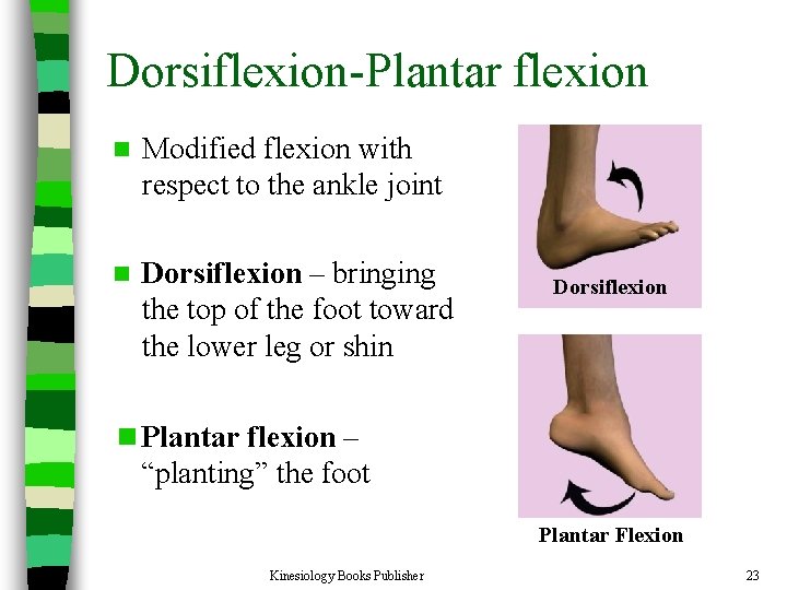
Dorsiflexion-Plantar flexion n Modified flexion with respect to the ankle joint n Dorsiflexion – bringing the top of the foot toward the lower leg or shin Dorsiflexion n Plantar flexion – “planting” the foot Plantar Flexion Kinesiology Books Publisher 23
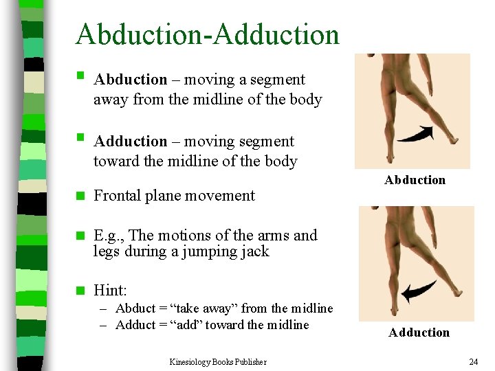
Abduction-Adduction § Abduction – moving a segment away from the midline of the body § Adduction – moving segment toward the midline of the body n Frontal plane movement n E. g. , The motions of the arms and legs during a jumping jack n Hint: – Abduct = “take away” from the midline – Adduct = “add” toward the midline Kinesiology Books Publisher Abduction Adduction 24
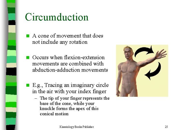
Circumduction n A cone of movement that does not include any rotation n Occurs when flexion-extension movements are combined with abduction-adduction movements n E. g. , Tracing an imaginary circle in the air with your index finger – The tip of your finger represents the base of the cone, while your knuckle forms the apex of this conical motion Kinesiology Books Publisher 25
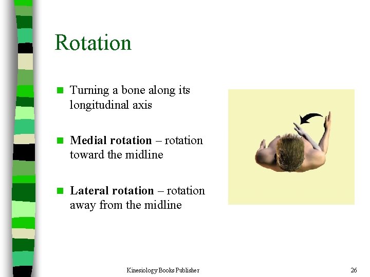
Rotation n Turning a bone along its longitudinal axis n Medial rotation – rotation toward the midline n Lateral rotation – rotation away from the midline Kinesiology Books Publisher 26
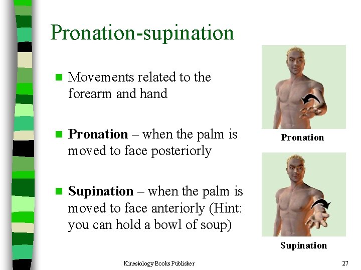
Pronation-supination n Movements related to the forearm and hand n Pronation – when the palm is moved to face posteriorly n Supination – when the palm is moved to face anteriorly (Hint: you can hold a bowl of soup) Pronation Supination Kinesiology Books Publisher 27
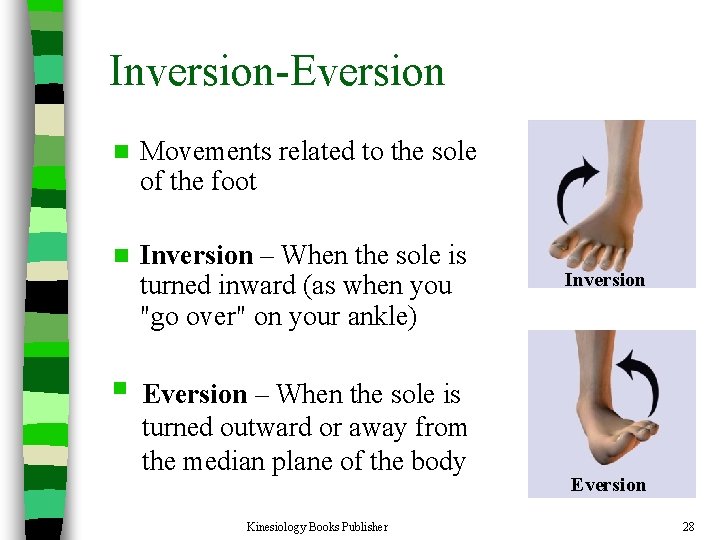
Inversion-Eversion n Movements related to the sole of the foot n Inversion – When the sole is turned inward (as when you "go over" on your ankle) Inversion § Eversion – When the sole is turned outward or away from the median plane of the body Kinesiology Books Publisher Eversion 28
The Musculoskeletal System
The Bare Bones of Human Anatomy Kinesiology Books Publisher 30
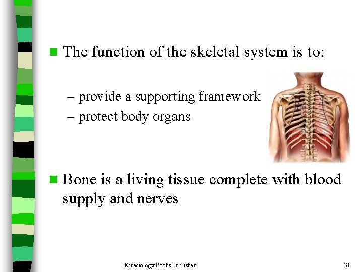
n The function of the skeletal system is to: – provide a supporting framework – protect body organs n Bone is a living tissue complete with blood supply and nerves Kinesiology Books Publisher 31
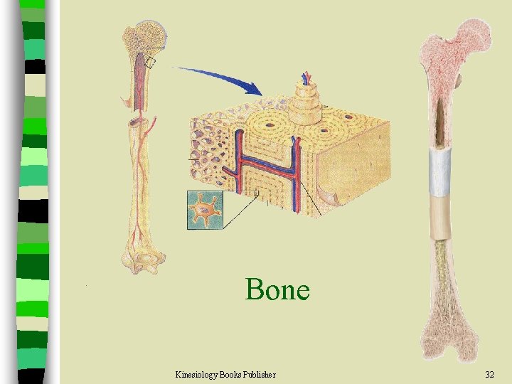
Bone Kinesiology Books Publisher 32
Bone Shape n The shapes of the bones allow them to perform specific functions more effectively Kinesiology Books Publisher 33
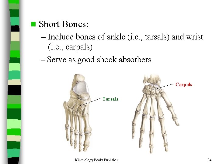
n Short Bones: – Include bones of ankle (i. e. , tarsals) and wrist (i. e. , carpals) – Serve as good shock absorbers Carpals Tarsals Kinesiology Books Publisher 34
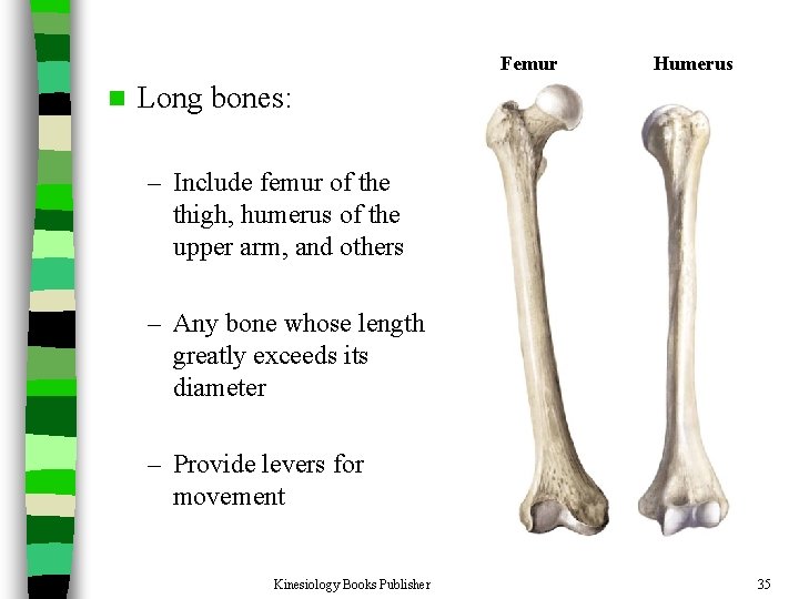
Femur n Humerus Long bones: – Include femur of the thigh, humerus of the upper arm, and others – Any bone whose length greatly exceeds its diameter – Provide levers for movement Kinesiology Books Publisher 35
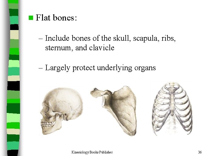
n Flat bones: – Include bones of the skull, scapula, ribs, sternum, and clavicle – Largely protect underlying organs Kinesiology Books Publisher 36
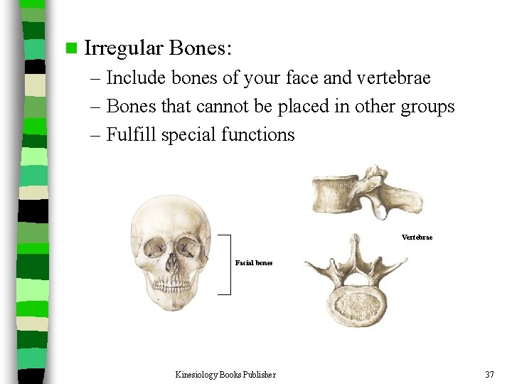
n Irregular Bones: – Include bones of your face and vertebrae – Bones that cannot be placed in other groups – Fulfill special functions Vertebrae Facial bones Kinesiology Books Publisher 37
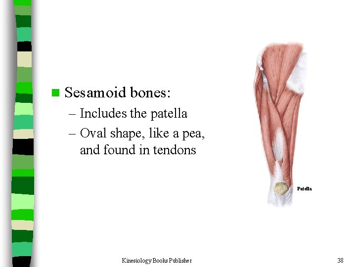
n Sesamoid bones: – Includes the patella – Oval shape, like a pea, and found in tendons Patella Kinesiology Books Publisher 38
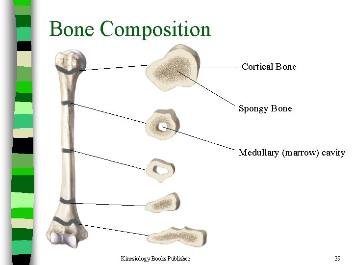
Bone Composition n Bone is very strong for its relatively light weight n The major components of bone are: – Calcium carbonate – Calcium phosphate – Collagen – Water Kinesiology Books Publisher Cortical Bone Spongy Bone Medullary (marrow) cavity 39
Bone Composition n Calcium carbonate and calcium phosphate: – Make up 60 to 70 percent of bone weight – Provide much of the bone’s stiffness and resistance to pressing or squeezing forces n Collagen (a protein): – Gives bone its characteristic flexibility and contributes to its ability to resist pulling and stretching forces – With aging, collagen is lost progressively and bone becomes more brittle. n Water – Bone consists of much smaller proportion of water than other body parts Kinesiology Books Publisher 40
Bone Classification n According to the degree of porosity, bone can be classified into two general categories: – Cortical bone (low porosity) – Spongy or cancellous bone (high porosity) Kinesiology Books Publisher 41
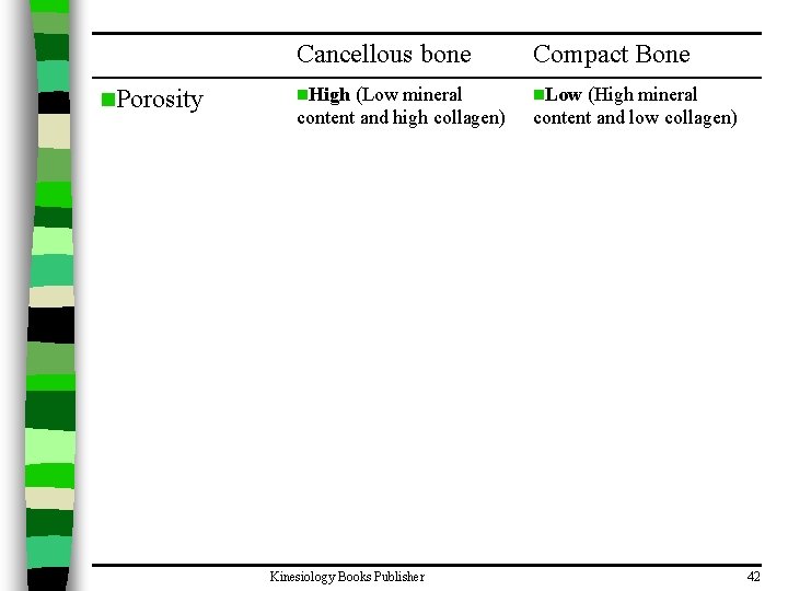
n. Porosity Cancellous bone Compact Bone n. High n. Low (Low mineral content and high collagen) Kinesiology Books Publisher (High mineral content and low collagen) 42
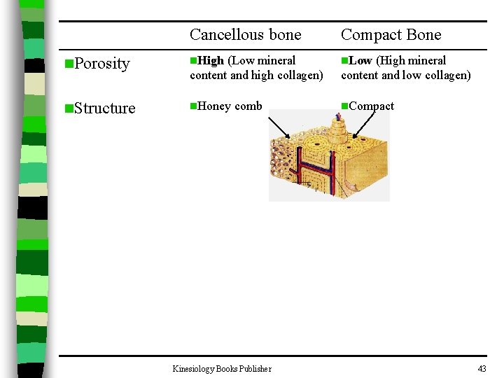
Cancellous bone Compact Bone n. Porosity n. High (Low mineral content and high collagen) n. Low n. Structure n. Honey n. Compact comb Kinesiology Books Publisher (High mineral content and low collagen) 43
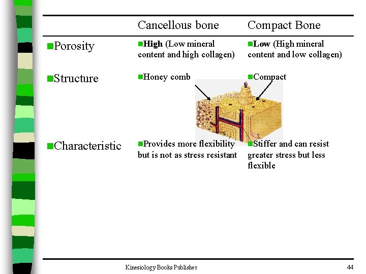
Cancellous bone Compact Bone n. Porosity n. High (Low mineral content and high collagen) n. Low n. Structure n. Honey n. Compact n. Characteristic n. Provides comb more flexibility but is not as stress resistant Kinesiology Books Publisher (High mineral content and low collagen) n. Stiffer and can resist greater stress but less flexible 44
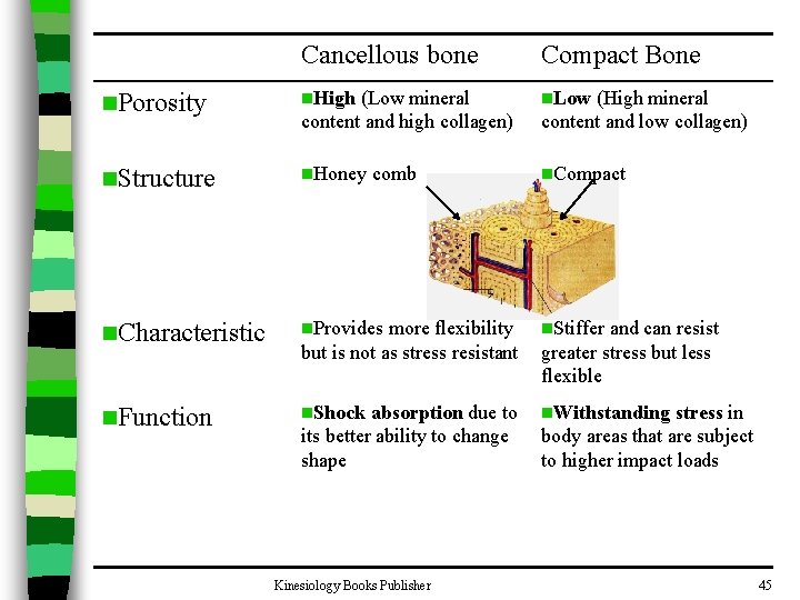
Cancellous bone Compact Bone n. Porosity n. High (Low mineral content and high collagen) n. Low n. Structure n. Honey n. Compact n. Characteristic n. Provides more flexibility but is not as stress resistant n. Stiffer n. Function n. Shock n. Withstanding comb absorption due to its better ability to change shape Kinesiology Books Publisher (High mineral content and low collagen) and can resist greater stress but less flexible stress in body areas that are subject to higher impact loads 45
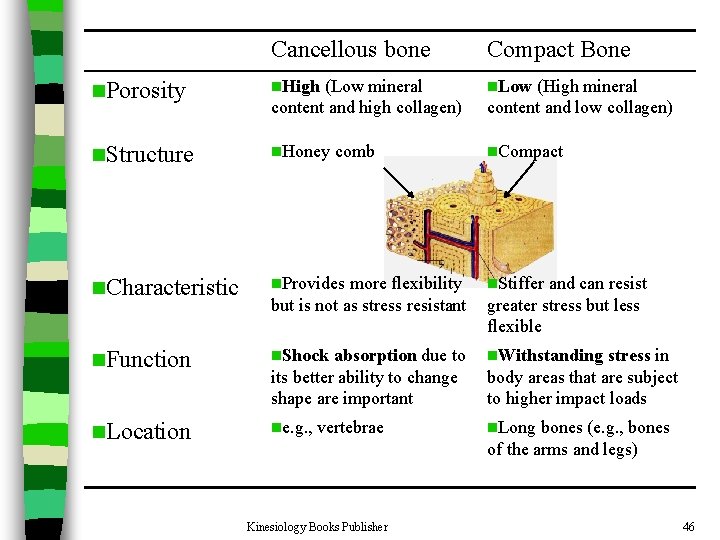
Cancellous bone Compact Bone n. Porosity n. High (Low mineral content and high collagen) n. Low n. Structure n. Honey n. Compact n. Characteristic n. Provides more flexibility but is not as stress resistant n. Stiffer n. Function n. Shock absorption due to its better ability to change shape are important n. Withstanding n. Location ne. g. , n. Long comb vertebrae Kinesiology Books Publisher (High mineral content and low collagen) and can resist greater stress but less flexible stress in body areas that are subject to higher impact loads bones (e. g. , bones of the arms and legs) 46
Effect of Fitness on Bone n When bones are subjected to regular physical activity and habitual loads, they tend to become more dense and more mineralized – E. g. , Right forearm bones of right-handed tennis players are more dense than their left ones due to more frequent use n Inactivity works in the opposite direction, leading to a decrease in weight and strength. – E. g. , Loss of bone mass has been noted in bed-ridden patients, inactive senior citizens, and astronauts Kinesiology Books Publisher 47
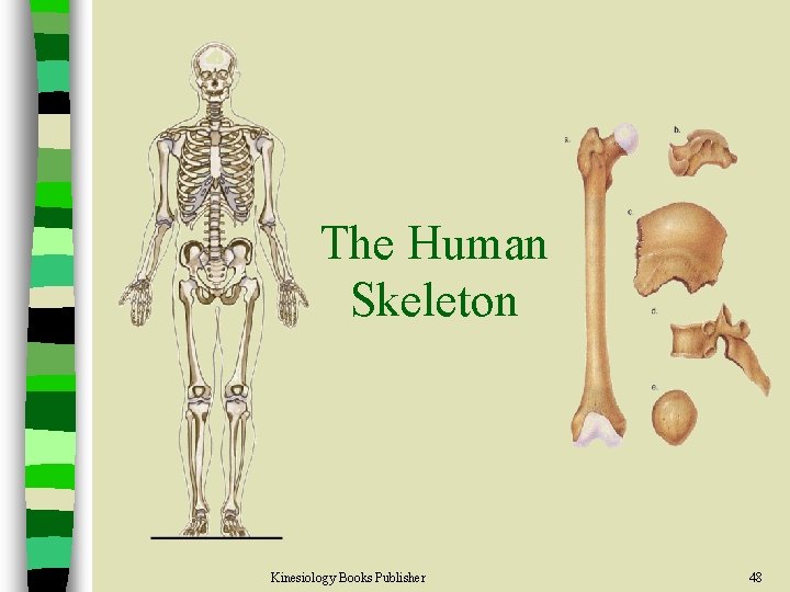
The Human Skeleton Kinesiology Books Publisher 48
Axial Skeleton Kinesiology Books Publisher 49
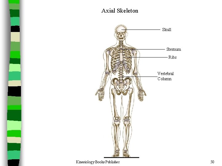
Axial Skeleton Skull Sternum Ribs Vertebral Column Kinesiology Books Publisher 50
Skull n Divided into two parts: a) Calvaria b) Face Kinesiology Books Publisher 51
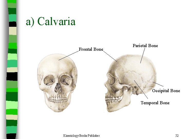
a) Calvaria Frontal Bone Parietal Bone Occipital Bone Temporal Bone Kinesiology Books Publisher 52
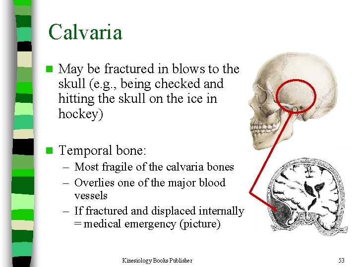
Calvaria n May be fractured in blows to the skull (e. g. , being checked and hitting the skull on the ice in hockey) n Temporal bone: – Most fragile of the calvaria bones – Overlies one of the major blood vessels – If fractured and displaced internally = medical emergency (picture) Kinesiology Books Publisher 53
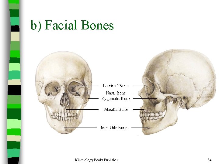
b) Facial Bones Lacrimal Bone Nasal Bone Zygomatic Bone Maxilla Bone Mandible Bone Kinesiology Books Publisher 54
Facial Bones n Often broken in contact sports due to rough impact n Some fractures of the maxilla (upper jaw) can leave the lower face separated from the upper face Kinesiology Books Publisher 55
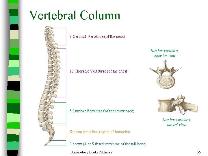
Vertebral Column 7 Cervical Vertebrae (of the neck) Lumbar vertebra, superior view 12 Thoracic Vertebrae (of the chest) 5 Lumbar Vertebrae (of the lower back) Lumbar vertebra, lateral view Sacrum (mid-line region of buttocks) Coccyx (4 or 5 fused vertebrae of the tail bone) Kinesiology Books Publisher 56
Vertebral Column n Vertebrae arranged in a cylindrical column interspersed with fibrocartilaginous (intervertebral) discs n Function: – Provides strong and flexible support for the body and the ability to keep the body erect – The point of attachment for muscles of the back – Protects the spinal cord and nerves – Absorbs shock through the intervertebral discs without causing damage to other vertebrae Kinesiology Books Publisher 57
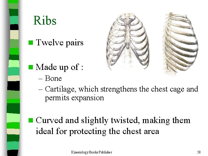
Ribs n Twelve n Made pairs up of : – Bone – Cartilage, which strengthens the chest cage and permits expansion n Curved and slightly twisted, making them ideal for protecting the chest area Kinesiology Books Publisher 58
Ribs All 12 pairs of ribs articulate with the twelve thoracic vertebrae posteriorly n Classified into three groups based on anterior attachment: n – True ribs • 1 -7 • attach to both the vertebrae and the sternum – False ribs • 8 -10 • attach only to the sternum indirectly, through 7 th rib – Floating ribs • 11 and 12 • attach only to the vertebral column Kinesiology Books Publisher 59
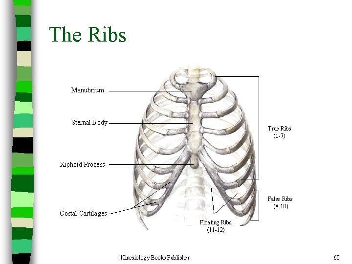
The Ribs Manubrium Sternal Body True Ribs (1 -7) Xiphoid Process False Ribs (8 -10) Costal Cartilages Floating Ribs (11 -12) Kinesiology Books Publisher 60
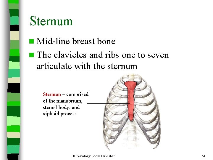
Sternum n Mid-line breast bone n The clavicles and ribs one to seven articulate with the sternum Sternum – comprised of the manubrium, sternal body, and xiphoid process Kinesiology Books Publisher 61
Appendicular Skeleton Kinesiology Books Publisher 62
Appendicular skeleton Consists of: § 1. The pectoral girdle (chest) § 2. The upper limb § 3. Pelvic girdle (hip) § 4. The lower limb Kinesiology Books Publisher 63
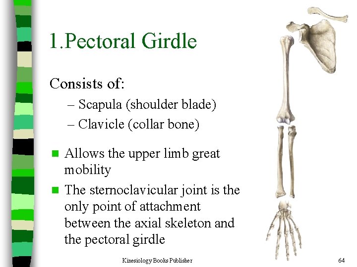
1. Pectoral Girdle Consists of: – Scapula (shoulder blade) – Clavicle (collar bone) Allows the upper limb great mobility n The sternoclavicular joint is the only point of attachment between the axial skeleton and the pectoral girdle n Kinesiology Books Publisher 64
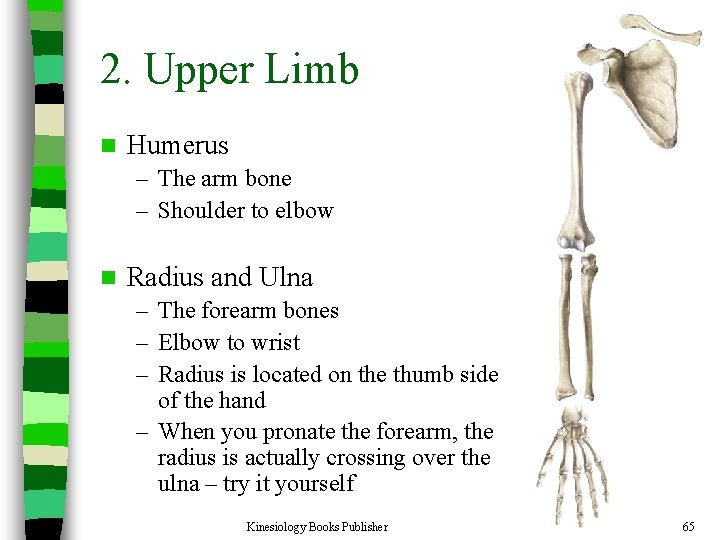
2. Upper Limb n Humerus – The arm bone – Shoulder to elbow n Radius and Ulna – The forearm bones – Elbow to wrist – Radius is located on the thumb side of the hand – When you pronate the forearm, the radius is actually crossing over the ulna – try it yourself Kinesiology Books Publisher 65
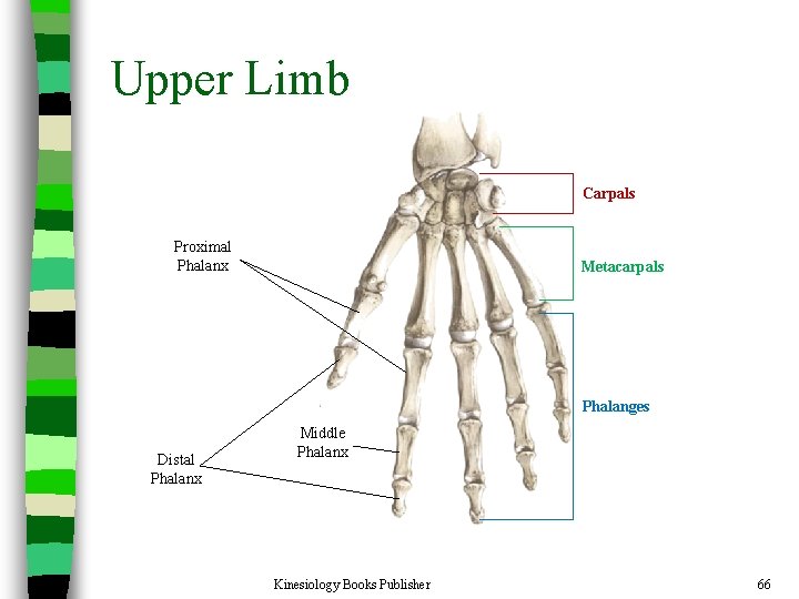
Upper Limb Carpals Proximal Phalanx Metacarpals Phalanges Distal Phalanx Middle Phalanx Kinesiology Books Publisher 66
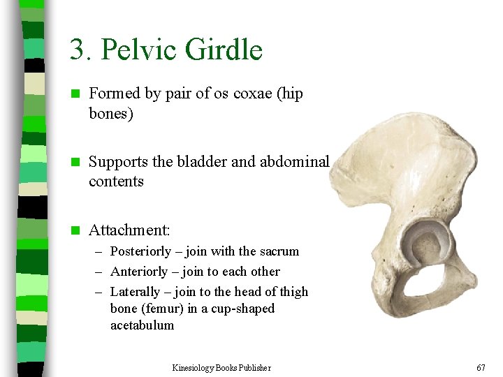
3. Pelvic Girdle n Formed by pair of os coxae (hip bones) n Supports the bladder and abdominal contents n Attachment: – Posteriorly – join with the sacrum – Anteriorly – join to each other – Laterally – join to the head of thigh bone (femur) in a cup-shaped acetabulum Kinesiology Books Publisher 67
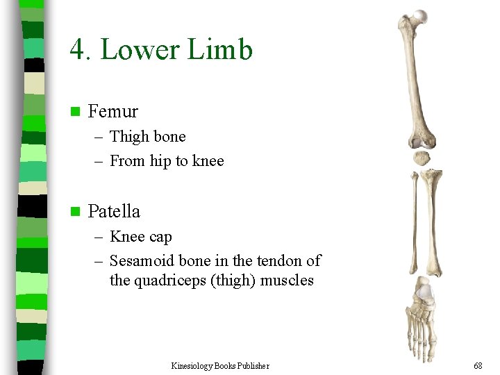
4. Lower Limb n Femur – Thigh bone – From hip to knee n Patella – Knee cap – Sesamoid bone in the tendon of the quadriceps (thigh) muscles Kinesiology Books Publisher 68
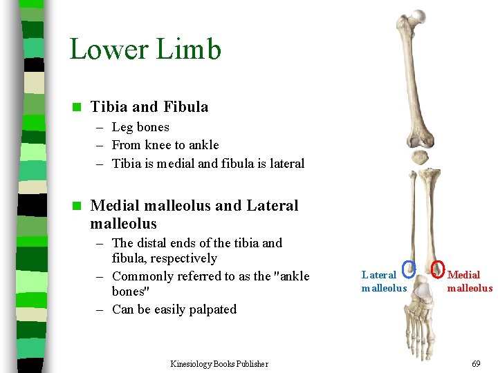
Lower Limb n Tibia and Fibula – Leg bones – From knee to ankle – Tibia is medial and fibula is lateral n Medial malleolus and Lateral malleolus – The distal ends of the tibia and fibula, respectively – Commonly referred to as the "ankle bones" – Can be easily palpated Kinesiology Books Publisher Lateral malleolus Medial malleolus 69
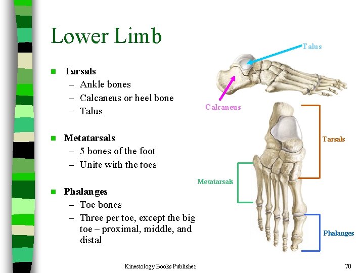
Lower Limb n n n Tarsals – Ankle bones – Calcaneus or heel bone – Talus Calcaneus Metatarsals – 5 bones of the foot – Unite with the toes Phalanges – Toe bones – Three per toe, except the big toe – proximal, middle, and distal Kinesiology Books Publisher Tarsals Metatarsals Phalanges 70
Joints of the Human Body Joint Classification n Synovial Joints n – Characteristics of synovial joints – Types of synovial joints Kinesiology Books Publisher n. Naming – – Joints: Pectoral Girdle Upper Limb Pelvic Girdle Lower Limb 71
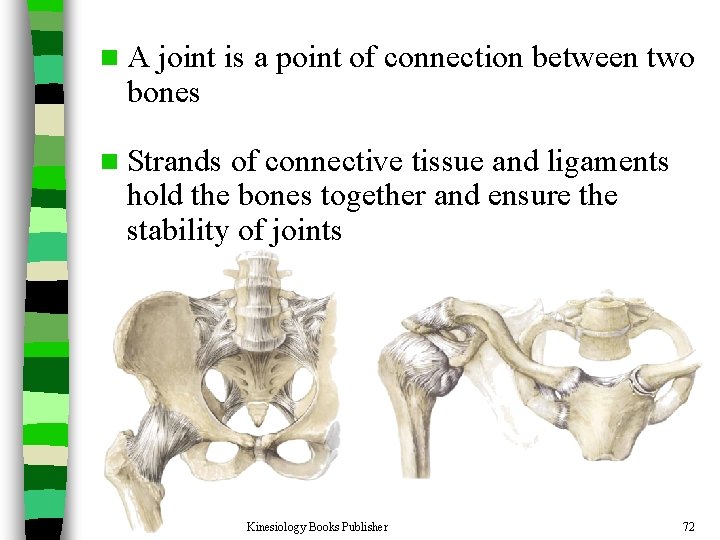
n. A joint is a point of connection between two bones n Strands of connective tissue and ligaments hold the bones together and ensure the stability of joints Kinesiology Books Publisher 72
Joint Classification n Joints are classified according to their motion capabilities: – Synarthroses • Immovable – Amphiarthroses • Slightly movable – Diarthroses • Allow the greatest amount of motion Kinesiology Books Publisher 73
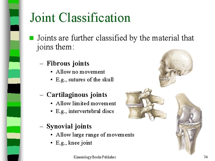
Joint Classification n Joints are further classified by the material that joins them: – Fibrous joints • Allow no movement • E. g. , sutures of the skull – Cartilaginous joints • Allow limited movement • E. g. , intervertebral discs – Synovial joints • Allow large range of movements • E. g. , knee joint Kinesiology Books Publisher 74
Characteristics of Synovial Joints n Hyaline cartilage – A protective layer of dense white connective tissue that covers the ends of the articulating bones Joint cavity n Synovial membrane n – Covers joint cavity, except over the surfaces of the articular cartilage – Secretes the lubrication fluid n Synovial fluid – Lubricates the joint n Capsule – May or may not have thickenings called intrinsic ligaments n Extrinsic ligaments – Support the joint and connect the articulating bones of the joint Kinesiology Books Publisher 75
Types of Synovial Joints n There are three basic types of synovial joints: – Unilateral (rotation about only one axis) – Biaxial joints (movement about two perpendicular axes) – Multiaxial joints (movement about all three perpendicular axes) Kinesiology Books Publisher 76
Types of Synovial Joints n Synovial are further classified into: 1. Hinge Joint 2. Pivot Joint 3. Condyloid Joint 4. Saddle-shaped joint 5. Ball and Socket Joint 6. Plane Joint Kinesiology Books Publisher 77
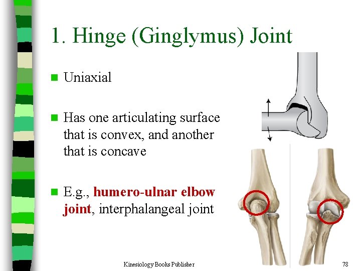
1. Hinge (Ginglymus) Joint n Uniaxial n Has one articulating surface that is convex, and another that is concave n E. g. , humero-ulnar elbow joint, interphalangeal joint Kinesiology Books Publisher 78
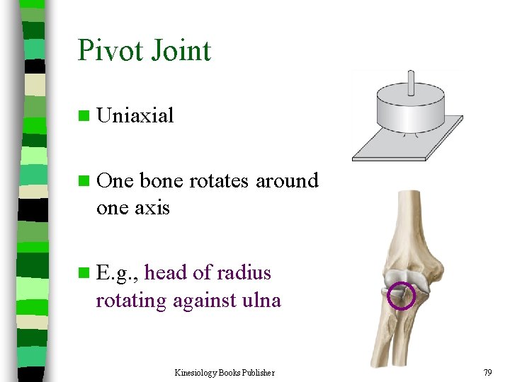
Pivot Joint n Uniaxial n One bone rotates around one axis n E. g. , head of radius rotating against ulna Kinesiology Books Publisher 79
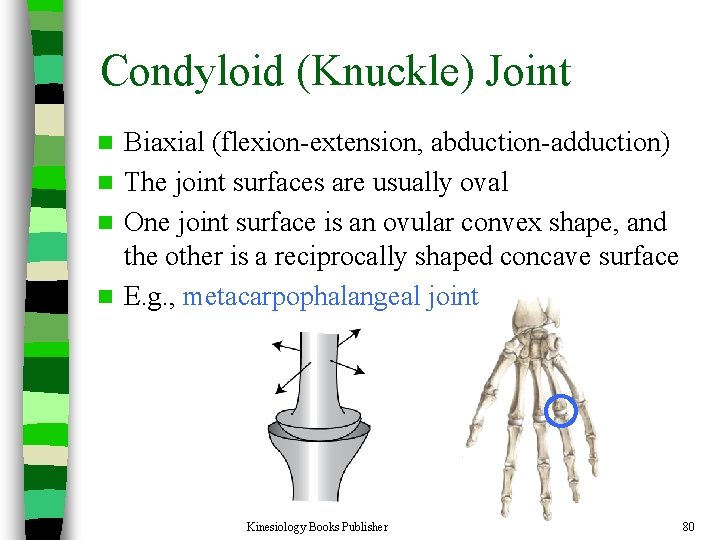
Condyloid (Knuckle) Joint Biaxial (flexion-extension, abduction-adduction) n The joint surfaces are usually oval n One joint surface is an ovular convex shape, and the other is a reciprocally shaped concave surface n E. g. , metacarpophalangeal joint n Kinesiology Books Publisher 80
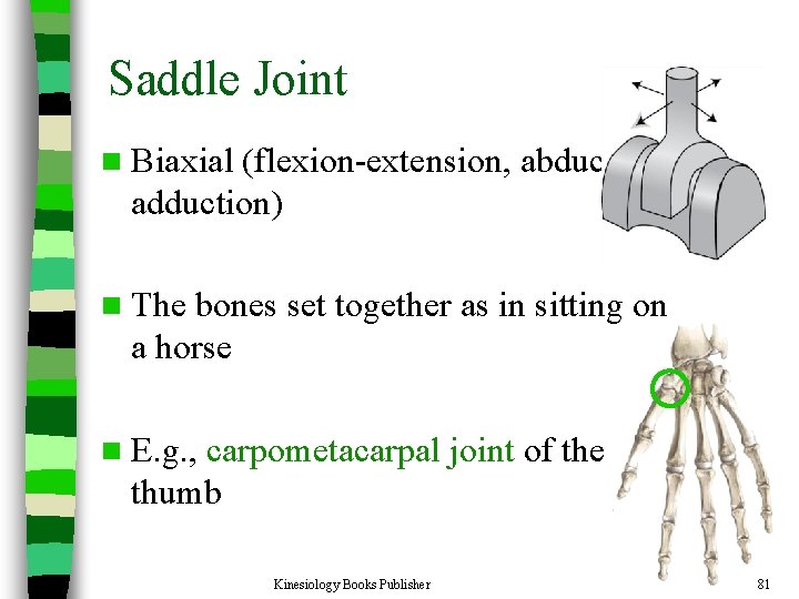
Saddle Joint n Biaxial (flexion-extension, abductionadduction) n The bones set together as in sitting on a horse n E. g. , carpometacarpal joint of the thumb Kinesiology Books Publisher 81
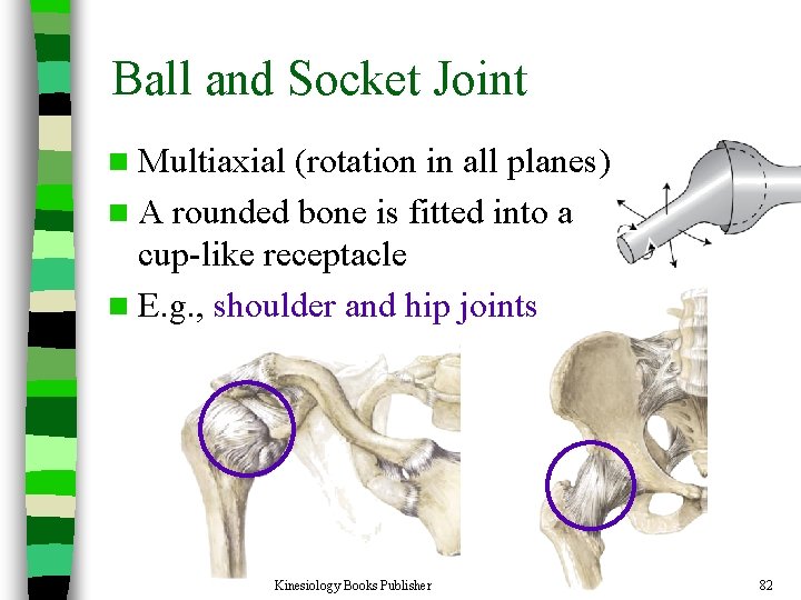
Ball and Socket Joint n Multiaxial (rotation in all planes) n A rounded bone is fitted into a cup-like receptacle n E. g. , shoulder and hip joints Kinesiology Books Publisher 82
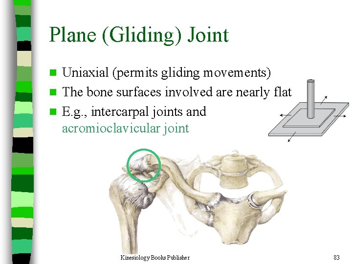
Plane (Gliding) Joint Uniaxial (permits gliding movements) n The bone surfaces involved are nearly flat n E. g. , intercarpal joints and acromioclavicular joint n Kinesiology Books Publisher 83
Joints of the Pectoral Girdle Kinesiology Books Publisher 84
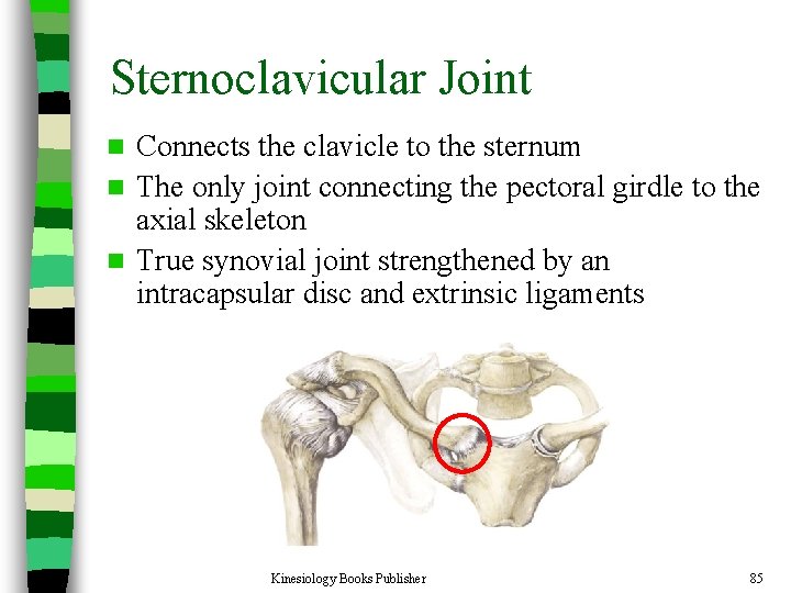
Sternoclavicular Joint Connects the clavicle to the sternum n The only joint connecting the pectoral girdle to the axial skeleton n True synovial joint strengthened by an intracapsular disc and extrinsic ligaments n Kinesiology Books Publisher 85
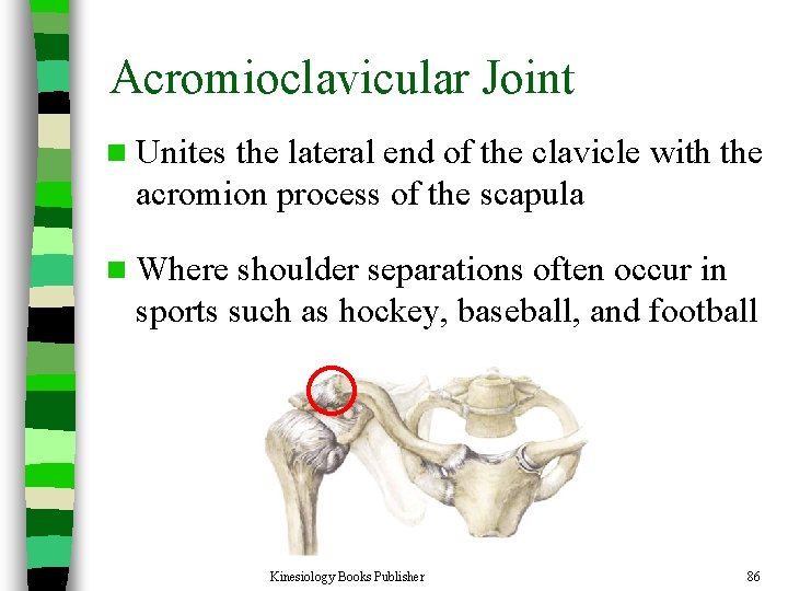
Acromioclavicular Joint n Unites the lateral end of the clavicle with the acromion process of the scapula n Where shoulder separations often occur in sports such as hockey, baseball, and football Kinesiology Books Publisher 86
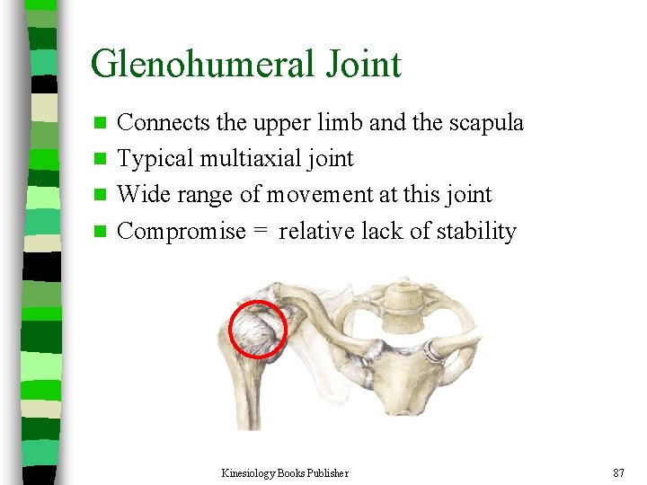
Glenohumeral Joint Connects the upper limb and the scapula n Typical multiaxial joint n Wide range of movement at this joint n Compromise = relative lack of stability n Kinesiology Books Publisher 87
Joints of the Upper Limb Kinesiology Books Publisher 88
Elbow Joint n There are three joints at the elbow: – Humero-ulnar joint • Medial • Between the trochlea of the humerus and the olecranon process of the ulna – Humero-radial joint • Lateral • Between the capitulum of the humerus and the head of the radius – Radio-ulnar joint • Between the radius and the ulna Kinesiology Books Publisher 89
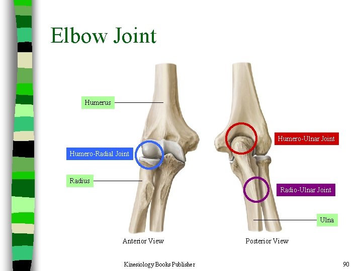
Elbow Joint Humerus Humero-Ulnar Joint Humero-Radial Joint Radius Radio-Ulnar Joint Ulna Anterior View Kinesiology Books Publisher Posterior View 90
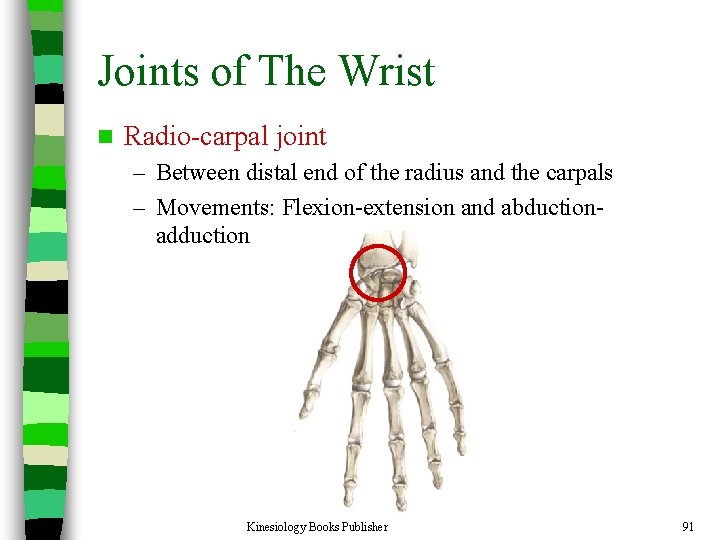
Joints of The Wrist n Radio-carpal joint – Between distal end of the radius and the carpals – Movements: Flexion-extension and abductionadduction Kinesiology Books Publisher 91
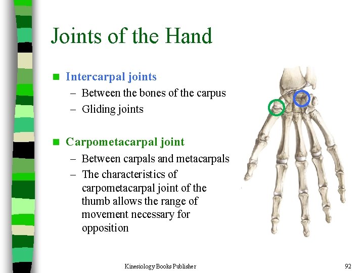
Joints of the Hand n Intercarpal joints – Between the bones of the carpus – Gliding joints n Carpometacarpal joint – Between carpals and metacarpals – The characteristics of carpometacarpal joint of the thumb allows the range of movement necessary for opposition Kinesiology Books Publisher 92
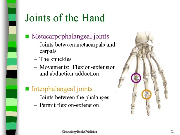
Joints of the Hand n Metacarpophalangeal joints – Joints between metacarpals and carpals – The knuckles – Movements: Flexion-extension and abduction-adduction n Interphalangeal joints – Joints between the phalanges – Permit flexion-extension Kinesiology Books Publisher 93
Joints of the Pelvic Girdle Kinesiology Books Publisher 94
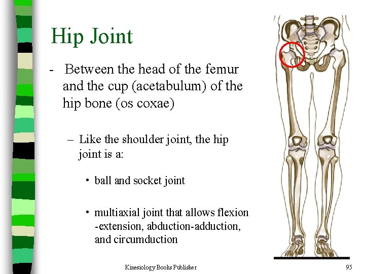
Hip Joint - Between the head of the femur and the cup (acetabulum) of the hip bone (os coxae) – Like the shoulder joint, the hip joint is a: • ball and socket joint • multiaxial joint that allows flexion -extension, abduction-adduction, and circumduction Kinesiology Books Publisher 95
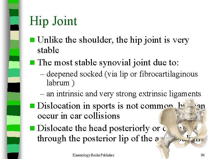
Hip Joint n Unlike the shoulder, the hip joint is very stable n The most stable synovial joint due to: – deepened socked (via lip or fibrocartilaginous labrum ) – an intrinsic and very strong extrinsic ligaments n Dislocation in sports is not common, but can occur in car collisions n Dislocate the head posteriorly or drive it through the posterior lip of the actetabulum Kinesiology Books Publisher 96
Lower Limb Joints Kinesiology Books Publisher 97
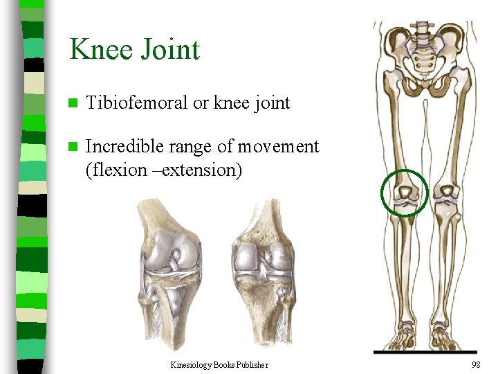
Knee Joint n Tibiofemoral or knee joint n Incredible range of movement (flexion –extension) Kinesiology Books Publisher 98
Knee Joint n The knee joint is relatively stable due to additional structural supports from: – Menisci • Shock-absorbing fibrocartilaginous discs – Anterior and posterior cruciate ligaments • Located in the center of the joint – Lateral and medial collateral ligaments • Extend from the sides of the femur to the tibia and fibula – Musculature that surrounds it Kinesiology Books Publisher 99
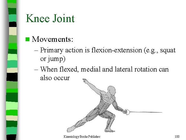
Knee Joint n Movements: – Primary action is flexion-extension (e. g. , squat or jump) – When flexed, medial and lateral rotation can also occur Kinesiology Books Publisher 100
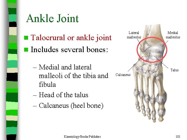
Ankle Joint n Talocrural or ankle joint n Includes several bones: – Medial and lateral malleoli of the tibia and fibula – Head of the talus – Calcaneus (heel bone) Kinesiology Books Publisher Lateral malleolus Medial malleolus Talus Calcaneus 101
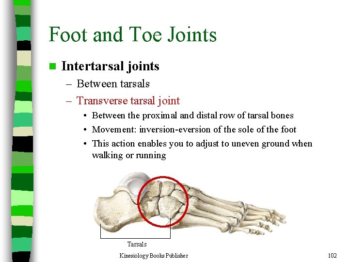
Foot and Toe Joints n Intertarsal joints – Between tarsals – Transverse tarsal joint • Between the proximal and distal row of tarsal bones • Movement: inversion-eversion of the sole of the foot • This action enables you to adjust to uneven ground when walking or running Tarsals Kinesiology Books Publisher 102
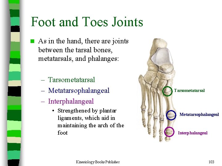
Foot and Toes Joints n As in the hand, there are joints between the tarsal bones, metatarsals, and phalanges: – Tarsometatarsal – Metatarsophalangeal – Interphalangeal • Strengthened by plantar ligaments, which aid in maintaining the arch of the foot Kinesiology Books Publisher Tarsometatarsal Metatarsophalangeal Interphalangeal 103
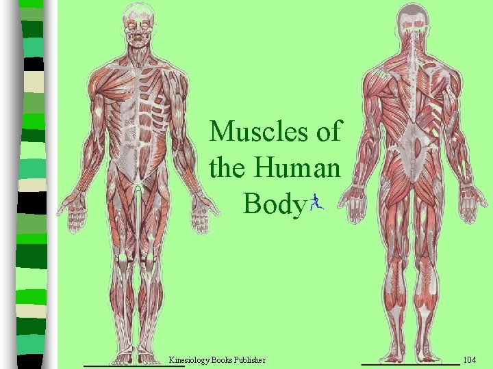
Muscles of the Human Body Kinesiology Books Publisher 104
n Over 600 muscles in the human body n Allow the skeleton to move n Both ends of a muscle must be attached to bone to create movement – Origin or proximal attachment: the attachment closer to the center of the body – Insertion or distal attachment: the attachment away from the center of the body Kinesiology Books Publisher 105
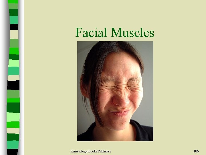
Facial Muscles Kinesiology Books Publisher 106
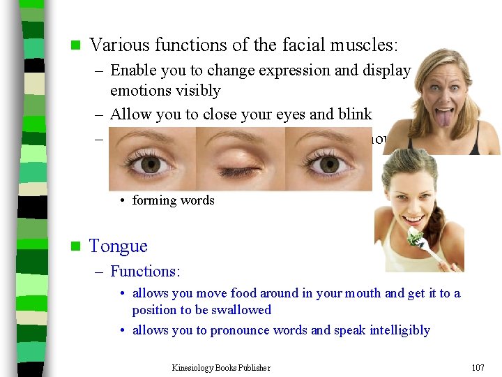
n Various functions of the facial muscles: – Enable you to change expression and display emotions visibly – Allow you to close your eyes and blink – Essential for opening and closing the mouth for: • keeping food in the mouth • chewing • forming words n Tongue – Functions: • allows you move food around in your mouth and get it to a position to be swallowed • allows you to pronounce words and speak intelligibly Kinesiology Books Publisher 107
Neck Muscles Kinesiology Books Publisher 108
Neck Muscles n There are muscles posterior, lateral, and anterior to the neck or cervical region n These muscles: – Maintain the position of the head on the 1 st cervical vertebra (atlas), i. e. , they hold up our head – Also permit a wide range of movement Kinesiology Books Publisher 109
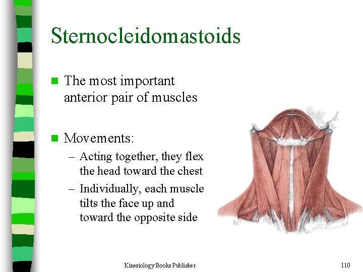
Sternocleidomastoids n The most important anterior pair of muscles n Movements: – Acting together, they flex the head toward the chest – Individually, each muscle tilts the face up and toward the opposite side Kinesiology Books Publisher 110
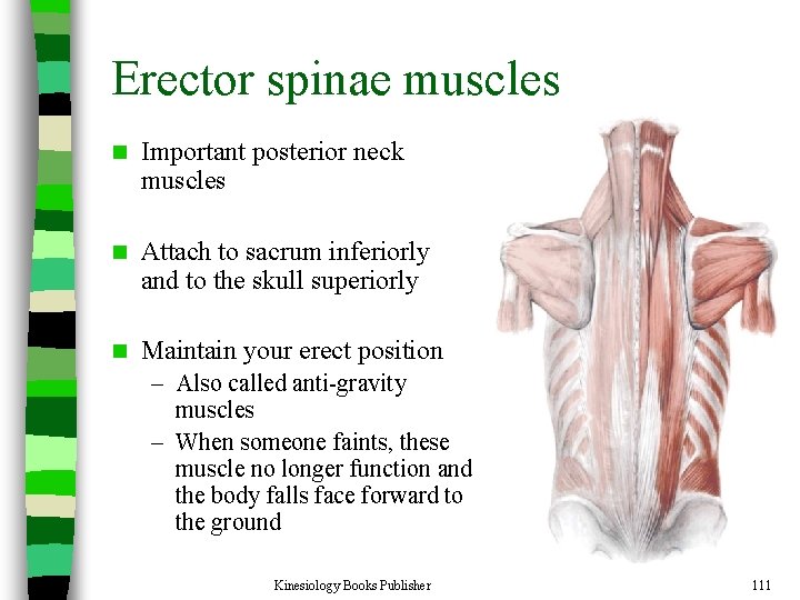
Erector spinae muscles n Important posterior neck muscles n Attach to sacrum inferiorly and to the skull superiorly n Maintain your erect position – Also called anti-gravity muscles – When someone faints, these muscle no longer function and the body falls face forward to the ground Kinesiology Books Publisher 111
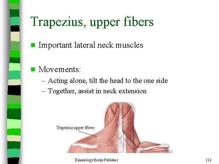
Trapezius, upper fibers n Important lateral neck muscles n Movements: – Acting alone, tilt the head to the one side – Together, assist in neck extension Trapezius upper fibers Kinesiology Books Publisher 112
Muscles of the Pectoral Girdle Kinesiology Books Publisher 113
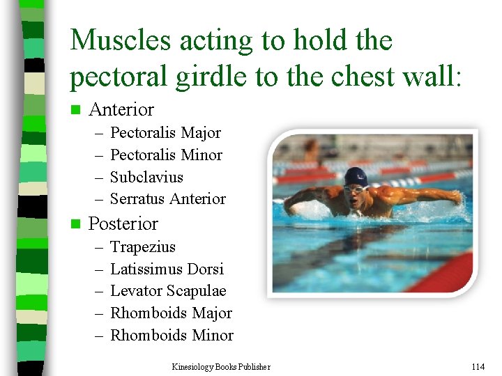
Muscles acting to hold the pectoral girdle to the chest wall: n Anterior – – n Pectoralis Major Pectoralis Minor Subclavius Serratus Anterior Posterior – – – Trapezius Latissimus Dorsi Levator Scapulae Rhomboids Major Rhomboids Minor Kinesiology Books Publisher 114
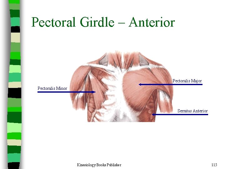
Pectoral Girdle – Anterior Pectoralis Major Pectoralis Minor Serratus Anterior Kinesiology Books Publisher 115
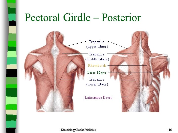
Pectoral Girdle – Posterior Trapezius (upper fibers) Trapezius (middle fibers) Rhomboids Teres Major Trapezius (lower fibers) Latissimus Dorsi Kinesiology Books Publisher 116
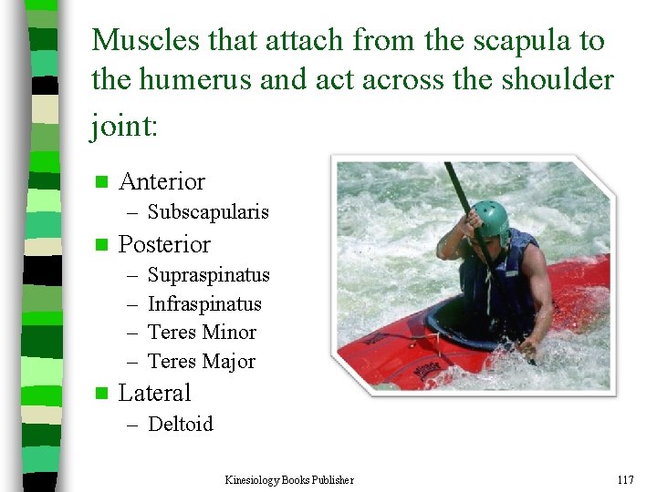
Muscles that attach from the scapula to the humerus and act across the shoulder joint: n Anterior – Subscapularis n Posterior – – n Supraspinatus Infraspinatus Teres Minor Teres Major Lateral – Deltoid Kinesiology Books Publisher 117
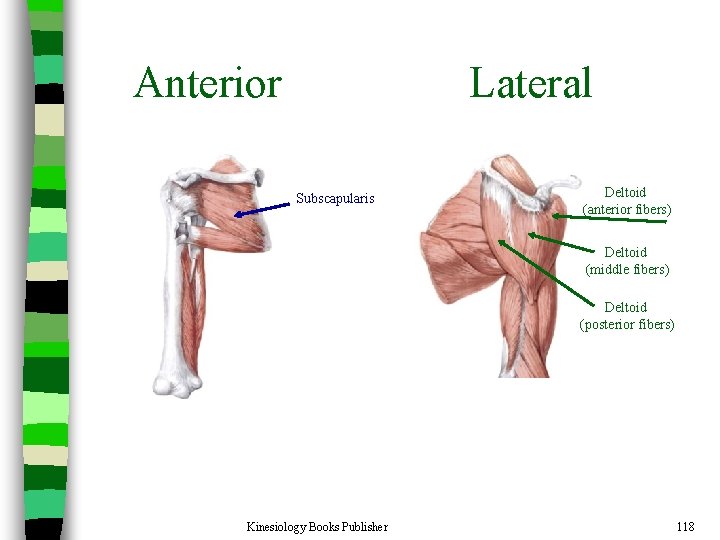
Anterior Lateral Subscapularis Deltoid (anterior fibers) Deltoid (middle fibers) Deltoid (posterior fibers) Kinesiology Books Publisher 118
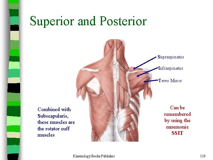
Superior and Posterior Supraspinatus Infraspinatus Teres Minor Combined with Subscapularis, these muscles are the rotator cuff muscles Kinesiology Books Publisher Can be remembered by using the mnemonic SSIT 119
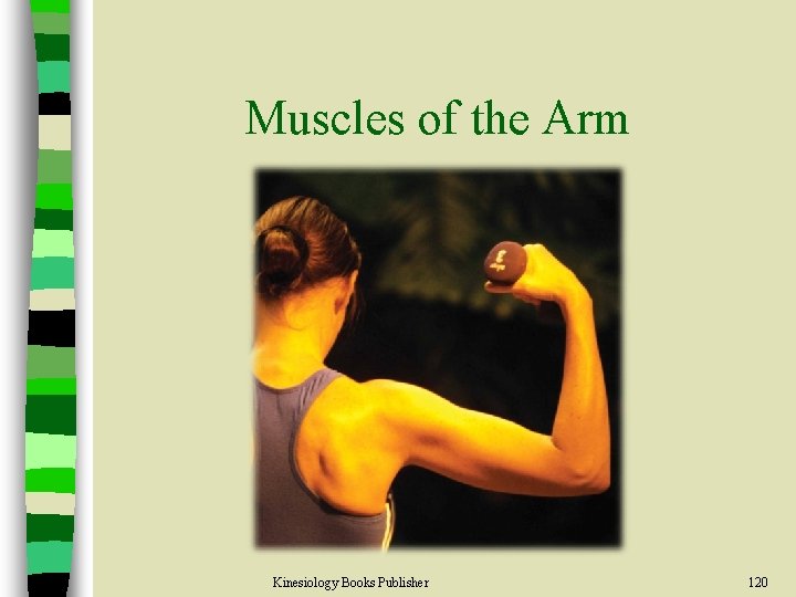
Muscles of the Arm Kinesiology Books Publisher 120
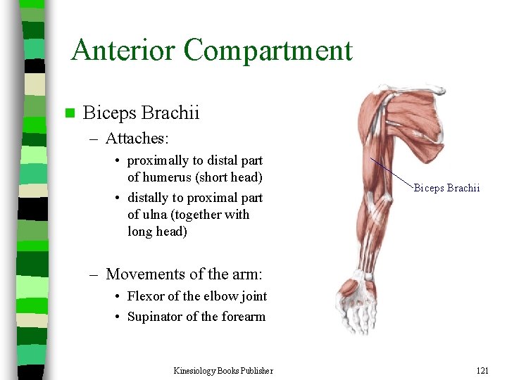
Anterior Compartment n Biceps Brachii – Attaches: • proximally to distal part of humerus (short head) • distally to proximal part of ulna (together with long head) Biceps Brachii – Movements of the arm: • Flexor of the elbow joint • Supinator of the forearm Kinesiology Books Publisher 121
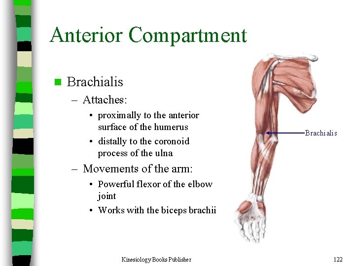
Anterior Compartment n Brachialis – Attaches: • proximally to the anterior surface of the humerus • distally to the coronoid process of the ulna Brachialis – Movements of the arm: • Powerful flexor of the elbow joint • Works with the biceps brachii Kinesiology Books Publisher 122
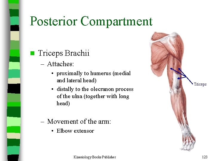
Posterior Compartment n Triceps Brachii – Attaches: • proximally to humerus (medial and lateral head) • distally to the olecranon process of the ulna (together with long head) Triceps – Movement of the arm: • Elbow extensor Kinesiology Books Publisher 123
Muscles of the Forearm Kinesiology Books Publisher 124
n Forearm muscles act on the elbow, wrist, and digits (fingers) n Flexor-pronator group – Attached to the medial epicondyle of the humerus n Extensor-supinator – Attached to the lateral epicondyle of the humerus Kinesiology Books Publisher 125
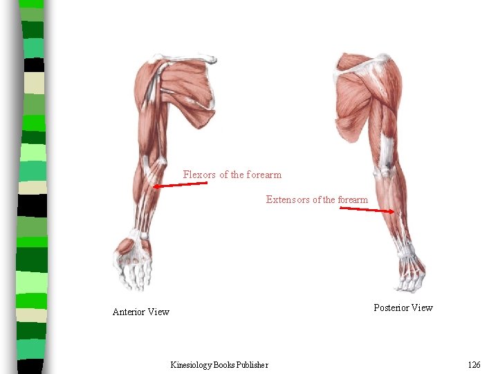
Flexors of the forearm Extensors of the forearm Posterior View Anterior View Kinesiology Books Publisher 126
Muscles of the Pelvic Girdle Kinesiology Books Publisher 127
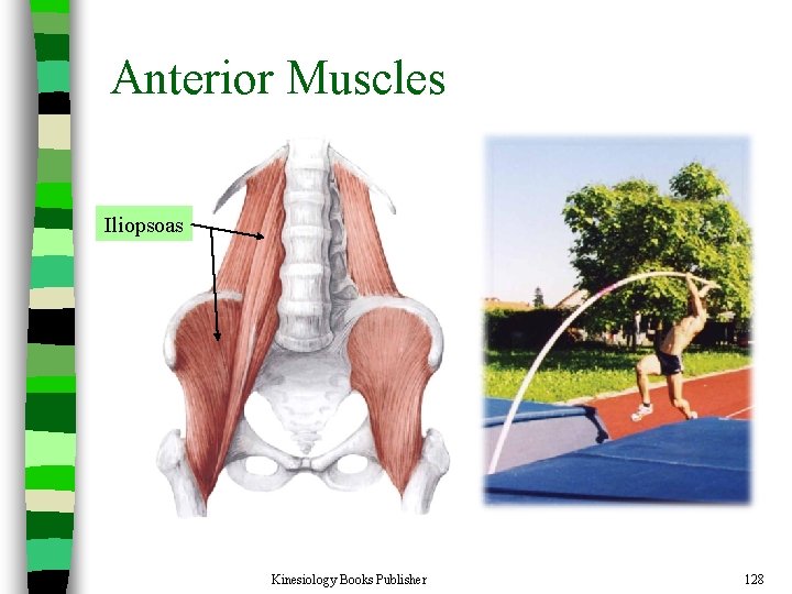
Anterior Muscles Iliopsoas Kinesiology Books Publisher 128
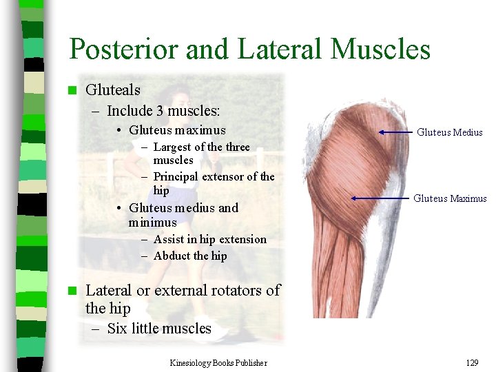
Posterior and Lateral Muscles n Gluteals – Include 3 muscles: • Gluteus maximus – Largest of the three muscles – Principal extensor of the hip • Gluteus medius and minimus Gluteus Medius Gluteus Maximus – Assist in hip extension – Abduct the hip n Lateral or external rotators of the hip – Six little muscles Kinesiology Books Publisher 129
Thigh Muscles Kinesiology Books Publisher 130
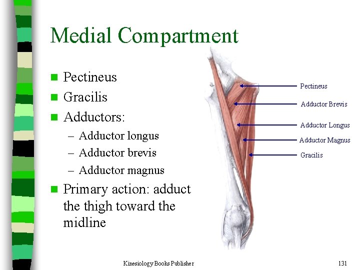
Medial Compartment Pectineus n Gracilis n Adductors: n – Adductor longus – Adductor brevis – Adductor magnus n Pectineus Adductor Brevis Adductor Longus Adductor Magnus Gracilis Primary action: adduct the thigh toward the midline Kinesiology Books Publisher 131
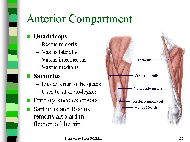
Anterior Compartment n Quadriceps – – n Rectus femoris Vastus lateralis Vastus intermedius Vastus medialis Sartorius Vastus Lateralis – Lies anterior to the quads – Used to sit cross-legged Primary knee extensors n Sartorius and Rectus femoris also aid in flexion of the hip n Kinesiology Books Publisher Vastus Intermedius Rectus Femoris (cut) Vastus Medialis 132
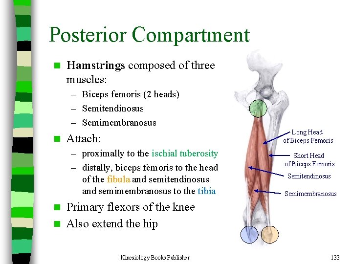
Posterior Compartment n Hamstrings composed of three muscles: – Biceps femoris (2 heads) – Semitendinosus – Semimembranosus n Long Head of Biceps Femoris Attach: – proximally to the ischial tuberosity – distally, biceps femoris to the head of the fibula and semitendinosus and semimembranosus to the tibia Short Head of Biceps Femoris Semitendinosus Semimembranosus Primary flexors of the knee n Also extend the hip n Kinesiology Books Publisher 133
Leg Muscles Kinesiology Books Publisher 134
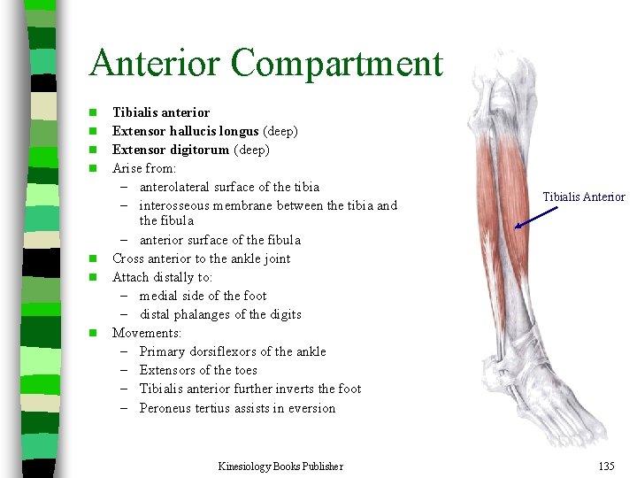
Anterior Compartment n n n n Tibialis anterior Extensor hallucis longus (deep) Extensor digitorum (deep) Arise from: – anterolateral surface of the tibia – interosseous membrane between the tibia and the fibula – anterior surface of the fibula Cross anterior to the ankle joint Attach distally to: – medial side of the foot – distal phalanges of the digits Movements: – Primary dorsiflexors of the ankle – Extensors of the toes – Tibialis anterior further inverts the foot – Peroneus tertius assists in eversion Kinesiology Books Publisher Tibialis Anterior 135
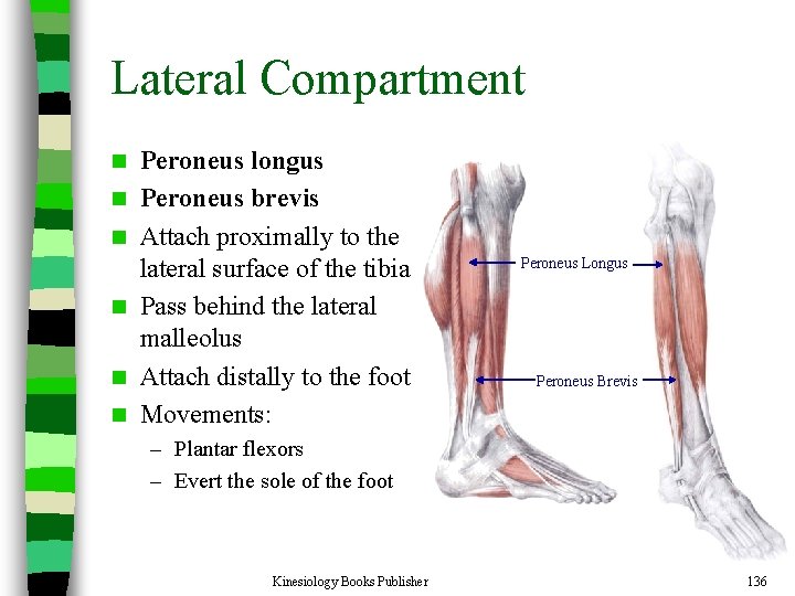
Lateral Compartment n n n Peroneus longus Peroneus brevis Attach proximally to the lateral surface of the tibia Pass behind the lateral malleolus Attach distally to the foot Movements: Peroneus Longus Peroneus Brevis – Plantar flexors – Evert the sole of the foot Kinesiology Books Publisher 136
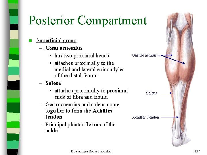
Posterior Compartment n Superficial group – Gastrocnemius • has two proximal heads • attaches proximally to the medial and lateral epicondyles of the distal femur – Soleus • attaches proximally to proximal ends of tibia and fibula – Gastrocnemius and soleus come together to form the Achilles tendon – Principal plantar flexors of the ankle Kinesiology Books Publisher Gastrocnemius Soleus Achilles Tendon 137
Posterior Compartment n Deep group – Primary role is flexion of the toes – Assist in plantar flexion of the ankle Kinesiology Books Publisher 138
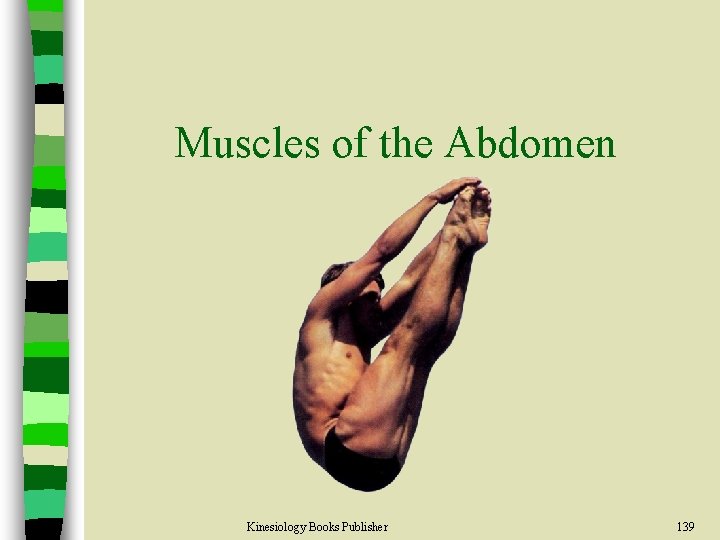
Muscles of the Abdomen Kinesiology Books Publisher 139
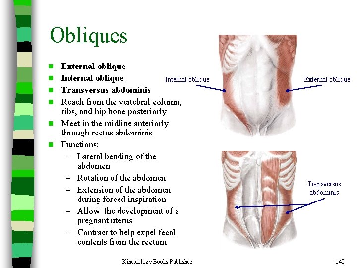
Obliques n n n External oblique Internal oblique Transversus abdominis Reach from the vertebral column, ribs, and hip bone posteriorly Meet in the midline anteriorly through rectus abdominis Functions: – Lateral bending of the abdomen – Rotation of the abdomen – Extension of the abdomen during forced inspiration – Allow the development of a pregnant uterus – Contract to help expel fecal contents from the rectum Kinesiology Books Publisher External oblique Transversus abdominis 140
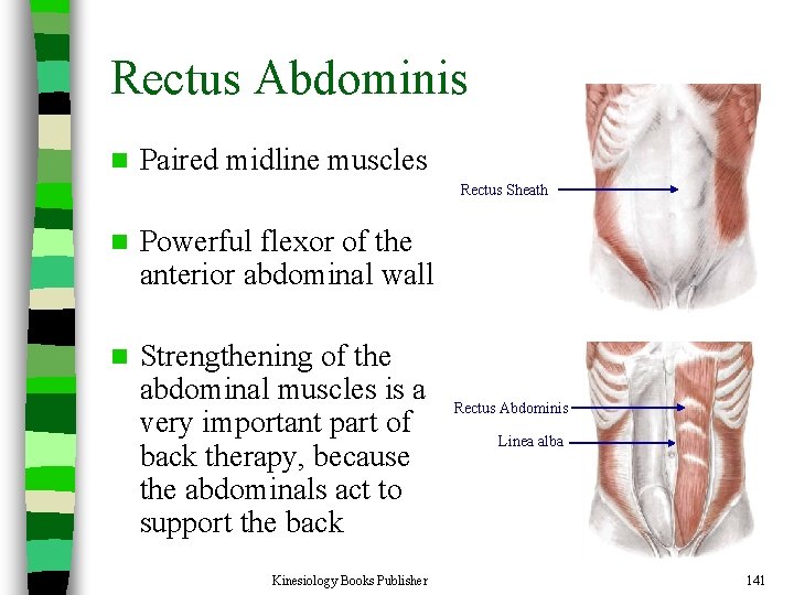
Rectus Abdominis n Paired midline muscles Rectus Sheath n Powerful flexor of the anterior abdominal wall n Strengthening of the abdominal muscles is a very important part of back therapy, because the abdominals act to support the back Kinesiology Books Publisher Rectus Abdominis Linea alba 141
Summary Human anatomy deals with the structures that make up the human body (structure determines function) n The bones, joints, and muscles that make up the musculoskeletal system allow numerous movements to occur, with varying degrees of: – Motion capabilities – Strength – Flexibility § Bones provide the structural framework necessary for support § Muscles supply the power § Joints supply the mechanism that allows human movement to occur n Kinesiology Books Publisher 142
Discussion Questions 1. Describe the anatomical position and discuss its relationship to the directional terms of the body. 2. What are the four major planes that bisect the body? Provide an example of a movement that occurs in each plane. 3. Define three types of movement and give an example of each. 4. Name three types of muscle. How do slow twitch and fast twitch fibbers differ? 5. Differentiate among agonists, antagonists, and synergists. What role is played by the fixators? 6. Which synovial joints allow the greatest amount of movement? The least? 7. Outline the components and roles of the axial and appendicular skeletons. 8. What structures present at the knee provide additional support to this joint? 9. What type of joint is the knee? 10. What muscles are primarily responsible for maintaining an upright posture? 11. The posterior group of leg muscles are commonly called the hamstrings. What three muscles combine to form the hamstrings? What role do they play? Kinesiology Books Publisher 143
- Slides: 143