Human Anatomy and Physiology II Special Senses All


transmit the vibratory motion from the eardrum")


 which is bent, straightens out,")


- Slides: 103
Human Anatomy and Physiology II Special Senses
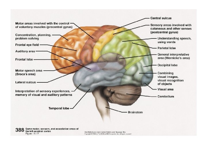
All senses work the same way: l Receptors collect information l stimulate neurons l information is sent to the brain l the cerebral cortex integrates the information with that from other senses l forms a perception (a person’s particular view of the stimulus)
Receptor types: l Pain receptors or nociceptors – respond to tissue damage due to mechanical, electrical, thermal or chemical energy l Thermoreceptors – respond to temperature change
Receptor types: l Mechanoreceptors – respond to mechanical forces, such as pressure or fluid movement; changes usually deform the receptor l Proprioceptors – sense changes in muscles and tendons l Baroreceptors – in blood vessels – detect changes in pressure l Stretch receptors – in lungs – sense degree of inflation
Receptor types: l Photoreceptors -respond to light – as little as one photon l Chemoreceptors – sensitive to chemical concentration of various substances
Receptors are structured in two basic ways: • receptors can be nerve endings • other kinds of cells which are associated with nerve endings • When these are stimulated, they produce graded potentials. If hit threshold, nerve fires.
A sensation or perception occurs when the brain interprets the incoming nerve impulses. All impulses coming into the brain are alike. The sensation depends on which part of the brain is stimulated. Synesthesia – tasting, colors, etc.
n Involuntary: synesthetes do not actively think about their perceptions; they just happen. n Projected: rather than experiencing something in the "mind's eye, " as might happen when you are asked to imagine a color, a synesthete often actually sees a color projected outside of the body. n Durable and generic: the perception must be the same every time; for example, if you taste chocolate when you hear Beethoven's Violin Concerto, you must always taste chocolate when you hear it; also, the perception must be generic -- that is, you may see colors or lines or shapes in response to a certain smell, but you would not see something complex such as a room with people and furniture and pictures on the wall. n Memorable: often, the secondary synesthetic perception is remembered better than the primary perception; for example, a synesthete who always associates the color purple with the name "Laura" will often remember that a woman's name is purple rather than actually remembering "Laura. " n Emotional: the perceptions may cause emotional reactions such as pleasurable feelings.
Who has synethesia n n Women: in the U. S. , studies show that three times as many women as men have synesthesia; in the U. K. , eight times as many women have been reported to have it. The reason for this difference is not known. Left-handed: synesthetes are more likely to be left-handed than the general population. Neurologically normal: synesthetes are of normal (or possibly above average) intelligence, and standard neurological exams are normal. In the same family: synesthesia appears to be inherited in some fashion; it seems to be a dominant trait and it may be on the X-chromosome.
7, 9, 4, 0, 3, 8, 2, 5, 1, 6.
Developmental Aspects of the Special Senses n n n Special sense organs are formed early in embryonic development. Maternal infections during the first five or six weeks of pregnancy may cause visual abnormalities as well as sensorineural deafness in the developing child. An important congenital eye problem is strabismus. The most important congenital ear problem is lack of the external auditory canal. Vision requires the most learning. The infant has poor visual acuity (is farsighted) and lacks color vision and depth perception at birth. The eye continues to grow and mature until the eighth or ninth year of life, Problems of aging associated with vision include presbyopia, glaucoma (the most common cause of blindness in the U. S. ), cataracts, and arteriosclerosis of the eye's blood vessels. The newborn infant can hear sounds, but initial responses are reflexive. By the toddler stage, the child is listening critically and beginning to imitate sounds as language development begins. Sensorineural deafness (presbycusis) is a normal consequence of aging. Taste and smell are most acute at birth and decrease in sensitivity after the age of 40 as the number of olfactory and gustatory receptors decreases.
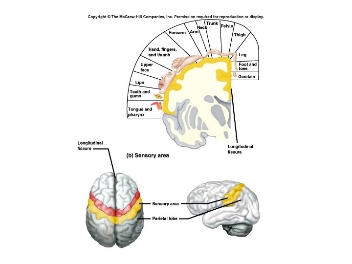
Sensory adaptation The only receptors that don’t adapt are: pain receptors Somatic Senses: Exteroceptive senses – changes at body surface Proprioceptive senses – changes in muscles and tendons and body position Visceroceptive senses – changes in viscera(The internal organs of the abdomen and thorax; specifically, the hollow Organs such as intestines, bladder, etc. )
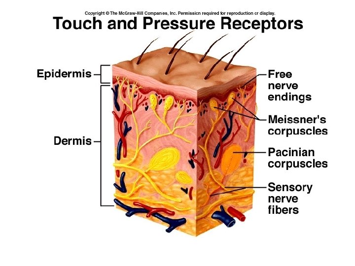
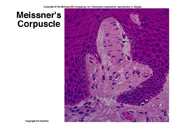
Touch and pressure senses: 1. Free nerve endings – touch and pressure 2. Meissner’s corpuscles – light touch receptors are connective tissue 3. Pacinian corpuscles – heavy pressure and vibrations receptors are connective tissue Itch and Tickle: Receptors are free nerve endings
Temperature senses: Free nerve endings in skin l Heat receptors – respond primarily between 25 – 45 o C or 77 – 113 o F unresponsive above, but pain receptors fire = burning l Cold receptors – respond primarily between 10 – 20 o C or 50 - 68 o F unresponsive below, but pain receptors fire = burning
Pain l Also free nerve endings l Most pain receptors can be stimulated by more than one stimulus, although some are more sensitive to mechanical damage, and others to extreme temperature, or chemicals. l Deficiency of blood flow (ischemia) and thus a deficiency of oxygen (hypoxia) can stimulate pain receptors.
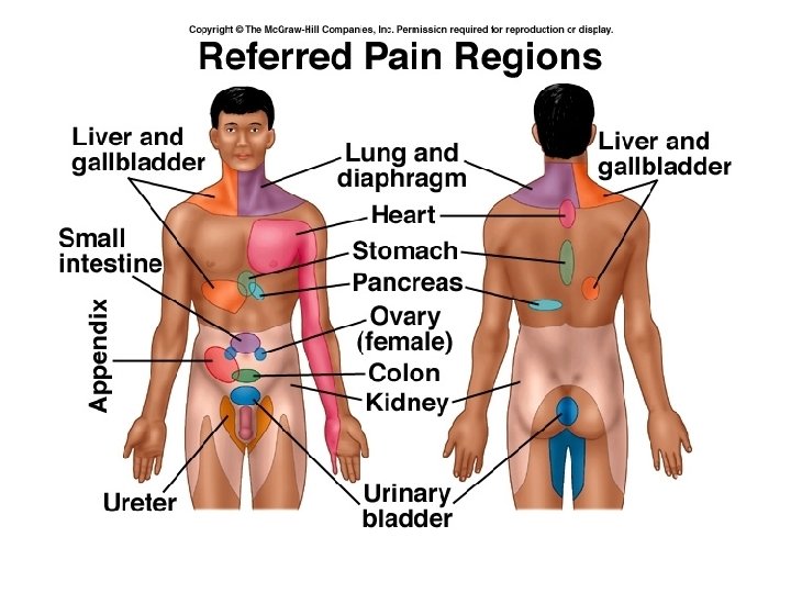
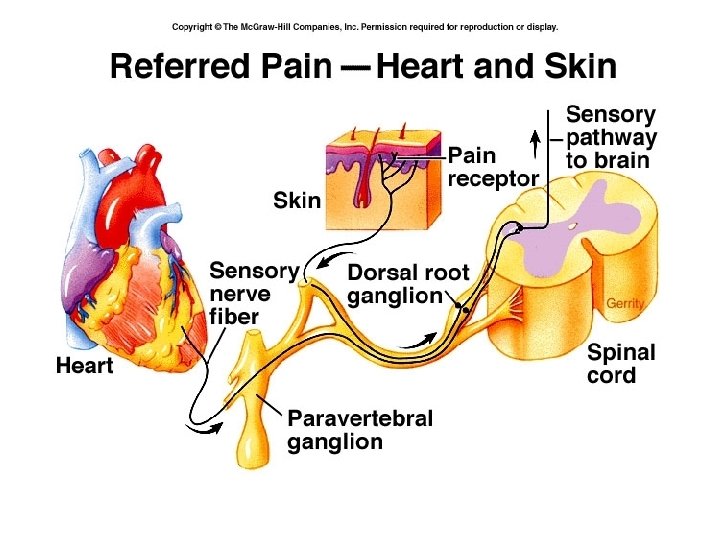
l Visceral pain: ¡Pain receptors are the only receptors in the viscera that produce sensations. ¡Tends to be referred pain – feels as though it comes from elsewhere – due to common nerve pathways
Phantom pain – comes from a limb that has been amputated. Pain fibers are of two types: Acute pain fibers ( A or delta fibers) – thin, myelinated fibers (Conducts up to 30 meters/sec) Sharp, localized pain Seldom continues after stimulus stops Chronic pain fibers (C fibers) – thin, unmyelinated fibers (conduct up to 2 meters per second) Dull, aching and widespread pain May continue for some time after stimulus
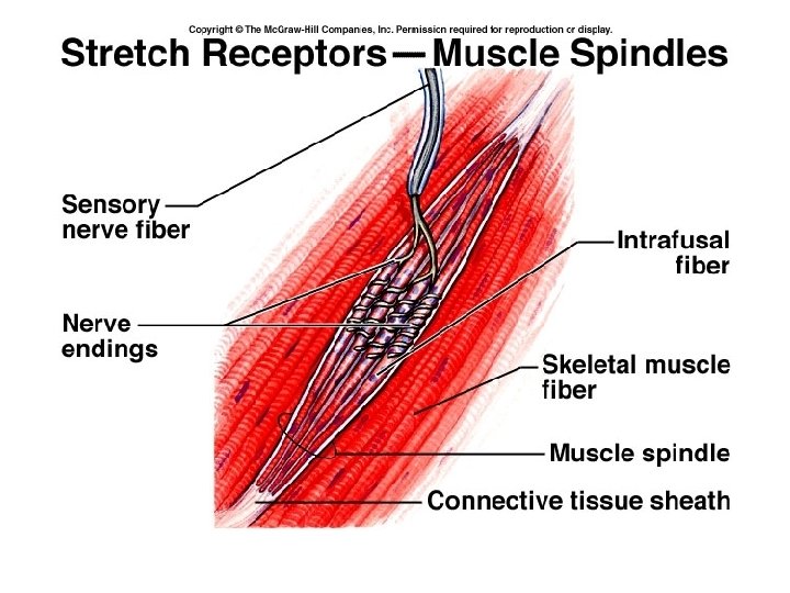
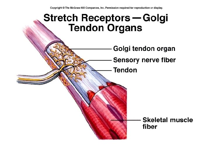
Stretch receptors: • We know how our body parts are moving through our proprioceptive or kinesthetic sense. • These receptors adapt only slightly • Keep brain informed of the status of body parts to insure coordination. • Use specialized receptors that sense tension in tendons and muscles. • No sensation occurs when these are stimulated.
Muscle spindles sense stretching of muscle, and cause contraction Of the muscle to maintain position. Golgi tendon organs sense stretching of tendons and cause the muscle to relax to prevent damage to the tendon.
Special Senses
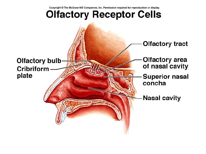
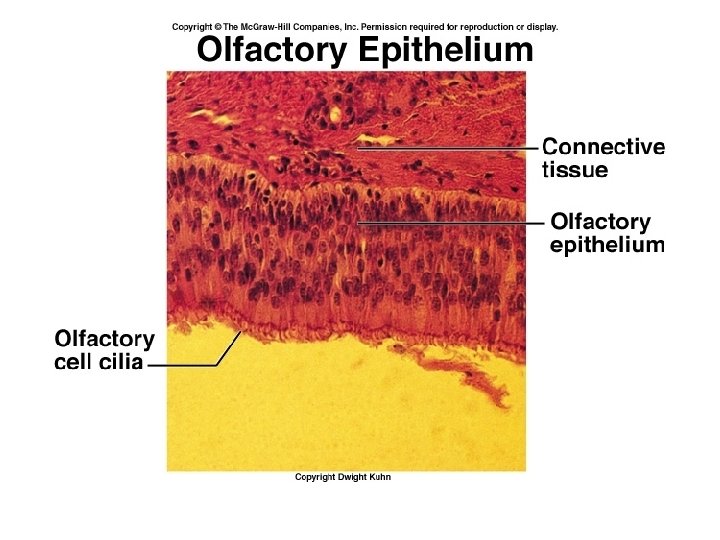
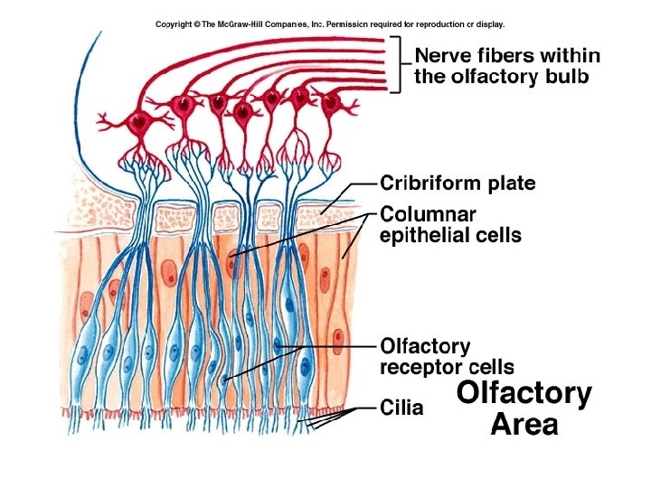
Olfactory sense – The sense of smell
Both smell and taste use chemoreceptors. Of all the senses, only smell and taste have fibers that run to both cortical areas And the limbic system.
Olfactory receptors are bipolar neurons. • Are replaced throughout lifetime, but lost at the rate of about 1 % per year. • The cilia, or olfactory hairs are the sensitive portions • Chemical must be dissolved in watery mucus to stimulate the receptor. • Combinations of primary scents allow us to recognize thousands of different odors.
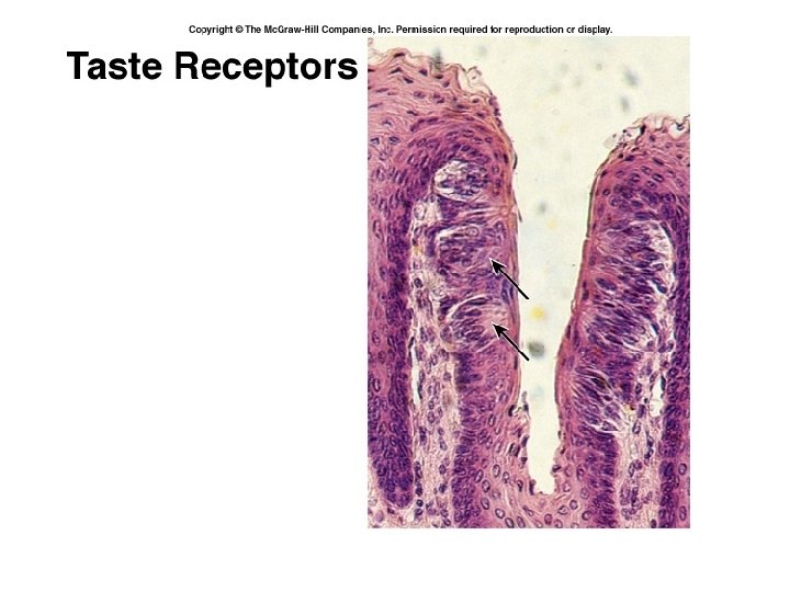
Gustatory sensations : Taste Uses chemoreceptors Substance must be dissolved before can be detected Detects 4 basic sensations: salty, sour, sweet and bitter Also may have receptors for alkaline, metallic, umami (Savoriness found in fermented and aged foods ) and water!
3, 6, 5, 9, 4, 1, 7, 0, 5, 2, 8.
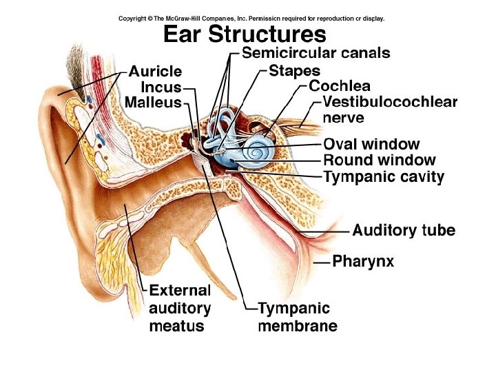
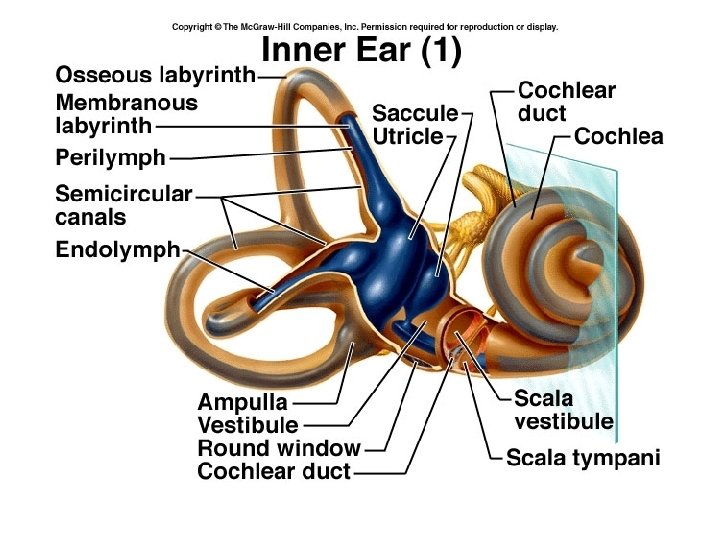
Auditory sensations and Equilibrium Hearing and equilibrium rely on mechanoreceptors The ear is divided into three parts: • Outer ear • Middle ear • Inner ear
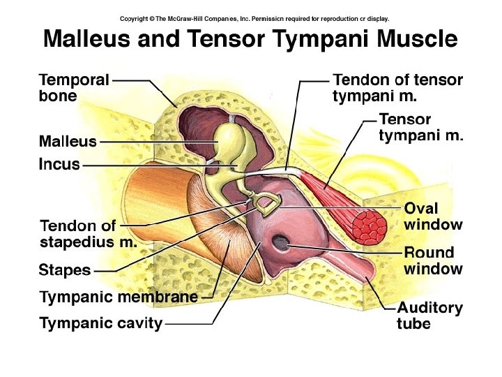
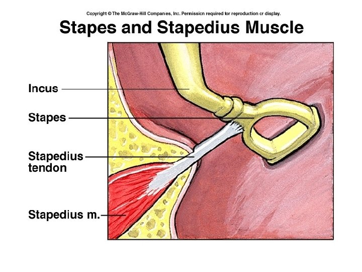
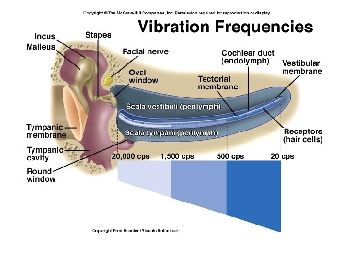
Outer ear: • Ceruminous glands – Cerumen - ear wax in external auditory meatus • Outer ear structures are the pinna (auricle), external auditory canal, and tympanic membrane (eardrum). Sound entering the external auditory canal sets the eardrum into vibration. These structures are involved with sound transmission only. Middle ear: • Tympanic antrum – opening into mastoid process • Auditory (Eustachian) Tube • Otitis media – inflammation of the MIDDLE ear • Auditory ossicles or ear bones • Tensor tympani muscle • Stapedius muscle • Tympanic reflex
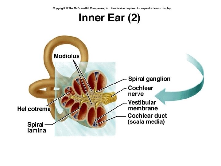
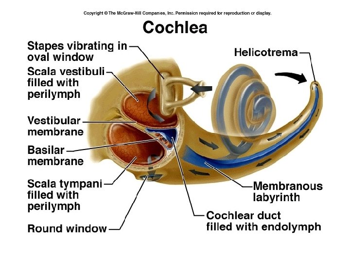
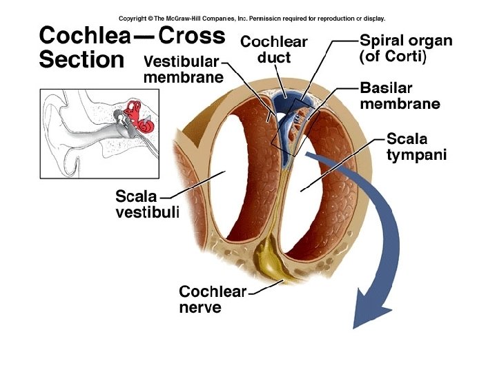
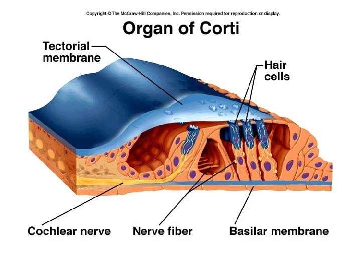
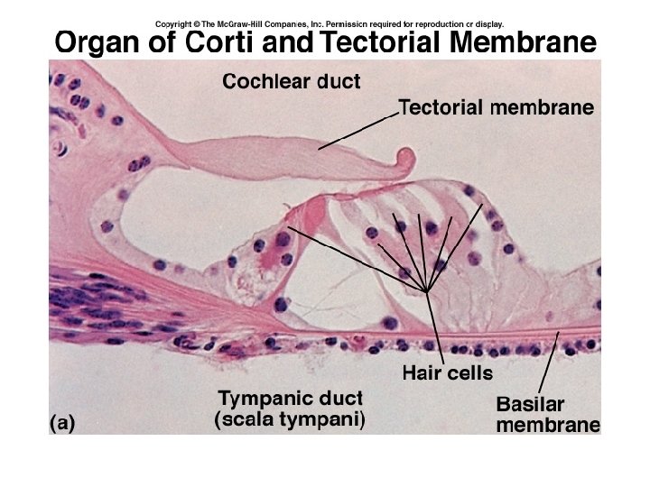
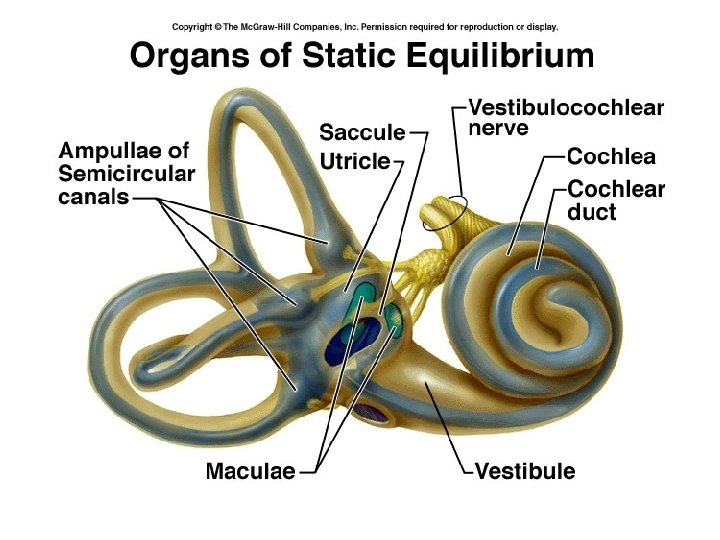
• The ossicles (malleus, incus, and stapes)transmit the vibratory motion from the eardrum to the oval window. The auditory tube allows pressure to be equalized on both sides of the eardrum. These structures are also involved with sound transmission only. INNER EAR : Bony chambers • Cochlea – hearing • Vestibule – static equilibrium • Semicircular canals – dynamic equilibrium • The bony labyrinth contains perilymph and membranous sacs filled with endolymph. Within the membranous sacs of the vestibule and semicircular canals are equilibrium receptors. Hearing receptors are found within the membranes of the cochlea.
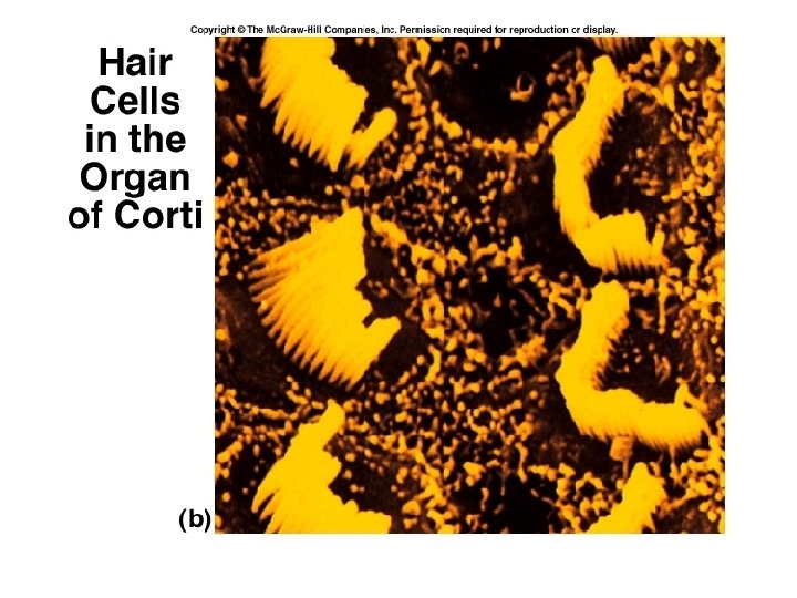
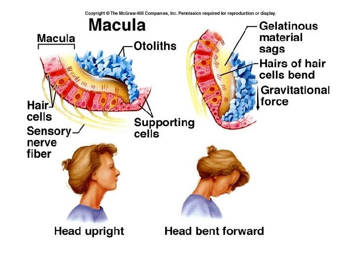
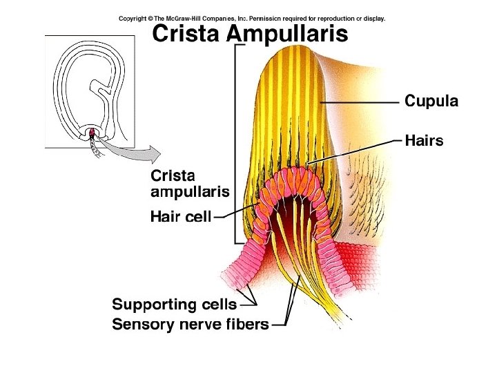
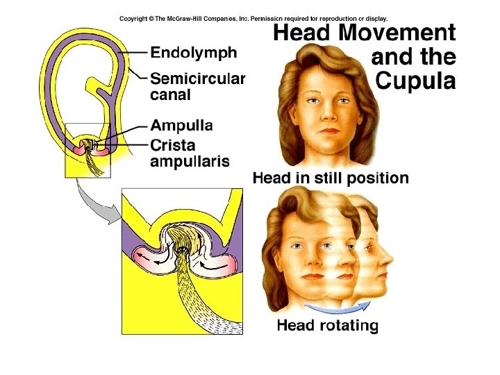
l Hair cells of the organ of Corti (the receptor for hearing within the cochlea) are stimulated by sound vibrations transmitted through air, membranes, and fluids l Deafness is any degree of hearing loss. Conduction deafness results when the transmission of sound vibrations through the external and middle ears is hindered. Sensorineural deafness occurs when there is damage to the nervous system structures involved in hearing. l Receptors of the semicircular canals (cristae) are dynamic equilibrium receptors, which respond to angular or rotational body movements. Receptors of the vestibule (maculae) are static equilibrium receptors, which respond to the pull of gravity and report on head position. Visual and proprioceptor input are also necessary for normal balance. l Symptoms of equilibrium apparatus problems include involuntary rolling of the eyes, nausea, vertigo, and an inability to stand erect.
Organ of Corti • Vestibulocochlear nerve – cranial nerve VIII • Audible range: 20 -- 20, 000 hertz • Ossicles amplify sound 22 X • Some nerve fibers cross over to opposite side of brain; some don’t. Why?
Equilibrium – Balance Static equilibrium – maintenance of body posture relative to gravity while the body is still. Dynamic equilibrium – maintenance of the body posture (mainly the head) in response to sudden movements. Tracking a moving object.
Static Equilibrium • Inside the vestibule are two chambers : utricle and saccule. • Regions of hair cells and supporting cells called maculae. • Otoliths – “ear rocks”
Dynamic Equilibrium • Semicircular canals • In ampulla is the crista ampullaris – contains hair cells and supporting cells covered by a gelatinous mass called the cupula. • Neurological connections between eyes and semicircular canals – for tracking • Nystagmus
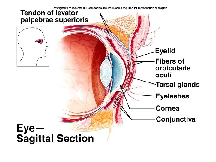
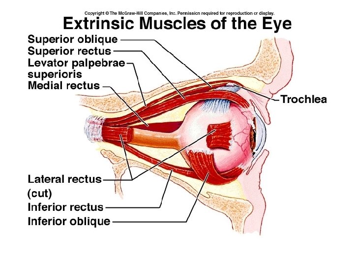
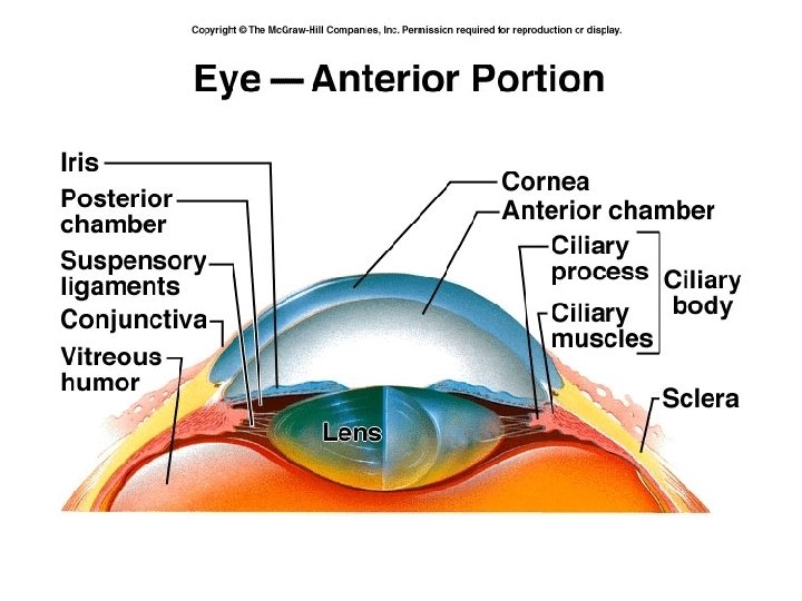
The Eye Accessory structures or Adnexa 4 layers: 1. Skin – thinnest in the body 2. muscle – orbicularis oculi and levator palpebrae superioris 3. Connective tissue – tarsal plate contains tarsal or Meibomian glands Chalazion
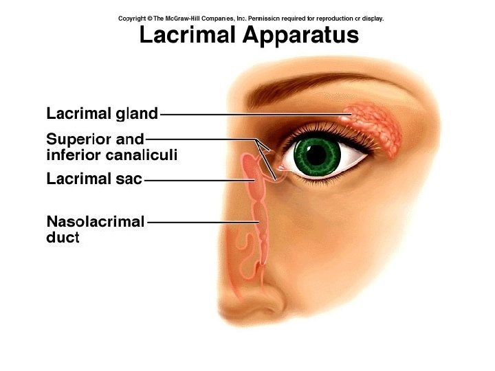
4. Conjunctiva – mucous membranpalpebral conjunctiva, bulbar conjunctiva Eyelashes sebaceous ciliary glands at base of hair follicles – hordeolum or stye Lacrimal apparatus – forming and draining tears.
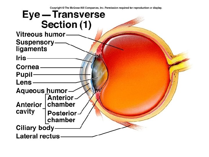
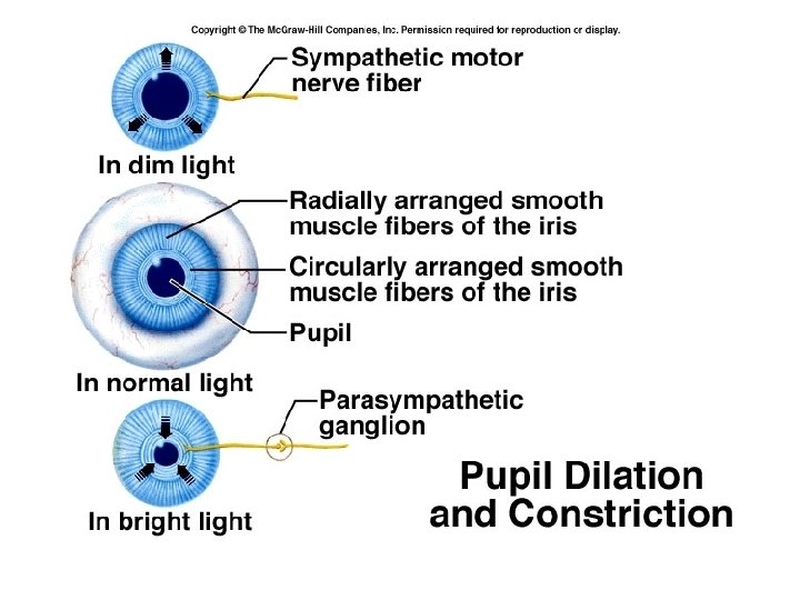
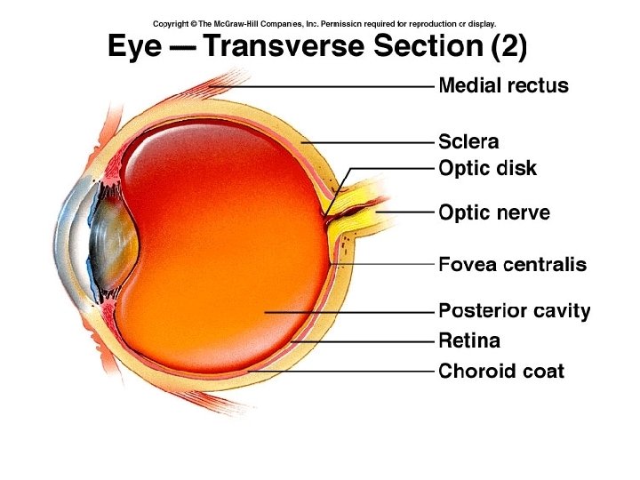
Strabismus – turned eye Phoria – weakness of eye muscles Eyeball – 3 layers or tunics: 1. Fibrous tunic (outer tunic) Cornea – clear - avascular Scleara – white –means “hard”
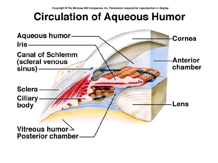
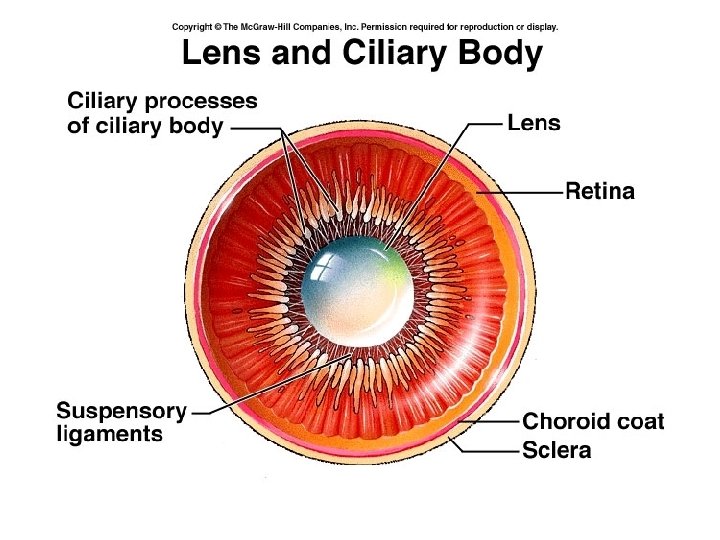
2. Vascular tunic : Uvea Choroid – blood vessels and pigment Ciliary body : ciliary processes make aqueous humor ciliary muscle - accomodation
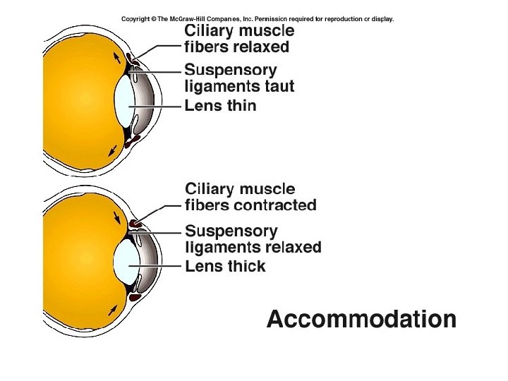
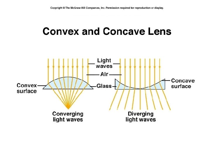
Accommodation: Focusing the eye to see close objects Lens is thin when stretched by suspensory ligaments (low power) When round ciliary muscle contracts, tension is released from lens and it becomes thicker (higher power lens).
Lens – avascular, clear, elastic contains proteins called crystallins becomes opaque = cataract Iris – pigmented, divides anterior and posterior chamber Aqueous humor – drains into scleral venous sinus (Schlemm’s canal) intraocular pressure - glaucoma
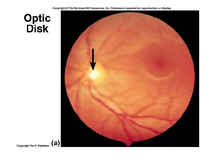
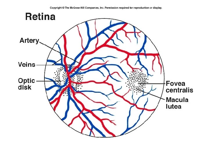
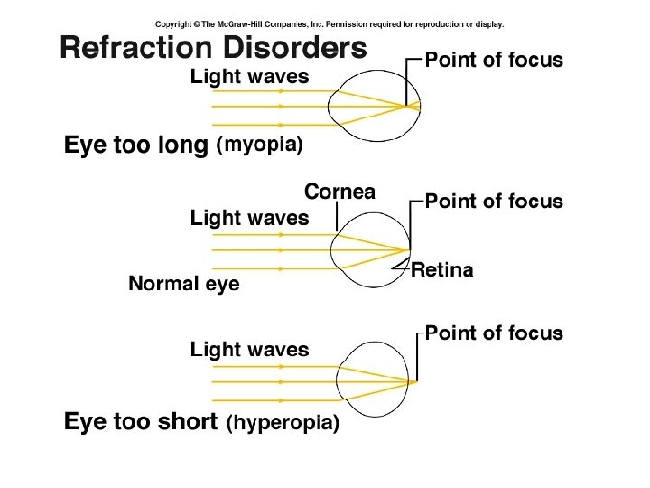
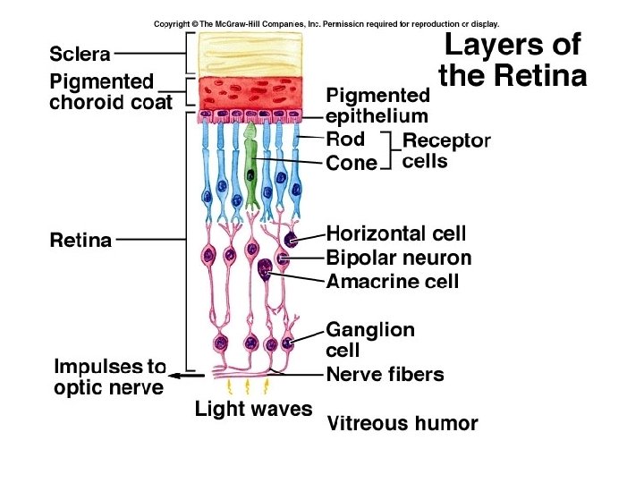
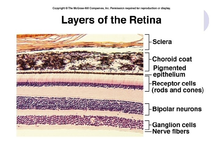
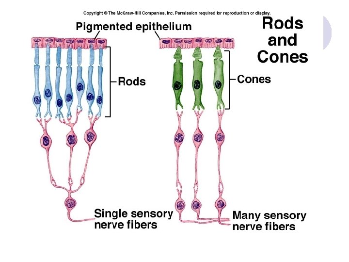
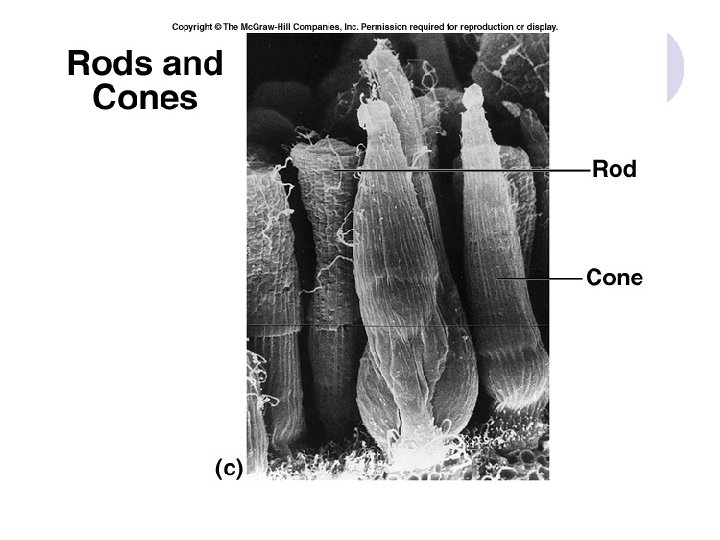
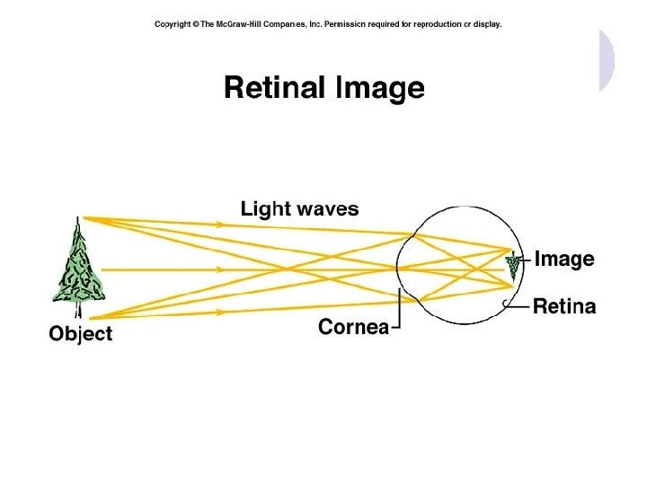
3. Nervous Tunic – Retina several layers “inside out” • Pigmented epithelium • Rods and Cones (photoreceptors) • Bipolar cells • Ganglion cells (vitreous humor) • Light is focused by cornea and lens on the Fovea centralis (“central pit”)which is in the center of the Macula lutea (“yellow spot”) The third refractive component is the length of the eyeball.
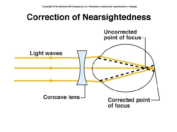
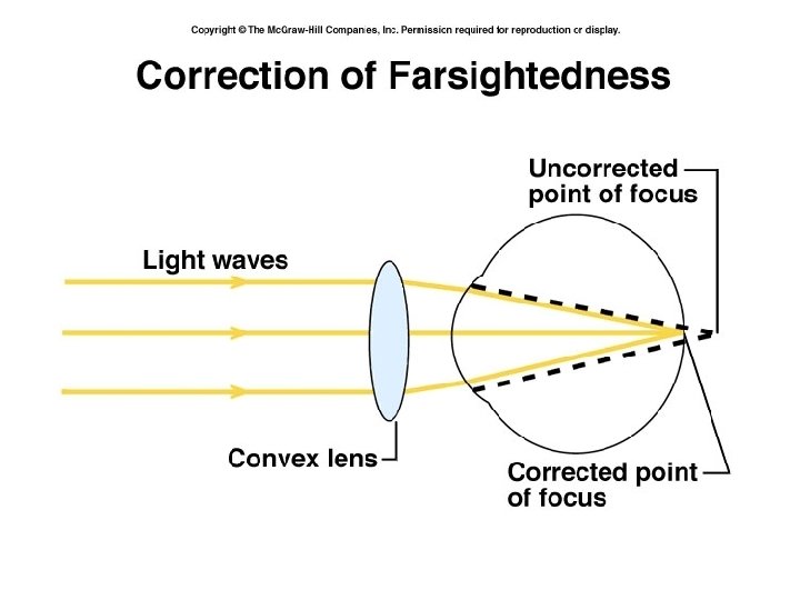
Refractive Disorders Emmetropia – good vision 20/20 Myopia – nearsightedness Hyperopia – farsightedness Astigmatism – light does not focus to a single point on the retina Presbyopia – “old sight” – loss of ability to accommodate or see up close
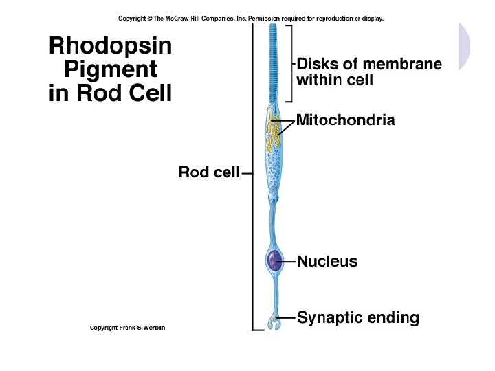
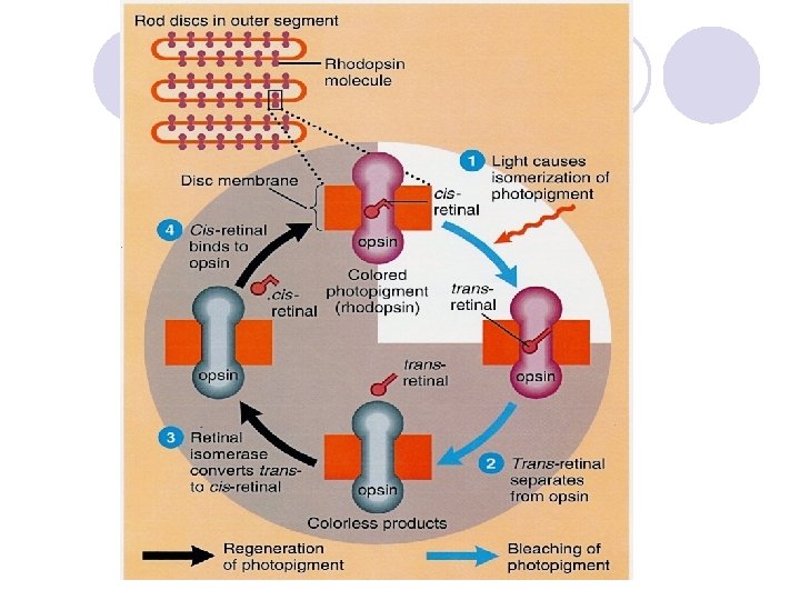
Physiology of vision In the dark, Na+ channels are held open by a nucleotide called cyclic GMP (guanosine monophosphate) Inflow of sodium (“dark current”) triggers the continual release of neurotransmitter. This neurotransmitter is inhibitory- it prevents bipolar cells from firing by hyperpolarizing them.
When light strikes the retina, retinal (from vitamin A) which is bent, straightens out, and no longer fits into the opsin. The two separate – this is called bleaching. The opsin becomes an active enzyme, that activates other enzymes that break down cyclic GMP. Without cyclic GMP, the Na+ channels close.
The receptor hyperpolarizes, stopping the release of inhibitory neurotransmitter. This decrease in inhibition allows the bipolar cells to fire, and information is sent to the visual cortex. Differentiation of color is assisted by horizontal cells. In darkness, retinal isomerase converts transretinal back to cis-retinal, which binds with opsin forming a functional photopigment.
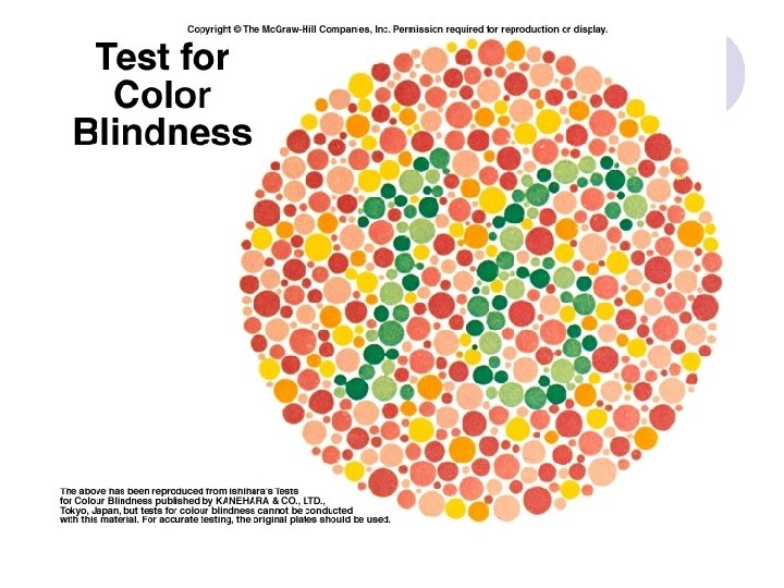
Color vision Uses three different photopigments : blue, green and red Wavelength of pigments may be shifted, causing color blindness Red – green color blindness most common Sex-linked trait carried on X chromosome (Males only have one gene for color vision)
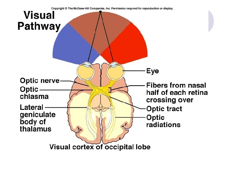
Stereopsis Use both eyes to perceive depth – “depth perception” Nasal fibers cross at the optic chiasm Temporal fibers do not cross over Each visual cortex (R &L) receives information from both eyes so it can compare what each eye sees.