Human Anatomy and Physiology Histology Slide Set 2


 Taste buds Taste bud Sublingual salivary glands")
 Cochlea Organ of Corti Cochlear duct Vestibular duct Tympanic duct Spiral ganglion")
 Organ of Corti Tectorial membrane Hair cells Basilar membrane Sensory neurons")
 Retina")
 Pacinian corpuscle")


 Adenohypophysis Neurohypophysis Pituitary stalk")
 Hypophysis cerebri (high power) Neurohypophysis Adenohypophysis")
 Thyroid gland")
 Parathyroid adipose tissue thyroid follicle")
 Adrenal gland Zona glomerulosa Zona fasciculata cortex Zona reticularis medulla")
 Pancreas islet of Langerhans acinar cells zymogen granules")

 Parotid salivary gland")
 Tongue")
 Esophagus muscle stratified squamous lumen submucosa muscle")

 Gastric mucosa")
")

 Islet of Langerhans (endocrine) (16) Pancreas (low power)")
 Jejunum")
 (18) Ileum")
 Appendix")
 Colon (large intestine)")
 Liver (hepatic lobule) central vein area of hepatic triad")

 Human blood erythrocytes neutrophil platelets lymphocyte")

 Neurovascular bundle vein artery nerve")
 and vein (above)")
 Artery (wall) smooth muscle connective tissue lumen w/ blood endothelium")
 Vein (showing valve) Which is the direction of normal blood flow?")
 Lymph node (cortical lymph nodule)")
 Lung bronchiole")
 Trachea")

 Kidney (renal cortex)")
 Ureter lumen transitional epithelium muscularis")
 Urinary bladder")

 Full urinary bladder")
 Empty urinary bladder")
 Ovary")
 Graffian follicle corona radiata primordial follicles oocyte antrum")
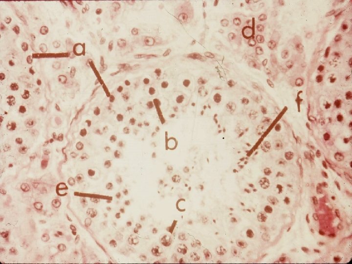
 Testis (seminiferous tubules) interstitial cells spermatozoa")
- Slides: 50
Human Anatomy and Physiology Histology Slide Set 2
Special Senses
papilla (1) Taste buds Taste bud Sublingual salivary glands
(2) Cochlea Organ of Corti Cochlear duct Vestibular duct Tympanic duct Spiral ganglion
(3) Organ of Corti Tectorial membrane Hair cells Basilar membrane Sensory neurons
(4) Retina
(5) Pacinian corpuscle
Pacinian corpuscle
Endocrine System
Hypopysis cerebri (low power) Adenohypophysis Neurohypophysis Pituitary stalk
(6) Hypophysis cerebri (high power) Neurohypophysis Adenohypophysis
thyroglobulin follicle (7) Thyroid gland
(8) Parathyroid adipose tissue thyroid follicle
(9) Adrenal gland Zona glomerulosa Zona fasciculata cortex Zona reticularis medulla
(10) Pancreas islet of Langerhans acinar cells zymogen granules
Digestive System
mucus acinus serous acinus (11) Parotid salivary gland
taste bud taste pore (12) Tongue
(13) Esophagus muscle stratified squamous lumen submucosa muscle
Stomach / Esophagus junction
gastric gland (14) Gastric mucosa
lumen muscularis crypts of Lieberkuhn inner circular outer longitudinal serosa submucosa (w/ Brunner’s glands) (15) Duodenum
Duodenum
acinar cells (exocrine) Islet of Langerhans (endocrine) (16) Pancreas (low power)
mucosa submucosa muscularis serosa (17) Jejunum
Peyer’s patch (lymph nodule) (18) Ileum
(19) Appendix
(20) Colon (large intestine)
(21) Liver (hepatic lobule) central vein area of hepatic triad
Cardiovascular & Respiratory Systems
(22) Human blood erythrocytes neutrophil platelets lymphocyte
Human blood
(23) Neurovascular bundle vein artery nerve
Artery (below) and vein (above)
(24) Artery (wall) smooth muscle connective tissue lumen w/ blood endothelium
(25) Vein (showing valve) Which is the direction of normal blood flow?
(26) Lymph node (cortical lymph nodule)
alveoli (27) Lung bronchiole
mucus glands hyaline cartilage pseudostratified ciliated columnar epithelium (28) Trachea
Urinary and Reproductive Systems
Bowman’s capsule glomerulus renal corpuscle (29) Kidney (renal cortex)
(30) Ureter lumen transitional epithelium muscularis
submucosa transitional epithelium detrusor muscle (31) Urinary bladder
Urinary bladder
(32) Full urinary bladder
(33) Empty urinary bladder
primary follicles cortex developing oocyte medulla (34) Ovary
(35) Graffian follicle corona radiata primordial follicles oocyte antrum
Testis (seminiferous (36) Testis (seminiferous tubules) interstitial cells spermatozoa