HUMAN ANATOMY AND PHYSIOLOGY 2 URINARY SYSTEM Consists


 in the ascending and descending")


 and")


 indicates abnormally high blood sugar")


")
 can warm the body")


 are located entirely within the CNS •")


 are surrounded")


 is used as a neurotransmitter")


 a hormone from the small intestine is also released")


 or Thyrotropin • Stimulates the thyroid gland to produce")


 It stimulates the kidneys to retain water Oxytocin Stimulates")


, the attachment of 2 iodines produces di-iodotyrosine (DIT) • Within the")

, which is important in the regulation of")


 in")

 secrete a")


- Slides: 179
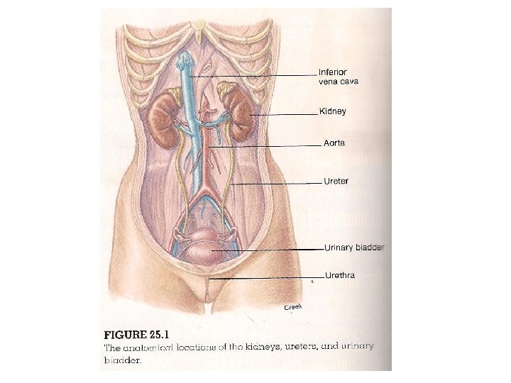
HUMAN ANATOMY AND PHYSIOLOGY 2 URINARY SYSTEM Consists of 2 kidneys, 2 ureters, the urinary bladder and the urethra Function of the human kidney : regulation of the extracellular fluid (plasma and interstitial fluid) environment in the human body This function is accomplished through the formation of urine During the formation of urine the kidneys regulate; 1. Regulate the volume of blood plasma- thus contributing to blood pressure 2. The concentration of waste products in the blood 3. The concentration of electrolytes- Na+, K+ HCO 3 - and other ions in the plasma 4. p. H of the blood plasma 1.
POSITION AND STRUCTURE OF THE KIDNEY • The kidneys lie on each lateral side of the vertebral column, between the levels of the 12 th thoracic and 3 rd lumbar vertebrae • The right kidney is usually slightly lower than the left kidney because of the area occupied by the liver • The kidneys are retroperitoneal ie they are positioned behind the parietal peritoneum • Thus strictly speaking they are not within the peritoneal cavity • They are bean-shaped • The hilum of the kidney is the depression through which the renal artery and nerves enter and the renal vein and ureter exit • Each kidney is embedded in a fatty fibrous pouch consisting of three layers:
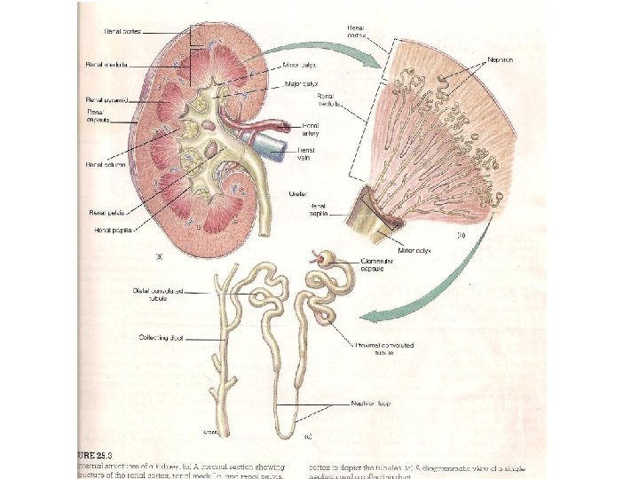
• Renal capsule: this the innermost layer that forms the strong transparent fibrous attachment to the kidney • It protects the kidney from trauma and the spread of infections • The second layer is formed by a tough protective layer of adipose tissue called the adipose capsule • The outermost layer is the renal fascia. It is a supportive layer that anchors the kidney to the peritoneum and the abdominal wall • Vertical Section of the Kidney • A vertical section of the kidney shows 2 distinct regions and a major cavity: • An outer renal cortex , in contact with the capsule. , this layer is reddish brown and granular because of its many capillaries
• The deeper region or renal medulla is darker in colour. , the presence of microscopic tubules and blood vessels gives this layer a striped appearance • The medulla is composed of 8 -15 conical renal pyramids which are separated by renal columns • The renal papillae are the apices of the renal pyramids • These projections are directed towards the large cavity of the kidney called the renal pelvis • The cavity of the kidney collects and transport urine from the kidney to the ureter •
Microscopic Structure of the Kidney • The nephron is the functional unit of the kidney that is responsible for urine production • A nephron consists of urinary tubules and associated blood vessels
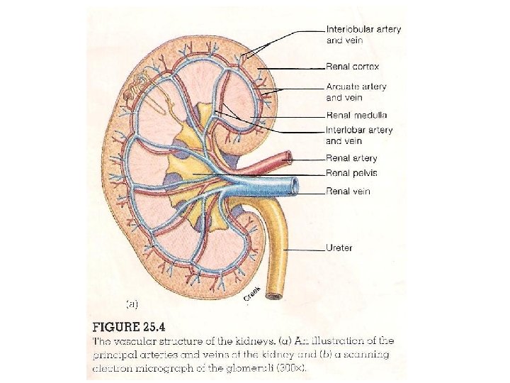
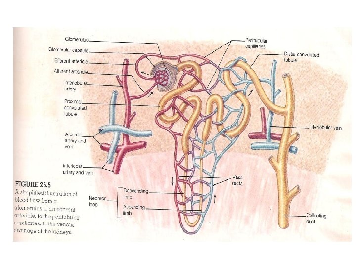
• • Renal Blood Vessels Arterial blood enters the kidney at the hilum through the renal artery It then divides into interlobar arteries that pass between the pyramids Arcuate arteries branch from the interlobar arteries at the boundary of the renal cortex and renal medulla Many interlobular arteries radiate from the arcuate arteries and subdivide into numerous afferent arterioles which are microscopic in size The afferent arterioles deliver blood into capillary networks called glomeruli The glomeruli produce a blood filtrate that enter the urinary tubules The blood remaining in the glomerulus leaves through the efferent arteriole which then delivers blood into another capillary network, the peritubular capillaries
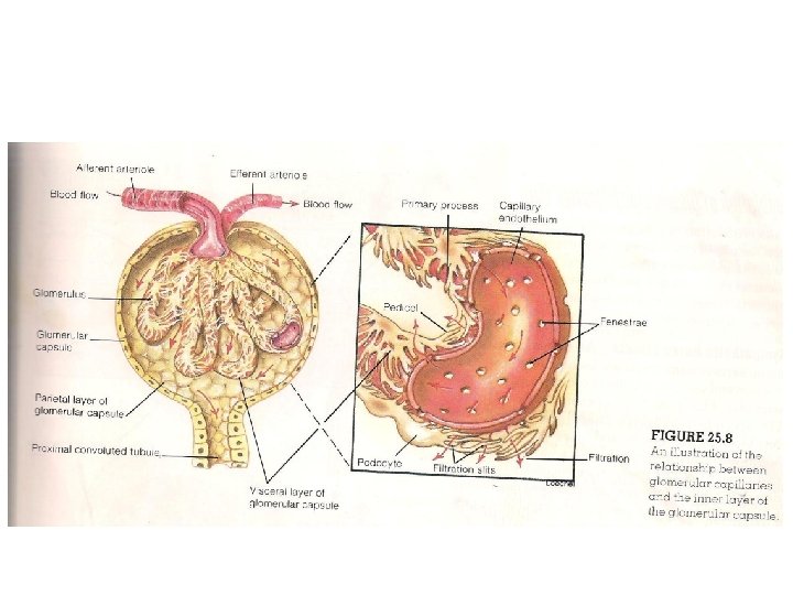
• Blood form the peritubular capillaries is drained into veins that • Run parallel to the course of the arteries in the kidney • These are the interlobular veins, arcuate veins and interlobar veins • The interlobar veins converge and leave the kidney as a single renal vein that empties into the inferior vena cava • Nephron tubules • The tubular portion of the nephron consists of a glomerular capsule, proximal convoluted tubule, descending limb of the loop of henle, ascending limb of nephron loop and distal convoluted tubule • The glomerular (Bowman’s) capsule surrounds the glomerulus • The Bowman’s capsule and the associated glomerulus are located in the renal capsule. , together they constitute the renal corpuscle
• The glomerular capsule contains an inner visceral layer of epithelium and an outer parietal layer • The space between these two layers is the capsular space which receives the glomerular filtrate • The filtrate in the glomerular capsule passes into the lumen of the proximal convoluted tubule • The wall of the proximal convoluted tubule consists of a single layer of cuboidal cells containing millions of microvilli • These serve to increase the surface area for reabsorption of useful substances • In the process of reabsorption, salt, water and other molecules needed by the body are transferred from the lumen into the tubular cell and into the surrounding peritubular capillaries • The glomerulus, glomerular capillaries and the proximal convoluted tubules are located in the renal cortex • Fluid then passes from the proximal convoluted tubule to the nephron loop
• The fluid is then carried into the renal medulla in the descending limb of the loop of Henle and returns to the renal cortex in the ascending limb of the loop • Back in the renal cortex, the tubule coils again as the distal convoluted tubule • The distal convoluted tubule is shorter than the proximal convoluted tubule and has fewer microvilli • It empties into the collecting duct • Glomerular Filtration • The glomerular capillaries have large pores called fenestrae and are thus said to be fenestrated • As a result of these large pores, glomerular capillaries are 100 to 400 times more permeable to plasma, water and other dissloved solutes than are the capillaries of skeletal muscles
• Although the pores of the glomerular capsule are large, they are still small enough to prevent the passage of blood corpuscles into the filtrate • Before the filtrate can enter the interior of the glomerular capsule it must pass through the capillary pores, the basement membrane and the inner visceral layer of the glomerular capsule • The inner layer of the glomerular capsule is composed of specialized cells called podocytes, with numerous cytoplasmic extensions known as pedicels • These interdigitate and wrap around the glomerular capillaries • The narrow slits between adjacent pedicels provide the passageways through which filtered molecules must pass to enter the interior of the glomerular capsule • Even though the glomerular capillary pores are large enough to permit the passage of proteins, the fluid that enters the capsular space is completely free of plasma proteins
• This is due to their negative charges which hinder their passage through the negatively charged glycoproteins in the basement membrane of the capillaries • Their large size together with the negative charges of the plasma proteins may also restrict their movement though the filtration slits between the pedicels • Glomerular Ultrafiltrate • The fliud that enters the glomerular capsule is called ultrafiltrate because it is formed under pressure • The volume of filtrate produced per minute by both kidneys is called the glomerular filtration rate (GFR) • GFR averages 115 ml per minute in women and 125 ml per minute in men • This is equivalent to about 7. 5 L per hour or 180 L per day (about 45 gallons)
• Since the total blood volume averages 5 L, it means that the total blood volume is filtered into the urinary tubules every 40 minutes • Most of the filtered water is immediately returned to the circulatory system otherwise a person would urinate to death within minutes • Reabsorption of salt and Water • Although about 180 L of glomerular ultrafiltrate are produced each day, the kidneys normally excrete only 1. 5 L of urine each day • Approximately 99% of the filtrate is thus returned to the circulatory system, while only 1% is excreted in urine • Urine however varies according to the needs of the body • When a well hydrated person drinks a litre or more of water, • Urine volume increases to 16 ml per minute (about 23 L /day. )
• In severe dehydration, when the body needs to conserve water, only 0. 3 ml of urine per minute or 400 ml per day are produced • A volume of 400 ml of urine per day is needed to excrete the amount of metabolic waste produced by the body • This is known as obligatory water loss • When water in excess of this amount is excreted, the urine volume is increased and its concentration is decreased • Regardless of the body’s state of hydration, most of the filtered water must be returned to the circulatory system to maintain blood volume and pressure • The return of filtered molecules from the tubules to the blood is called selective reabsorption
Reabsoption in the proximal convoluted tubule • The concentration of Na+ in the glomerular filtrate is the same as that in the plasma • The epithelial cells of the tubule, however have a much lower Na+ concentration • This is due in part to the low permeability of the cell membrane to Na+ and in part to the active transport of Na+ out of the cell by Na+/K+ pumps • As a result of the action of these active transport pumps, a concentration gradient is created that favours the diffusion of Na+ from the tubular fluid into the cells of the proximal convoluted tubule • The Na+ is then extruded into the surrounding tissue fluid by the Na+/K+ pumps
• The transport of Na+ from the tubular fluid into the interstitial fluid creates a potential difference across the wall of the tubule • This electrical gradient favours the passive transport of Cltowards the higher Na+ concentration in the tissue fluid • Chloride ions therefore passively follow Na+ out of the filtrate into the interstitial fluid • As a result of the accumulation of Na. Cl, the osmolarity and the osmotic pressure values of the interstitial fluid surrounding the epithelial cells increase to a value above those of the tubular fluid • An osmotic gradient is thus created between the tubular fluid and the interstitial fluid surrounding the proximal convoluted tubule • Water moves by osmosis from the tubular fluid into the epithelial cells and then into the interstitial fluid
• The salt and water which were reabsorbed form the tubular fluid then moves passively into the surrounding peritubular capillaries • Significant of P. C. T Reabsorption • Approximately 65% of the salt and water in the original glomerular ultrafiltrate is reabsorbed in the proximal convoluted tubule and returned to the vascular system • The volume of tubular fluid is reduced, but this is still isoosmotic with the blood • An additional small amount of salt and water is returned to the vascular system by reabsorption in the nephron loop • This reabsorption, like that in the proximal convoluted tubule occurs constantly regardless of the person’s state of hydration and is not subjected to hormonal regulation
• Approximately 85% of salt and water are reabsorbed in a constant unregulated fashion in the proximal convoluted tubule and the nephron loop • This reabsorption is costly in terms of energy expenditures, accounting for 6% of the calories consumed by the body at rest • Only 15% of the initial filtrate remains to enter the distal convoluted tubule and collecting duct • This still large volume of fluid • 15% of GFR , 15% × 180 L per day = 27 L/day • This must be reabsorbed to varying degrees in accordance with the body’s state of hydration
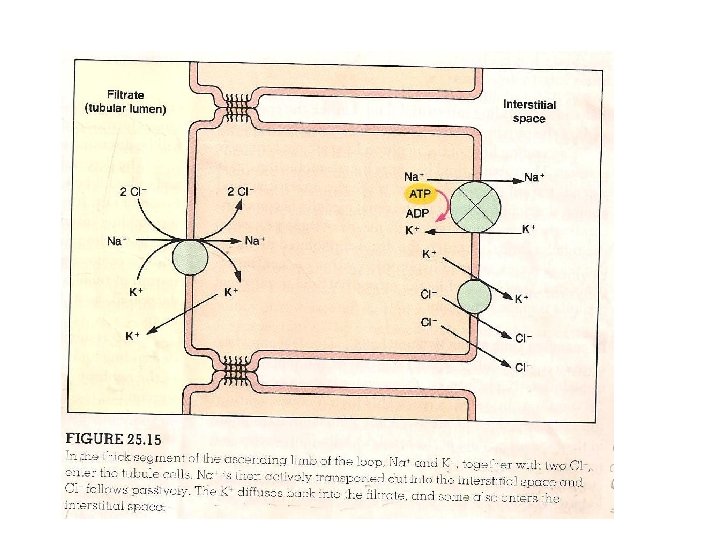
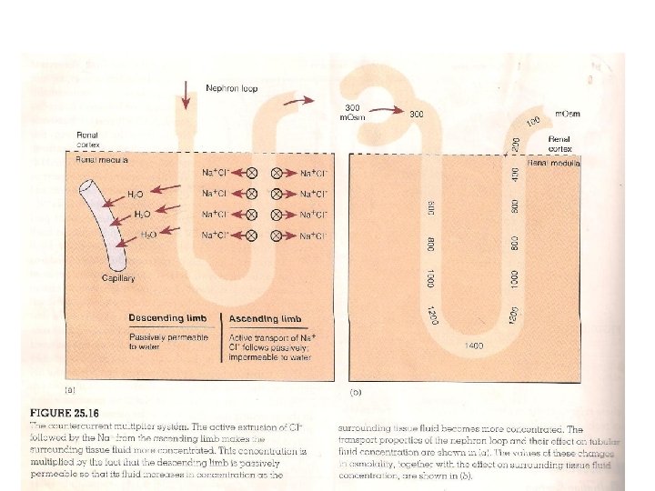
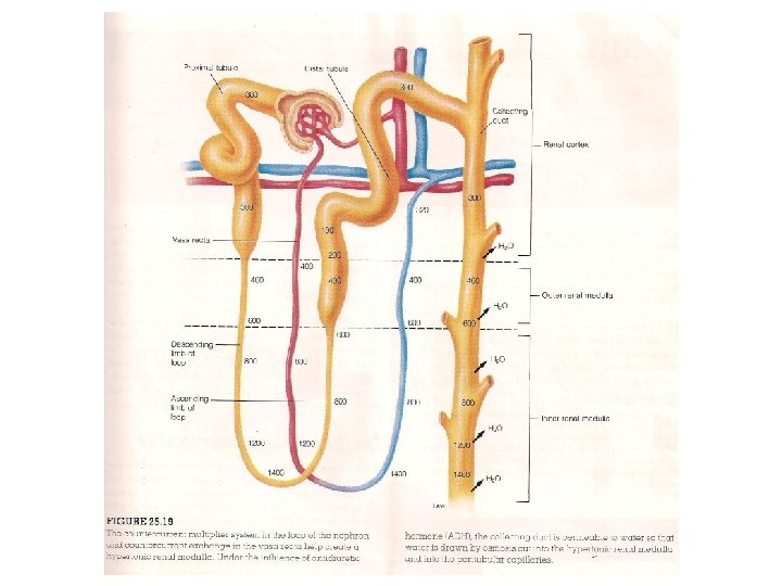
Counter current Multiplier System • In order for water to be reabsorbed by osmosis, the surrounding interstitial fluid must be hypertonic • The osmotic pressure of the interstitial fluid in the renal medulla is raised to over four times that of the plasma • This result party because the tubule bends, allowing for the interaction between the ascending and descending limbs of the nephron loops • Ascending limb of nephron • Salt (Na. Cl) is actively extruded from the ascending limb into the surrounding interstitial fluid • This is not accompanied by the same process that occurs in the proximal convoluted tubule
• Instead Na+, K+ and Cl- passively diffuse from the filtrate into the ascending limb cells • Na+ is then actively transported to the tissue fluid by the Na+/K+ pump • Cl- ion follows the Na+ passively and K+ diffuses back into the filtrate • The ascending limb is structurally divisible into 2 regions: a thin segment, nearest to the tip of the nephron loop • And a thick segment which carries the filtrate into the renal cortex and into the distal convoluted tubule • The thin segment is composed of simple squamous epithelium, whereas the thick segment is composed of simple cuboidal epithelium • It is believed that only the cells of the thick segment of the ascending limb are capable of actively transporting Na. Cl from the filtrate into the tissue fluid
• Unlike the epithelial walls of the proximal convoluted tubule, however, the walls of the ascending limb of the nephron loop are not permeable to water • Thus the tubular fluid becomes increasingly dilute as it ascends towards the renal cortex, whilst the interstitial fluid around the nephron loops in the renal medulla becomes increasingly more concentrated • By means of these processes, the tubular fluid that enters the distal convoluted tubule in the renal cortex is made hypotonic, while the interstitial fluid in the renal medulla is made hypertonic • Descending limb of nephron loop • The descending limb of the nephron does not actively transport salt and is impermeable the passive diffusion of salt • It is however permeable to water
• Since the surrounding interstitial fluid is hypertonic to the filtrate in the descending limb, water is drawn out of the descending limb by osmosis and enters the blood capillaries • The concentration of the tubular fluid is thus increased and its volume is decreased as it descends towards the tips of the nephron loops • As a result of these passive transport processes in the descending limb, the fluid that ‘rounds the bend’ at the tip of the nephron loop has the same osmolarity as that of the surrounding tissue fluid • There is therefore a higher salt concentration arriving in the descending limb • Salt transport by the ascending limb is increased accordingly, so that the ‘saltness’ of the interstitial fluid is multiplied
Counter-current Multiplication • Counter-current flow (flow in opposite direction) in the ascending and descending limbs and the close proximity of the two limbs allow for interaction • Since the concentration of fluid in the descending limb reflects the concentration of the surrounding fluid, and since the concentration of this interstitial fluid is raised by the extrusion of salt from the ascending limb, a positive feedback mechanism is created. • The more salt the ascending limb extrudes, the more concentrated will be the fluid that returns to it from the descending limb • This positive feedback mechanism that multiplies the concentration of the interstitial fluid and descending limb is called the counter-current multiplier system
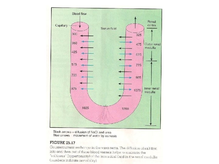
Effects of urea • Urea also contributes significantly to the total osmolarity of the interstitial fluid • It has been shown that the ascending limb of the nephron loop and the collecting duct are permeable to urea • Urea can thus diffuse out of the collecting duct and into the ascending limb • In this way, a certain amount of urea is recycled through these two segments of the nephron and trapped in the interstitial fluid • Collecting duct: Effect of Antidiuretic hormone • As a result of the recycling of salt between the ascending and descending limbs and the recycling of urea between the collecting duct and the nephron loop, the medullary interstitial fluid is made very hypertonic
• The collecting duct must transport their fluid through this hypertonic environment • While the fluid surrounding the collecting duct in the renal medulla is hypertonic, the fluid that passes into the collecting ducts in the renal cortex is hypotonic, due to the active extrusion of salt by the ascending limb of the nephron loops • The walls of the collecting ducts are permeable to water but not to salt • Since the surrounding interstitial fluid in the renal medulla is very hypertonic, water is drawn out of the collecting duct by osmosis • This water does not dilute the surrounding interstitial fluid because it is transported by capillaries to the general circulation • In this way, most of the water remaining in the filtrate is returned to the circulatory system
• It must be noted that the osmotic gradient created by the countercurrent multiplier system provides the force for water absorption through the collecting ducts • The rate at which this osmotic movement occurs is determined by the permeability of the collecting duct cell membranes to water • This also depends on the number of water channels in the cell membranes of the collecting duct epithelial cells • The water channels are proteins within the membranes of vesicles that branch from the Golgi apparatus • Anti-diuretic hormone (ADH) acting as a messenger stimulates the fusion of these vesicles with the cell membranes in response to ADH, so that the collecting duct becomes more permeable to water • When ADH is no longer secreted and no longer binds to its membrane receptors in the collecting duct, the water channels are removed from the cell membrane by the process of endocytosis
• When ADH concentration is increased, the collecting duct becomes more permeable and more water is reabsorbed • A decrease in ADH results in less reabsorption of water leading to excretion of larger volumes of more dilute urine • ADH is produced by neurons in the hypothalamus and is secreted from the posterior lobe of the pituitary gland • The secretion of ADH is stimulated when osmoreceptors in the hypothalamus respond to an increase in blood osmotic pressure • During dehydration, therefore when the plasma becomes more concentrated, increased secretion of ADH promotes increased permeability of the collecting ducts to water • In severe dehydration, only minimal amount of water needed to eliminate the body’s waste is ecreted • This minimum, about 400 ml/day is limited by the fact that urine cannot become more concentrated than the medullary • Tissue fluid surrounding the collecting ducts
• Under these conditions about 99. 8% of the original glomerular filtrate is reabsorbed • A person in a normal state of hydration excretes about 1. 5 L of urine per day, indicating that 99. 2% of the glomerular filtrate volume is reabsorbed • Reabsorptionof Glucose and Amino Acids • Glucose and amino acids in the blood are easily filtered by the glomeruli into the renal tubules • These molecules are however usually not present in the urine • This means that the filtered glucose and amino acids are normally completely reabsorbed by the nephrons • This reabsorption occurs by carrier-mediated active transport process • One characteristic of carrier mediated transport is saturation
• This means that when a transport molecule is present in sufficiently high concentration, all of the carriers are occupied and transport rate reaches a maximal value known as Transport maximum (Tm) • Beyond this rate, further increases in concentration will not increase the transport rate further • The carriers for glucose and amino acids in the renal tubules are not saturated and so are able to remove the filtered molecules completely • The Tm for glucose averages 375 mg/min, which is well above the rate at which glucose is delivered to the tubules • The rate of glucose delivery can be calculated by multiplying • The plasma glucose concentration(about 1 mg/ml) by the Glomerular Filtration Rate ( about 125/min) • Approximately 125 mg/min are thus delivered to the tubules whereas a rate of 375 mg/min is required to reach saturation
Glycosuria • When more glucose passes through the tubules than can be reabsorbed, glucose appears in the urine in a condition called glycosuria • This condition occurs when the plasma glucose concentration reaches 180 -200 mg/100 ml • Since the rate of glucose delivery is still below the average Tm for glucose , one can conclude that some nephrons are have considerably lower Tm values than the average • The renal plasma threshold is the minimum plasma concentration of a substance that results in the excretion of that substance in the urine • The renal plasma threshold for glucose is 180 -200 mg/ 100 ml • Glucose is normally absent from urine because glucose concentration remains below this threshold value
• Glycosuria occurs only when plasma glucose concentration is abnormally high (hyperglycemia) and exceeds renal plasma threshold • Fasting hyperglycemia is caused by inadequate secretion or action of insulin • When hyperglycemia results in glycosuria, the disease is called diabetes mellitus • A person with uncontrolled diabetes mellitus excretes large volumes of urine because the excreted glucose carries with it water as a result of the osmotic pressure it generates in the tubules • This condition is different from diabetes insipidus, in which a large volume of dilute urine is excreted as a result of inadequate ADH secreton
Renal Control of Electrolyte Balance • The kidneys help to regulate the concentration of plasma electrolytes such as Na+, K+, Cl-, HCO 3 - PO 4 - etc • This it does by matching the urinary excretion of these ions to the amounts ingested • The control of plasma Na+ is important in the regulation of blood volume and pressure • Control of K+ is required to maintain proper functioning of cardiac and skeletal muscles • The hormone aldosterone stimulates the reabsorption of Na+ in exchange for K+ in the distal convoluted tubule • It therefore promotes the renal retention of Na+ and excretion of K+
Role of Aldosterone in Na+/K+ Balance • Approximately 90% of the filtered Na+ and K+ are reabsorbed by the proximal convoluted tubule cells before the filtrate reaches the distal convoluted tubules • This early reabsorption occurs at a constant rate and not subjected to hormonal regulation • The final concentration of Na+ and K+ in the urine is varied according to the needs of the body by processes that occur in the distal convoluted tubule and in the cortical region of the collecting duct • Renal excretion and retention of Na+ and K+ are regulated by aldosterone, a hormone secreted by the adrenal cortex
Sodium ion Reabsorption • About 90% of Na+ present in the filtrate is reabsorbed in the proximal convoluted tubule • The amount left that is delivered to the distal convoluted tubule is still quite large • In the absence of aldosterone, 80% of this amount is automatically reabsorbed through the wall of the distal convoluted tubule into the peritubular blood • This is equivalent to 8% of the amount filtered • The amount of Na+ excreted without aldosterone is 2% of the amount filtered • This represents about 30 g /day excreted in the urine • However when aldosterone is secreted in maximal amounts all of the Na+ delivered to the distal convoluted tubule is reabsorbed • Under these conditions, urine contains no Na+ at all
Potassium ion Secretion • About 90% of the filtered K+ is reabsorbed in the early regions of the nephron, mainly in the proximal convoluted tubule • When aldosterone is absent all of the remaining filtered K+ is reabsorbed in the distal convoluted tubule • Thus in the absence of aldosterone no K+ is excreted in the urine • The presence of aldosterone stimulates the secretion of K+ from the peritubular blood into the distal convoluted tubule and cortical region of the collecting duct • This aldosterone-induced secretion is the only means by which K+ can be found in the urine • When aldosterone secretion is at its maximum, as much as 50 times more K+ is excreted in the urine because of secretion into the distal convoluted tubule than was filtered through the glomeruli
• Thus aldosterone promotes Na+ retention and K+ ion loss from the blood by stimulating the reabsorption and the secretion of K+ across the wall of the distal convoluted tubules and cortical portions of the collecting ducts • Since aldosterone promotes the retention of Na+ ion it contributes to an increased blood volume and pressure • Control of Aldosterone Secretion • Since aldosterone promotes Na+ retention and K+ ion loss, one might predict that aldosterone secretion will be increased when there is a low or high K+ concentration in the blood. , this indeed is the case • A rise in blood K+ directly stimulates the secretion of aldosterone from the adrenal cortex. , decrease in plasma Na+ concentrations also promotes aldosterone secretion
Juxtaglomerular Apparatus • The juxtaglomerular apparatus is the region in each nephron where the affarent arteriole and distal convoluted tubule come into contact • The micoscopic appearance of the afferent arteriole and distal convoluted tubule in this region differs from their appearance in other regions • Modified cells in the walls of the afferent arteriole , called granular cells contain the enzyme renin • When this enzyme is released in to the blood it catalyses the conversion of angiotensinogen into angiotensin I • Secretion of renin into the blood results in the formation of angiotensin I which is converted to angiotensin II • Angiotensin II stimulates the adrenal cortex to secrete aldosterone • Conditions that result in renin secretion cause increased aldosterone secretion and thus promotes the reabsorption of Na+ in the distal convoluted tubule
Regulation of renin secretion • A fall in plasma Na+ concentration is always accompanied by a fall in blood volume • This is because ADH secretion is inhibited by the decreased plasma concentration(osmolarity) • With less ADH, les water is reabsorbed through the collecting ducts and more water is excreted in urine • The fall in blood volume and the fall in renal blood flow causes increased renin secretion • Renin secretion is also stimulated by sympathetic nerve activity, which is increased when the blood volume and pressure fall • An increased secretion of renin as a result of increased production of angiotensin II acts to stimulate aldosterone production • Consequently less Na+ is excreted in the urine and more is retained in the blood –a negative feedback mechanism
Renal control of Acid-Base balance • Through the excretion of H+ and their reabsorption of HCO 3 -, the kidneys are responsible for the metabolic component of acid-base regulatory systems in the body • The regulation of acid-base balance has both respiratory and metabolic components • The respiratory component refers to the effect of ventilation on arterial PCO 2 and therefore the production of carbonic acid • The metabolic component refers to the effect of nonvolatile metabolic acids such as lactic acids and fatty acids on blood p. H • A normal arterial blood p. H is obtained when both the lungs and kidneys are functioning correctly and when the ratio of bicarbonate and carbon dioxide is normal
• The p. H of the blood can be calculated using the Henderson. Hasselbalch equation • p. H = 6. 1+ log {HCO 3 -} • {CO 2} • A normal p. H is obtained when the ratio of HCO 3 - : CO 2 is 20: 1 • A change in this ratio results in an abnormal blood p. H leading to acidosis or alkalosis • Pure respiratory acidosis or alkalosis occurs when the HCO 3 concentration is normal but the PCO 2 and H 2 CO 3 are altered • Pure metabolic acidosis or alkalosis occurs when the PCO 2 and H 2 CO 3 are normal but the HCO 3 - concentration is abnormal
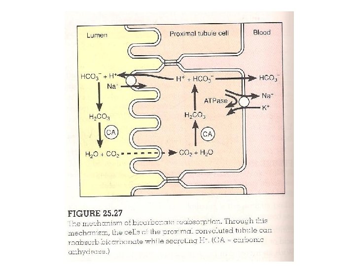
Mechanism of Acid-Base Balance • The kidneys help to regulate the blood p. H by their excretion of H+ in the urine and by their reabsorption of HCO 3 • These two mechanisms are independent • Reabsorption of HCO 3 - occurs as a result of filtration and secretion of H+ • The kidneys normally reabsorb all of the filtered HCO 3 - and excrete H+ • Normal urine therefore is free of bicarbonate ion and is slightly acidic (p. H 5 -7) • Reabsorption of HCO 3 - in the P. C. T. • The apical membranes of the tubules are impermeable to bicarbonate ion. , reabsorption of HCO 3 - occurs indirectly • When the filtrate is acidic, HCO 3 - combines with H+ to form carbonic acid, H 2 CO 3
• Carbonic acid in the filtrate dissociates to form carbon dioxide and water by the action of carbonic anhydrase located in the apical cell membranes of the proximal convoluted tubule • Under the conditions of high carbon dioxide that prevails within the cytoplasm, carbonic anhydrase catalyses the reverse • The carbon dioxide that entered the tubule cell is converted to carbonic acid, which then dissociates to H+ and HCO 3 - within the tubule cell • The HCO 3 - within the tubule cell then diffuse through the basolateral membrane and enters the blood • Under normal conditions the same amount of HCO 3 - passes into the blood as was removed from the filtrate • The H+ which was produced at the same time as the HCO 3 - in the tubule cell can either pass back into the filtrate or pass into the blood
• Under acidotic conditions almost al of the H+ goes back into the filtrate and used to help reabsorb all the filtered bicarbonate • During alkalosis, less H+ is secreted into the filtrate • Since the reabsorption of filtered bicarbonate requires its combination with H+ to form carbonic acid, less HCO 3 - is reabsorbed • This results in urinary excretion of HCO 3 - which partially compensates for alkalosis • In this way disturbances in acid-base balance caused by respiratory problems can be partially compensated for by changes in plasma bicarbonate concentration
ACIDOSIS AND ALKALOSIS • Acidosis occurs when the blood p. H falls below 7. 35 • The nervous system malfunctions. , the person becomes disoriented • Acidosis is separated into 2: respiratory and metabolic acidosis • Respiratory acidosis results when the respiratory system is unable to eliminate adequate amounts of carbon dioxide • Carbon dioxide accumulates in the circulatory system causing the p. H of the body fluids to decline • Metabolic acidosis results from excess production of acidic substances such as lactic acid as a result of increased metabolism or decreased ability of the kidneys to eliminate H+ in urine
• Alkalosis occurs when the blood p. H increases above 7. 45 • A major effect of alkalosis is hyperexcitability of the nervous system • Peripheral nerves are affected first, resulting in spontaneous stimulation of muscles • Tetanic contraction of muscles result; tetany of respiratory muscles can cause death • Respiratory alkalosis results from hyperventilation in response to stress • Metabolic alkalosis usually results from rapid elimination of H+ from the body, such as during severe vomiting or when excess aldosterone is secreted by the adrenal cortex
Urinary bladder, Ureter and Urethra • • Ureter Like the kidney, the ureters are retroperitoneal Each ureter is a tubular organ about 25 cm long It is thickest in diameter (1. 7 cm) near where it enters the urinary bladder The wall of the ureter consists of three layers: The innermost mucosa is continuous with the linings of the renal tubule and the urinary bladder The mucosa consists of transitional epithelium The cells of this layer secrete mucus that lines the walls of the ureter with a protective film
• The middle layer is called muscularis. , consisting of an inner longitudinal and outer circular layer of smooth muscle • Additionally, the lower 1/3 of the ureter contains another longitudinal layer to the outside of the circular layer • The outer layer of the ureter is the adventitia- composed of loose connective tissue, with extensions that anchor it in place • Urinary Bladder • It is a storage bag for urine • In males the prostate gland is positioned below the urinary bladder • In females however, the urinary bladder is in contact with the uterus and vagina
• When empty, the urinary bladder is pyramidal in shape, but as the urinary bladder fills, it becomes ovoid and bulges upwards in to the abdominal cavity • The apex of the urinary bladder is secured to the median umbilical ligament by a fibrous cord called urachus • The wall of the urinary bladder consists of 4 layers ; mucosa, submucosa, muscularis and serosa (adventitia) • The mucosa is composed of transitional epithelium which decreases in thickness as the urinary bladder distends and the cells are stretched • Further distention is permitted by folds of mucosa called rugae • The second layer, the submucosa supports the mucosa
• The muscularis consists of 3 intercalated smooth muscle layers referred to as detrusor muscle • At the neck of the urinary bladder, the detrusor muscle is modified to form the upper portion of two muscular sphincters that surround the urethra • The outer covering of the urinary bladder is the adventitia - it appears only on the superior surface of the urinary bladder • URETHRA • Conveys urine from the urinary bladder to the outside of the body • The urethral wall has an inside lining of mucous membrane, surrounded by a thick layer of smooth muscle
• Specialised glands called urethral glands embedded in the urethral wall secrete mucus into the urethral canal • There are two muscular sphincters that surround the urethra 1. The upper involuntary smooth muscle sphincter forms the internal urethral sphincter • This is formed from the detrusor muscle of the urinary bladder 2. The lower sphincter is made up of voluntary skeletal muscle fibre and is called the external urethral sphincter • In females, the urethra is a simple tube about 4 cm long. , it empties urine through the urethral orifice into the vestibule located between the labia minora
• In males the urethra serves both the urinary and reproductive systems • It is about 20 cm (8 in) long, S-shaped , and three regions can be identified 1. Prostatic urethra-this is the proximal portion, about 2. 5 cm long that passes through the prostate 2. Membranous urethra- a short portion of the urethra that passes through the urogenital diaphragm 3. Spongy urethra- this is the longest portion (15 cm) extending from the outer edge of the urogenital diaphragm to the external urethral orifice an the glands penis • This portion is surrounded by erectile tissue as it passes through the corpus spongiosum of the penis
MICTURITION • Commonly called urination/voiding and is a reflex action that expels urine from the urinary bladder • It requires a stimulus from the urinary bladder and a combination of voluntary and involuntary nerve impulses to appropriate muscular structures of the urinary bladder and the urethra • In young children, micturition is a simple reflex action that occurs when the urinary bladder becomes distended • However, voluntary control of micturition is established when the child is 2 or 3 years old. • This voluntary control requires the development of inhibitory functioning by the cerebral cortex and portions of the spinal cord
• Urine produced by an adult averages 1. 2 l per day. , but can vary from 600 ml to 2500 ml • The average capacity of the urinary bladder is 700 -800 ml • A volume of 200 -300 ml will distend the urinary bladder to stimulate stretch receptors and trigger micturition reflex • Micturition reflex is located in the 2 nd- 4 th sacral segments of the spinal cord • Following stimulation by impulses from the stretch receptors in the urinary bladder, parasympathetic nerves that stimulate the detrusor muscle and internal urethral sphincter are activated • Stimulation of these muscles causes a rhythmic contraction of the urinary bladder wall and relaxation of the internal urethral sphincter
• At this point, a sensation of urgency is perceived in the brain, but there is still a voluntary control over the external urethral sphincter • At the appropriate time, the conscious activity of the brain activates the motor nerve fibres to the external urethral sphincter • This causes it to relax for urination to occur • INCONTINENCE • Incontinence is the inability to control urination voluntarily • Trauma to the external and internal urethral sphincters can contribute to incontinence in healthy adults • Some others develop stress incontinence if childbirth overstretches and damages the sphincter muscles
• In this condition elevated abdominal pressures caused by a cough or sneeze can affect the sphincter muscles, causing urine to leak out • Incontinence can also develop in older people due to general loss of muscle tone • Damage to the central nervous system, or nerve supply to the urinary bladder or the external urethral sphincter can also produce incontinence • For example, incontinence commonly accompanies Alzheimer’s disease or spinal cord damage
Characteristics of Urine • Freshly voided urine is clear and pale yellow to amber • This normal yellow colour is due to urochrome, a pigment metabolite arising from the destruction of haemoglobin • Colour variations from pale to deeper amber indicate relative concentration of solutes to water in urine • The greater the solute concentration the deeper the colour • Odour • The odour of freshly voided urine is characteristic and slightly aromatic, but bacterial action gives it an ammonialike odour • Some drugs, vegetables and disease conditions (such as diabetes mellitus) alter the odour of urine. Eg the urine of a person with uncontrolled diabetes smells fruity
• The p. H of urine ranges from 4. 5 to 8. 0, but is normally slightly acidic at 6. 0 • Diets may influence the p. H of urine. Diet high in protein increases the acidity of urine • On the other hand, vegetation diet increases the alkalinity of urine • Bacterial infection of the urinary tract may also result in urine with high p. H • Normal constituents of urine(in order of decreasing concentration) include water, urea, sodium, potassium phosphate sulphate, creatinine and uric acid • Abnormal Urinary constituents • These are substances that normally present in the urine when the body is operating normally
Glucose • The presence of glucose in urine (glycosuria) indicates abnormally high blood sugar levels • Normal blood glucose levels are maintained between 80 and 120 mg/100 ml of blood • At this level, all glucose in the filtrate is reabsorbed and returned to the blood • Glycosuria may result from carbohydrate intake so excessive that normal physiologic of hormonal mechanisms cannot clear it from the blood quickly • In such a case the active transport reabsorption mechanisms of renal tubules for glucose are exceeded, but only temporarily
• Pathologic glycosuria occurs in conditions such as diabetes mellitus, in which the body cells are unable to absorb glucose from the blood because of inadequate secretion of insulin or some abnormality of the insulin receptors • Under such conditions the body cells increase their metabolism of fats and the excess glucose spills out in the urine • Albuminuria is the presence of albumin in urine • Albumin, like other blood proteins is normally too large to pass through the glomerular membrane • Certain non-pathologic conditions like excessive exertion, pregnancy and overabundant protein intake can temporarily increase membrane permeability leading to pathologic albuminuria
• Pathologic conditions resulting in albuminuria include damage to the glomerular membrane, caused by kidney trauma due to blows, ingestion of heavy metals, bacterial toxin and hypertension • Red blood cells • Haematuria is the appearance of red blood cells in the urine and it indicates pathology of the urinary tract because erythrocytes are too large to pass through the glomerular pores • Possible causes include irritation of the urinary tract by kidney stones (calculi) which produce bleeding, infections or physical trauma to the urinary organs • In healthy menstruating females it may be due to accidental contamination of the urine samples with the menstrual flow
• Haemoglobinuria is the presence of haemoglobin in the urine • It occurs as a result of fragmentation (haemolysis) of red blood cells • As a result, haemoglobin is liberated into the plasma and subsequently appears in the filtrate • Haemoglobinuria indicates various pathologic conditions including transfusion reactions, burns or renal disease, haemolytic anaemia • Ketone Bodies • These include acetoacetic acid, beta-hydroxybutyric acid and acetone • These normally appear in the urine in very small amounts
• Ketonuria is the presence of these intermediate products of fat metabolism in excessive amount • It normally indicates that abnormal metabolic processes are occurring. , the result is acidosis • Ketonuria is an expected finding during starvation when inadequate food intake forces the body to use its fat stores • Ketonuria, coupled with a finding of glycosuria is generally diagnostic for diabetes mellitus
REGULATION OF BODY TEMPERATURE • Animals capable of maintaining their body temperature within very narrow limits are termed homeothermic animals • However, the maintenance of a relatively high body temperature imposes a requirement for regulatory mechanisms • This due to the fact that large elevations of temperature cause nerve malfunction and protein denaturation
• Some important generalizations about normal human body temperature include the following: • 1. Oral temperature averages about 0. 5 o. C less than rectal temperature. , thus not all parts of the body have the same temperature • 2. Internal temperature varies several degrees in response to activity pattern and changes in external temperature • 3. There is a characteristic circadian fluctuation of about 10 C. Temperature being lowest at night and highest during the day • 4. Women tend to have a higher body temperature during the second half of the menstrual cycle
• Mechanisms of heat loss and gain • The surface of the body can lose heat to the environment by radiation, conduction, convection and by the evaporation of water • Radiation, conduction and convection , under certain circumstances lead to heat gain instead of water loss • Radiation is the process whereby the surfaces of all objects constantly emit heat in the form of electromagnetic waves • In this case the rate of emission is determined by the temperature of the radiating surface
• Thus the higher the temperature the higher the rate of emission • Thus if the body surface is warmer than the average of the various surfaces in the environment, net heat is lost from the body • The rate of heat loss is directly dependent upon the temperature difference between the surfaces • Conduction • This is the loss or gain of heat by the transfer of thermal energy during collision between adjacent molecules • During this process, heat is conducted from molecule to molecule
• The body surface loses or gains heat by conduction through direct contact with cooler or warmer substances, including air and water • Convection • This is the process whereby conductive heat loss or gain is aided by the movement of air or water next to the body • For example, the air next to the body is heated by conduction, moves away and carries off the heat taken from the body • The air that moved away is replaced by cooler air, which in turn follows the same pattern
• Convection is always occurring because warm air is less dense and therefore rises • This process is enhanced by external forces such as the wind and fans • Evaporation of water from the skin and membranes lining the respiratory tract is the other major process for heat loss from the body • A large amount of energy (600 kcal/L) is required to transform water from liquid to the gaseous state
• Temperature Regulation • Temperature regulation is an example of a biological control system • The balance between heat production and heat loss is being disturbed, either by changes in metabolic rate or by changes in the external environment that alter heat loss or gain • The resulting changes in body temperature are detected by thermoreceptors • They initiate reflexes that change the output of various effectors so that heat production and/or loss are changed and the body temperature is restored toward normal
• There are two categories of thermoreceptors: one in the skin (peripheral thermoreceptors) and the other is the central thermoreceptors - deep in the body structures • The central thermoreceptors include the hypothalamus, spinal cord and abdominal organs • Since it is the internal temperature and not the skin temperature that is being maintained relatively constant, the central thermoreceptors provide the essential negative feedback component of the reflexes to maintain a constant body temperature
• Heat Production • In mammals, heat production (thermiogenesis) can warm the body • Changes in muscle activity constitutes the major control of heat production for temperature regulation • The first muscle changes in response to cold are gradual and the general increase in skeletal muscle contraction • This may lead to shivering and because no external work is done during shivering, virtually all the energy liberated appears as internal heat • This is known as shivering thermiogenesis
• Sometimes exposure to cold induces an increase in metabolic rate or heat production that is not due to an increased muscular activity- this is termed nonshivering thermiogenesis • The causes of non-shivering thermiogenesis are increased adrenal secretion of epinephrine and increased sympathetic activity to adipose tissue, with some contribution by thyroxine • Non-shivering thermiogenesis is quite minimal in adult human being because there is no increased secretion of thyroid hormone in response to cold • Non-shivering thermiogenesis does occur in infants
• Control of heat loss by radiation and conduction • For the purposes of temperature control, it is convenient to view the body as a central core surrounded by a shell consisting of a skin and subcutaneous tissue • It is the temperature of the central core that is being regulated at 37 o. C; the temperature of the outer surface of the skin changes markedly • If the skin were a perfect insulator, no heat will be lost from the core • The temperature of the outer skin would then be equal to that of the environment and net conduction and radiation would be zero
• The skin is however not a perfect insulator and so the temperature of its outer surface is between that of the external environment and that of the core • The effectiveness of the skin as an insulator is subject to physiological control by a change in the flow of blood to it • The more blood reaching the skin from the core, the more closely the skin‘s temperature approaches that of the core • This means that blood vessels reduce the insulating capacity of the skin by carrying heat to the surface to be lost to the environment
• These blood vessels are controlled by vasoconstrictor sympathetic nerves, whose rate increase in response to cold and decrease in response to heat • There is also a number of sympathetic neurons to the skin whose neurotransmitters cause vasodilation of blood vessels • Behavioural mechanisms for reducing heat loss include the following: • 1. Changes in surface area: curling into a ball, or hunching the shoulders in response to cold to reduce the surface area exposed to the environment , thereby decreasing heat loss by radiation and conduction
• 2. Clothing is an important component of temperature regulation in humans, substituting for the insulating effects of feathers in birds and fur in other mammals • In this case the outer surface of the clothes form the true exterior surface of the body • The skin loses heat directly to the air spaces trapped in the clothes which in turn pick up heat from the inner air layer and transfer it to the external environment • The insulating ability of the clothing is determined by the thickness of the trapped layer
• Clothing is also important at very high temperatures • When the environmental temperature is higher than that of the body temperature, radiation and conduction favour heat gain rather than heat loss • In this situation, people insulate themselves by wearing clothes; but this must be loose enough to allow adequate movement of air to permit evaporation • White clothing is cooler since it reflects more radiant energy, while dark colours absorb radiant energy • Loose fitting, light coloured clothes are far more cooling than going nude during exposure to the sun
• 3. The third behavioural mechanism for altering heat loss is to seek warmer or cooler surroundings e. g. By moving away from a shady spot into sunlight, turning on the air conditioner or the fan • Control of heat loss by evaporation • Even in the absence of sweating there is loss of water by diffusion through the skin • A similar amount of water is lost from the respiratory lining during expiration • These two losses are called insensible water loss and amounts to approximately 600 ml/day in human beings. , evaporation of this water accounts for significant heat loss
• Fever and Hyperthemia • Fever is an elevation of body temperature due to the resetting of the ‘thermostat’ in the hypothalamus • A person with a fever still regulates body temperature in response to heat or cold but at a higher set point • The most common cause of fever is infection. , however, physical trauma can also induce fever • The onset of fever during infection is gradual, but may occur rapidly in the form of a chill • The brain’s thermostat is suddenly raised, the person feels cold and marked vasoconstriction and shivering occurs
• When the body temperature is above the set point it is termed hyperthemia • The most common cause of hyperthemia in normal people is exercise • The rise in body temperature above the set point is a physical consequence of internal heat generated by the exercising muscles • Heat exhaustion and heat stroke • Heat exhaustion is a state of collapse, often taking the form of fainting due to hypotension • This is brought about by depletion of plasma volume due to sweating or extreme dilation of skin blood vessels
• Heat exhaustion occurs as a direct consequence of heat loss mechanisms • Because these mechanisms are active, the body temperature is slightly elevated • Heat exhaustion is a safety valve, in that by forcing cessation of work in a hot environment it prevents a larger rise in body temperature • Heat stroke represents a complete breakdown in heatregulating systems so that the body temperature keeps going up and up • It is a very dangerous situation characterised by collapse or prolonged unconsciousness
• It always occurs in association with exposure to or overexertion in hot and humid environment • What triggers the transition to heat stroke is not clear • It is believed that reduced circulation to the brain due to dehydration
• NERVOUS SYSTEM • The nervous system is divided into the central nervous system i. e. brain and spinal cord and the peripheral nervous system i. e. the cranial nerves which arise from the brain and the spinal nerves, which arise from the spinal cord • The nervous system is composed of only 2 principal types of cell: neurons and neuroglia • The neuron is the basic structural and functional unit of the nervous system • They are specialized to respond to physical and chemical stimuli, conduct electrochemical impulses and released specific chemical regulators
• Through these activities, neurons perform such functions as storing memory, thinking and controlling muscles and glands • Neuroglia or glial cells are supportive cells in the nervous system that aid in the function of the neurons • Neuroglia are about 5 times more abundant than neurons but have limited mitotic activity • Brain tumours are usually composed of neuroglia rather than neurons
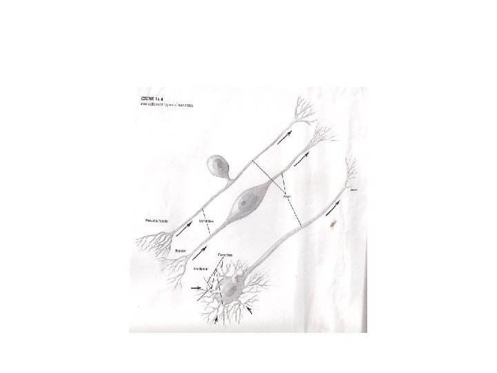
• Neurons • A neuron has 3 principal regions: cell body, dendrites and axon • Dendrites and axons are processes or extensions from the cell body • The cell body or perykaryon is the enlarged portion of the neuron that contains the nucleus • It is the nutritional centre of the neuron where macromolecules are produced • It also contains chromatophilic substances (Nissl’s bodies) composed of granular (rough) endoplasmic reticulum
• Cell bodies within the CNS are frequently clusted into groups called nuclei • Cell bodies within the PNS usually occur in clusters called ganglia • Dendrites are thin branched processes that extend from the cytoplasm of the cell body • They serve to receive stimuli and transmit impulses to the cell body • The axon is a longer process that conducts impulses away from the cell body • The origin of the axon, near the cell body is called the axon hillock
• Side branches that may extend from the axon are called collateral axons or collateral branches • Axon terminals are the bulbular endings of an axon • Classification of neurons and nerves • Neurons are classified according to their structure or function • Functional classification is based on the direction in which they conduct impulses • Sensory or afferent neurons conduct impulses from sensory receptors to the central nervous system • Motor or efferent neurons conduct impulses out of the CNS to effector organs (muscles and glands)
• Association neurons (or interneurons/relay neurons) are located entirely within the CNS • They serve the associative and integrative functions of the nervous system • There are two types of motor neurons: somatic and autonomic • Somatic motor neurons are responsible for both reflex and voluntary control of skeletal muscles • Autonomic motor neurons innervate the involuntary effectors i. e smooth muscles, cardiac muscle and glands • There are 2 subdivisions of autonomic neurons: sympathetic and parasympathetic divisions
• Autonomic motor neurons together with their central control centres constitute the autonomic nervous system • Structural classification of neurons is based on the processes that extend from the cell body of the neuron • Bipolar neurons have 2 processes, one at both ends. This type of neuron occurs in the retina of the eye • Multipolar neurons are characterised by several dendrites and one axon extending from the cell body. This is the most common type of neuron. • Motor neurons are good examples of this type
• Pseudounipolar/unipolar neuron has a single short process that divides like a T to form a longer process • Sensory neurons are pseudounipolar. , one end receives sensory stimuli and produces an impulse • The other end delivers these impulses to synapses within the brain and spinal cord • A Nerve • A nerve is a bundle of fibres located outside the CNS • Most nerves are composed of both motor and sensory fibres and are called mixed fibres. Some cranial nerves however contain sensory fibres only
• These are the nerves that serve the special senses of smell, sight, hearing and equilibrium • Neuroglial cells are also derived from the ectoderm • There are 6 categories of neuroglial cells: • 1. Schwann cell- these form the myelin sheaths around peripheral axons • 2. Oligodendrocytes- they form myelin sheaths around axons of the central nervous system • 3. Microglia- these are phagocytic cells that migrate through the CNS and remove foreign and degenerated material
• 4. Astrocytes- they help to regulate the passage of molecules from the blood to the brain • 5. Ependymal cells- they line the ventricles of the brain and central canal of the spinal cord • 6. Satellite cells- they support neuron cell bodies within the ganglia of the peripheral nervous system • Sheath of Schwann and Myelin Sheath • Some axons in the PNS and CNS are surrounded by a myelin sheath. Such axons are known as myelinated axons • Those that do not have myelin sheath are called unmyelinated axons
• All axons in the PNS (but not in the CNS) are surrounded by a living sheath of Schwann cell • The outer membrane of the Schwann cell is covered by a glycoprotein basement membrane • The basement membrane and the cell membrane of the Schwann cell is known as the neurilemma • Axons of the CNS lack a sheath of Schwann and also lack a continuous basement membrane • Axons that are smaller than 2 microns in diameter are usually unmyelinated • Larger axons are generally surrounded by a myelin sheath composed of Schwann cell or oligodendrocytes
• Schwann cells form myelin sheath in the PNS, whereas oligodendrocytes form myelin sheath in the CNS • Action Potentials in Neurons • All cells in the body maintain a potential difference across the membrane i. e the resting membrane potential • The inside is negatively charged in comparison to the outside of the cell (-65 m. V) • This potential difference is due to the permeability properties of the cell membrane • The membrane traps large negatively charged organic molecules within the cell and permits only
• limited diffusion of positively charged inorganic ions • This differential permeability results in an unequal distribution of Na+ and K+ across the membrane • The action of Na +/K + pumps also help to maintain a potential difference because they pump out 3 Na+ for every K+ that they transport into the cell • Na+ is thus highly concentrated in the extracellular fluid than in the cell, whereas K+ is more concentrated within the cell
• Ion Gating in Axons • The permeability of the membrane to Na+, K+ and other ions is regulated by ion channels through the membrane called gates • Gates are composed of polypeptide chains that can open or close according to specific conditions • When a gated channel for a specific ion is closed, the membrane is not permeable to that ion • When opened, the permeability to that ion can be greatly increased • There are 2 types of channels for K+, one type lacks gates and is always open, whereas the other type has gates that are closed in a resting cell
• The channels for Na+ always have gates and these gates are closed in the resting cell • The resting cell is thus more permeable to K+ than Na+ • Whether the gates for Na+ and K+ are open or closed depends on the membrane potential • The gated channels are closed at the resting membrane potential of -65 m. V, but open when the membrane is depolarized to a certain threshold level • Since the opening and closing of these gates is regulated by the membrane voltage, the gates are said to be voltage regulated
• When the axon is stimulated, the membrane is depolarized • If a certain level of depolarization is achieved( from -65 m. V to -55 m. V) by artificial stimulation, a sudden and rapid change in the membrane potential will be observed • This is because depolarization to a certain threshold level causes the Na+ gates to open • The permeability properties of the membrane are changed and Na+ diffuses down its concentration gradient into the cell • A fraction of a second after the Na+ gates open, they close again
• At this time , the depolarization stimulus causes the K+ gates to open • This makes the membrane more permeable to K+ than it is at rest and K+ then diffuses down its concentration gradient out of the cell • The K+ gates will then close and the permeability of the membrane will return to what they were at rest • Action Potential • When the axon membrane has been depolarized to a certain threshold, the Na+ gates open and the membrane becomes permeable to Na+ • This permits Na+ to enter the axon by diffusion, which further depolarizes the membrane
• This makes the inside less negative or more positive • Since the Na+ gates are voltage regulated, this further depolarization makes the membrane even more permeable to Na+ so that even more Na+ can enter the cell • After a slight time delay, depolarizarion of the axon membrane also causes the opening of the voltage-regulated K+ gates and the diffusion of K+ out of the cell • Since K+ are positively charged, the diffusion of K+ out of the cell makes the inside of the cell less positive or more negative and acts to restore the original resting membrane potential
• This process is called repolarization • The large increase in Na+ diffusion causes rapid depolarization to 0 m. V • The inside of the cell actually becomes positively charged (almost +40 m. V) compared to the outside • The Na+ permeability rapidly decreases as the diffusion of K+ increases, resulting in repolarization to the membrane potential • These changes in Na+ and K+ diffusion and the resulting changes in the membrane potential constitutes the action potential or nerve impulse
• Once an action potential has been completed, the Na+/K+ pumps will extrude the extra Na+ that entered the axon and recover the K+ that has diffused out of the axon • This active transport of ions occur very quickly, because the action potential occurs across a small area of the membrane and so only small amounts of Na+ and K+ actually diffuse through the membrane during the production of an action potential • The total concentration of Na+ and K+ in the axon and in the extracellular fluid are not significantly changed during an action potential
• It must be noticed that active transport processes are not directly involved in the production of action potential • Both depolarization and repolarization are produced by diffusion of ions down their concentration gradients • A neuron poisoned with cyanide such that it cannot produce ATP can still produce an action potential for a period of time • After a while, the lack of ATP for active transport by the Na+/K+ pump will result in a decline in the ability of the axon to produce action potentials
• This demonstrates that the Na+/K+ pumps are not directly involved but are required to maintain the concentration gradients needed for the diffusion of Na+ and K+ during action potentials • All-or- none Law • Once a region of an axon has been depolarized to a certain threshold value, the membrane potential shoots to about +40 m. V • It does not become more positive because the Na+ gates quickly close and the K+ gates open • The length of time that the Na+ and K+ gates remain open is independent of the strength of the depolarization stimulus
• The amplitude of action potentials is therefore all-ornone • When depolarization is below a threshold, the voltage-regulated gates are closed • When depolarization reaches a threshold, an action potential is produced • Since the change from -65 m. V to +40 m. V and back to -65 m. V lasts only about 3 msec, the image of an action potential on an oscilloscope screen looks like a spike • Action potentials are therefore called spike potentials
• Refractory Periods • If a stimulus of a given intensity is maintained at one point and depolarizes it to a threshold, action potentials will be produced • During the time that a section of an axon membrane is producing an action potential, it is incapable of responding to further stimulation and is said to be refractory • If a second stimulus is applied (for example, while the Na+ gates are open) the second stimulus cannot have any effect because the gates are already open • During the time, the Na+ gates are open, the membrane is in an absolute refractory period and
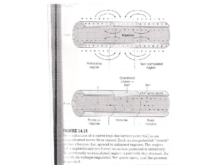
• cannot respond to any subsequent stimulation • If a second stimulus is applied whilst the K+ gates are open (and the membrane is in the process of repolarizing) the membrane is said to be in a relative refractory period • During this time, only a very strong stimulus can depolarize the membrane and produce a second action potential • Conduction of Nerve Impulse • When one point of an axon is stimulated to a threshold, the voltage-regulated gates are open and an action potential is produced at that small region of the axon
• When the membrane voltage changes from -65 m. V to +40 m. V, a current of Na+ enters the cell by diffusion • Each action potential injects positive charges into the axon • These positively charged ion (Na+) are conducted to an adjacent region that still has a membrane potential of -65 m. V • This conduction helps to depolarize the adjacent region of the axon membrane • When this adjacent region of membrane reaches a threshold of depolarization, it too produces an action potential
• Each action potential acts as a stimulus for the production of another action potential at the next region of the membrane • Conduction in an unmyelinated axon • In an unmyelinated axon, every patch of membrane that contains Na+ and K+ gates can produce an action potential • Action potentials are thus produced at locations only a fraction of micrometer all along the length of the axon and spreads in a cable-like manner • The cable-like spread of depolization induced by Na+ influx during an action potential helps to depolarize adjacent regions of the membrane
• Action potentials are passed in one direction only because the area in the opposite direction has just produced an action potential and cannot produce a new one. , it is still in its refractory period • Each action potential is a separate complete event that is repeated or regenerated along the axon • Action potential produced at the end of the axon is a completely new event and it occurs in response to depolarisation from the previous action potential • The last action potential has the same amplitude as the first • Thus action potentials are said to be conducted without decrement
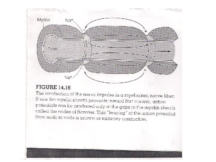
• Conduction in a Myelinated Axon • The myelin sheath provides insulation for the axon, preventing the movement of Na+ and K+ through the membrane • If the myelin sheath were continuous, action potentials could not be produced • Fortunately, there are interruptions in the myelin sheath known as the nodes of Ranvier • Studies have shown that Na+ channels are highly concentrated at the nodes and almost absent in the region of the axon between the nodes • Action potentials therefore occur only at the nodes of Ranvier and seem to leap from node to node
• This type of conduction is called saltatory conduction • Saltatory conduction permits a faster rate of conduction because depolarization between the node is faster and fewer action potentials need to be produced per given length of axon • THE SYNAPSE • The synapse is the functional connection between a neuron and a second cell • In the CNS, this other cell is also a neuron • In the PNS, the other cell may either be a neuron or an effector cell within a muscle or gland
• The neuron-muscle synapse is often called myoneural or neuromuscular junctions • Neuron-neuron synapses usually involve a connection between axon terminal of one neuron and the dendrites, cell body or axon of a second neuron • These are called respectively axodendritic, axosomatic or axoaxonic synapses • In almost all synapses, transmission is in one direction only i. e from the axon of the first(presynaptic) cell to the second (postsynaptic) cell
• There are tiny gaps in the synapses • It has been demonstrated that actions of the autonomic nerves could be duplicated by certain chemicals • This led to the hypothesis that synaptic transmission might be chemical • The presynaptic axon terminals release chemical neurotransmitters that stimulate action potentials in the postsynaptic cells
• The axon of the presynaptic neuron ends in a slight swelling called the axon terminal • The postsynaptic membrane appears denser • A small extracellular space, the synaptic cleft separates the pre- and postsynaptic neurons and prevents direct propagation of the current from the presynaptic neuron to the postsynaptic cell • Signals are transmitted across the synaptic cleft by means of a chemical messenger, a neurotransmitter • The neurotransmitter is stored in synaptic vesicles
• Synaptic Transmission • The chemical acetylcholine (Ach) is used as a neurotransmitter by some neurons in the CNS, somatic motor neurons at the neuromuscular junctions and by certain autonomic nerve endings • The effects of this chemical are excitatory in the first two synapses and either excitatory or inhibitory in the third • Axon terminals have synaptic vesicles that contain about 10, 000 molecules of acetylcholine each • Once released, they quickly diffuse across the synaptic cleft to the membrane of the postsynaptic cell
• Here, they are chemically bound to receptor proteins in the postsynaptic membrane • The receptor proteins combine with acetylcholine in a specific manner • Acetylcholine is not transported into the postsynaptic cell after diffusing across the synaptic cleft • Instead the bonding of acetylcholine to its receptor protein causes changes in the membrane structure that results in the opening of the ion gates for Na+ and K+ • These gates are located only in the postsynaptic membrane
• Unlike the voltage-regulated gates in the axon, the chemically-regulated gates do not respond to changes in the membrane potential • Also, chemically-regulated gates permit the simultaneous diffusion of Na+ and K+ • However, the depolarizing effect of Na+ diffusion predominates because the electrochemical gradient for Na+ is greater than that for K+ • Because of the characteristics of chemicallyregulated gates, neurotransmitters do not directly produce action potentials • They can only produce depolarization, which may stimulate the opening of the voltage-regulated gates
• and produce action potentials a short distance away from the site of the synapse • Excitatory Postsynaptic Potential(EPSP) • The interaction of acetylcholine with its receptors in the postsynaptic membrane depolarises that region of the membrane • Depolarisations produced in response to acetylcholine are known as excitatory postsynaptic potential when they occur in postsynaptic neurons • Unlike action poptentials, EPSP’s have no threshold • Thus when more Ach are released, depolarisation is correspondingly greater
• EPSP’s are graded in magnitude unlike the all-ornone nature of action potentials • Acetylcholinesterase • The bond between acetylcholine and its receptor protein exist for only a brief period • The Ach-receptor complex usually dissociates • In order for the activity in the postsynaptic cell to be controlled, free Ach must be inactivated soon after it is released • The inactivation of Ach is achieved by an enzyme called acetylcholinesterase. This enzyme is present on the postsynaptic membrane or immediately outside the membrane
• Stimulation of Skeletal Muscles • The synapse of a motor axon with a skeletal muscle is known as a motor end plate • The release of Ach from the motor axon produces an end-plate potential • This is identical in nature to EPSP in a postsynaptic neuron • This depolarisation opens the voltage-regulated gates serving as a stimulus for production of action potentials in the muscle fibre • If any stage in the process of neuromuscular transmission is blocked, muscle weakness, sometimes leading to paralysis and death results
• For example the drug curare competes with Ach for attachment to the receptor proteins and reduces the size of the end-plate potential • The drug is clinically used as muscle relaxant during anaesthesia to prevent muscle damage • Neurotransmitters of the CNS • Acetylcholine is not the only neurotransmitter of somatic motor neurons of the PNS • It is also an important neurotransmitter within the CNS • In addition to Ach a number of other molecules serve as neurotransmitters in the CNS
• Neurotransmitters • These are substances which act as chemical messengers to enable nerve cells communicate with each other or with effectors • One nerve cell alters the activity of the other by releasing from its ending a neurotransmitter that acts upon the second cell • Neuromodulators • These often modify the postsynaptic cell’s response to specific neurotransmitters by modifying or dampening(reducing) the effectiveness of synaptic activity
• The effect of neuromodulators are to elicit responses that are described as EPSP’s or IPSP’s • Acetylcholine: Synthesized from choline and acetyl coenzyme A in the cytoplasm of synaptic terminals • It is stored in the synaptic vesicles • After activating the receptors in the postsynaptic membrane it is reduced by the enzyme acetylcholinesterase • The choline is then transported back into the axon terminals where it is reused in the synthesis of Ach • Even though Ach is a major neurotransmitter in the PNS it is also present in the brain
• Fibres that release Acetylcholine are called cholinergic fibres • One cholinergic system in the brain plays a major role in attention, learning and memory • When neurons associated with this system degenerate it leads to the disease called Alzheimers disease • This is a brain disease that is age-related • In addition to acetylcholine, a number of molecules serve as neurotransmitters in the central nervous system
• Catecholamine neurotransmitters • These are made up of a group of molecules derived from the amino acid tyrosine • These include dopamine, norepinephrine and epinephrine • Dopamine and norepinephrine function as neurotransmitters only • Epinephrine and norepinephrine additionally serve as hormones secreted by the adrenal medulla • Neurons that use dopamine as neurotransmitter are dopaminergic • They are highly concentrated in the substantia nigra
• Medical research findings strongly indicate that Parkinson’s disease is caused by the degeneration of the dopaminergic neurons in the substantia nigra • Patients are treated with L-Dopa to increase the production of dopamine in the brain • Norepinephrine, like Ach is used as neurotranmitter in both the PNS and CNS • Sympathetic neurons of the PNS use norepinephrine as neurotransmitter at their synapse with smooth muscles, cardiac muscles and glands • Some neurons in the CNS also use norepinephrine as a neurotransmitter
• Amino Acid Neurotransmitters • The amino acids glutamic acid and aspartic acid function as excitatory neurotransmitters in the CNS eg. glutamic acid is the major excitatory neurotransmitter in the brain • Gamma-amino butyric acid (GABA) a derivative of glutamic acid is the most prevalent neurotransmitter in the brain • As many as 1/3 of the neurons in the brain use GABA as neurotransmitter • GABA is inhibitory because it hyperpolarises the postsynaptic membrane, making the membrane potential even negative than it is at rest
• In this case, the membrane potential changes from 65 m. V to -85 m. V • Such hyperpolarisation is known as inhibitory postsynaptic potential (IPSP) • The action of the amino acid glycine is also inhibitory • Polypeptide neurotransmitters • Many polypeptides are found in the brain and are believed to function as neurotransmitters • Some of the polypeptides that function as hormones secreted by the wall of the small intestine and other endocrine glands are also produced in the brain
• For example, cholecystokinin(CCK) a hormone from the small intestine is also released from neurons and used as neurotransmitter in the brain • Evidence suggests that CCK, acting as a neurotransmitter may promote the feeling of satiety in the brain following meals
• ENDOCRINE SYSTEM • The endocrine system consists of all those glands termed endocrine glands • Endocrine glands are ductless and secrete biologically active chemicals called hormones directly into the blood • The cells influenced by a particular hormone are the target cells for that hormone • Hormones affect the metabolism of their organs and by this means they help regulate • 1. Total body metabolism • 2. Growth 3. Reproduction
• Chemical Classification of Hormones • All hormones can be grouped into 3 broad chemical categories: • 1. Catecholamines (epinephrine and norepinephrine • 2. Polypeptides and glycoproteins e. g. ADH, insulin, thyroid stimulating hormone • 3. Steroids e. g. cortisol and testosterone • Steroid hormones which are derived from cholesterol are lipids and are not water soluble • The gonads i. e. the testes and ovaries produce sex steroids
• Prohormones and Prehormones • Hormone molecules that affect the metabolism of target cells are often derived from less active parent or precursor molecules • In the case of polypeptide hormones the precursor may be a longer chained prohormone that is cut and put together to make a hormone • For example, insulin is produced from proinsulin • Prehormone- molecules secreted by an endocrine glands that are inactive until they are converted by their target cells into the active form
• Hormone Interactions • The responsiveness of a target tissue to a particular hormone may not only be affected by the concentration of that hormone, but also by the effects of other hormones on that tissue • Terms used to describe hormone interactions include synergistic, permissive and antagonistic • Synergistic effects occur when two or more hormones work together to produce a particular result • The results they produce may be additive or complementary
• The action of epinephrine and norepinephrine on the heart is a good example of a additive effect • Each of these hormones, acting alone produces an increase in the cardiac rate • Acting together in the same concentrations, they stimulate an even greater increase in cardiac rate • The synergistic action of FSH and testosterone is an example of complementary effect • Each hormone, acting separately stimulates a different stage of spermatogenesis during puberty • Both hormones together are needed at that time to complete sperm development
• A hormone is said to have a permissive effect on the action of a second hormone when it enhances the responsiveness of a target organ to the second hormone or it increases the activity of the second hormone • For example, prior exposure of the uterus to oestrogen induces the formation of receptor proteins for progesterone which improves the response of the uterus when it is exposed to progesterone • Antagonistic Effects • In some situations, the actions of one hormone antagonizes the effects of another
• Lactation during pregnancy is prevented because the high concentration of oestrogen inhibits the secretion of prolactin • Another example of antagonism is the action of insulin and glucagon • Pituitary Gland • The pituitary gland, also known as hypophysis is located in the diencephalon • Structurally and functionally, it is divided into an anterior lobe or adenohypophysis and the posterior lobe or neurohypophysis
• The adenohypophysis consists of 3 parts: • 1. Pars distalis- this is the major endocrine part of the gland • 2. Pars tuberalis- a thin extension which is in contact with the infundibulum • 3. Pars intermedia- located between the anterior and posterior parts of the pituitary • The neurohypophysis is the neural part of the pituitary gland • It consists of the pars nevosa which is in contact with the pars intermedia of adenohypophysis and the infundibulum
Pituitary Hormones The hormones secreted by the anterior pituitary are called trophic hormones High levels of anterior pituitary hormones make their target organs hypertrophy, whereas low amounts cause their target organs to atrophy Hormones of the Pars distalis are: 1. Growth Hormone(GH) or somatotropin- this hormone promotes the movement of amino acids into tissues and incorporate these amino acids into tissue protein It therefore promotes the growth of organs
• 2. Thyroid-stimulating hormone(TSH) or Thyrotropin • Stimulates the thyroid gland to produce and secretes thyroxine • 3. Adrenocorticotropic hormone(ACTH)- Stimulates the adrenal cortex to secrete glucocorticoids such as hydrocortisone • 4. Follicle stimulating hormone(FSH) or Follicotropinstimulates the growth and secretion of ovarian follicles in females and production of sperm in the testes of males • 5. Luteinizing Hormone(LH) or Luteotropin- this hormone, together with FSH are collectively called gonadotropic hormones
• In females, LH stimulates ovulation and conversion of the ovulated ovarian follicle into the corpus luteum • In males (sometimes called ICSH) it stimulates the interstitial cells of the testes to secrete the male sex hormones, mainly testosterone • 6. Prolactin • Secreted in both males and females • It stimulates the production of milk from the mammary glands of women after parturition • It plays a supporting role in the regulation of the male reproductive system by FSH and LH, acts on the kidneys to regulate water and electrolyte balance
• Pars intermedia • This part of the pituitary is not well defined in an adult human • It produces different forms of melanocytestimulating hormone (MSH) which in lower vertebrates cause a darkening of the skin to provide a camouflage against a dark background • Pars nervosa • The posterior pituitary secretes only 2 hormones, both of which are produced in the hypothalamus and stored in the posterior lobe of the pituitary
• • Antidiuretic hormone(Vasopressin) It stimulates the kidneys to retain water Oxytocin Stimulates the contractions of the uterus during labour. , thus needed for parturition • It also stimulates the contraction of the mammary gland alveoli and ducts which result in milk ejection reflex in a lactating woman • In men a rise in oxytocin has been observed at the time of ejaculation • However its physiological significance remains unresolved
• Hypothalamic Control of Neurohypophysis • Although the posterior pituitary secretes ADH and oxytocin, these hormones are actually produced in neuron cell bodies of the supra-optic nuclei and paraventricular nuclei of the hypothalamus • These nuclei within the hypothalamus are endocrine glands • The hormones they produce are transported along the axons of the hypothalamo-hypophyseal tract to the posterior pituitary which stores and later releases these hormones
• Hypothalamic Control of Adenohypophysis • The anterior pituitary was called the ‘master gland’ because it secretes hormones that regulate some endocrine glands • For example ACTH simulate the adrenal cortex to secrete glucocorticoids • Thyroid stimulating hormone stimulates the thyroid gland to secrete thyroxine • FSH and LH act on the gonads • Secretion of hormones of the anterior pituitary is controlled by hormones secreted by the hypothalamus
• Feedback Control of Adenohypophysis • Anterior pituitary secretion of ACTH, TSH and the gonadotrophins (LH and FSH) is controlled by a negative feedback inhibition from the target gland hormones • Secretion of ACTH is inhibited by a rise in corticosteroid secretion • TSH secretion is inhibited by a rise in the secretion from the thyroid • Castration (surgical removal of the gonads) produces a rise in the secretion of FSH and LH • In the same way, removal of the adrenals or the thyroid results in an abnormal rise in ACTH or TSH secretion from the anterior pituitary.
• These demonstrate that these target glands exert an inhibitory effect on the anterior pituitary • Adrenal Glands • The adrenal glands are paired organs located on the superior boarders of the kidneys • Each consists of an outer adrenal cortex and inner adrenal medulla • The 2 regions function as separate organs • These differences are related to their different embryonic origins • The adrenal medulla is derived from neural crest
• ectoderm, whereas the adrenal cortex is derived from the embryonic mesoderm • The adrenal medulla secretes catecholamine hormones (mainly epinephrine(80%) with lesser amounts of norepinephrine) • These are secreted in response to stimulation by preganglionic sympathetic nerve fibres • The adrenal cortex does not receive neural innervation • It is stimulated hormonally by ACTH from the anterior pituitary gland • The adrenal cortex makes up the bulk of the adrenal gland
• Histologically the adrenal cortex is divided into 3 zones: • An outer zona glomerulosa, an intermediate zona fasciculata and inner zona reticularis • The adrenal medulla is composed of clusters of chromaffin cells • Functions of the Adrenal cortex • The adrenal glands secretes steroid hormones called corticosteroids • There are three functional categories of cortocosteroids: • 1. Mineralocorticoids- these regulate Na+ and K+ balance
• 2. Glucocorticoids- regulate the metabolism of glucose and other organic molecules • 3. Sex steroids- weak androgens and lesser amounts of oestrogens. These supplements sex steroids secreted by the gonads • Aldosterone is the most potent mineralocorticoid • Mineralocorticoids are produced in the zona glomerulosa • The most predominant glucocortocoid in humans is corticol (hydocortisone) • This is secreted by the zona fascuculata and the zona reticularis • Secretion of cortisol is stimulated by ACTH
• Functions of the Adrenal Medulla • The chromaffin cells of the adrenal medulla produce epinephrine and norepinephrine in a ratio of 4: 1 respectively • These hormones are derived from the amino acid tyrosine • The effects of the hormones are similar to those produced by the stimulation of the sympathetic nervous system • The hormones from the adrenal medulla increase cardiac output and heart rate, dilate coronary blood vessels, enhance mental alertness, increases respiratory rate and increases metabolic rate
• • • Thyroid and Parathyroid Glands Structure of the Thyroid Gland The thyroid gland is located just inferior to the larynx It consists of two lobes positioned on either side lateral side The thyroid gland is the largest endocrine gland, weighing between 20 and 25 g • Microscopically the thyroid gland consists of spherical hollow sacs called thyroid follicles • These follicles are lined with simple cuboidal epithelium composed of follicle cells that synthesize thyroid hormones
• The interior of the follicles contain colloid, a protein-rich fluid • Between the follicles are epithelial cells called parafollicular cells that produce the hormone calcitonin • Production and Action of Thyroid Hormones • The thyroid follicles actively accumulate iodide (I-from the blood and secrete it into the colloid • Once in the colloid, it is oxidized into iodine and attached to specific amino acid (such as tyrosine) within the polypeptide chain of a protein called thyroglobulin • The attachement of one iodine to tyrosine produces
• monoiodotyrosine(MIT), the attachment of 2 iodines produces di-iodotyrosine (DIT) • Within the follicle cells, enzymes modify the structure of MIT and DIT and couple them together • When 2 DIT molecules are coupled together, a molecule of tetraiodothyrosine, T 4 or thyronine is produced • The combination of MIT and DIT forms triiodothyrosine, T 3 • Upon activation by genes, thyroid hormones stimulate protein synthesis, promote maturation of the nervous system and increase the rate of energy utilization in the body
• The development of the central nervous system depends on the thyroid hormones • Deficiency of the hormones during development an cause mental retardation • The basal metabolic rate (BMR), which is the minimum rate of calorific expenditure is determined largely by the level of thyroid hormones in the blood • Parathyroid Glands • These are located on the posterior surface of the lateral lobes of the thyroid gland • There are four parathyroids: a superior and inferior pair
• They secrete parathyroid hormone (PTH), which is important in the regulation of calcium levels in body fluids • Bone, the kidneys and the intestines are the major target tissues • PTH stimulates osteoclast activity in bone and can cause the number of osteoclasts to increase • Increased osteoclast activity results in bone resorption and the release of calcium and phosphates, causing an increase in the blood calcium • PTH also inducescalcium reabsorption within kidneys so that less calcium leaves the body in urine • It also induces the formation of active vitamin D in
• the kidneys • Inactive parathyroid glands result in hypocalcemia • Reduced extracellular calcium levels cause voltage – gated Na+ channels to open • This increases the permeability of cell membranes to Na+. , Na+ diffuse into cell and cause depolarization • Symptoms of hypocalcemia include nervousness, muscle spasms and convulsions
• Pancreas • The pancreas is both an endocrine and exocrine gland • The exocrine portion consists of acini, which produce pancreatic juice, and a duct system which carries pancreatic juice to the small intestine • The endocrine portion consists of pancreatic islet (Islets of Langerharns) which produce hormones • Between 500, 000 and 1000000 pancreatic islets are dispersed among the ducts and acini of the pancreas • Each islet is composed of alpha cells (20%) which secrete glucagon and beta cell (75%) which secrete insulin and other types of cells
• Nerves from both divisions of the autonomic system innervate the pancreatic islets • The alpha cells secrete glucagon in response to a fall in blood glucose concentration • Glucagon stimulates the liver to hydrolyse glycogen to glucose (glycogenolysis) • Glucagon also stimulates the hydrolysis of stored fats into fatty acids (lypolysis) • This helps to provide energy substrates for the body during fasting when blood glucose levels fall • Glucagon, together with other hormones stimulate the conversion of fatty acids into ketone bodies
• These are secreted by the liver into the blood to serve as energy source for many organs • The beta cells secrete insulin in response to a rise in blood glucose concentration • It promotes the entry of glucose into tissue cells and its conversion into fat and glycogen for storage • Insulin also aids the entry of amino acids into cells and the production of cellular protein • The action of insulin and glucagon are antagonistic • After a meal, insulin secretion is increased and glucagon secretion is decreased • Fasting causes a rise in glucagon and a decline in
• Pineal body • Located in the roof of the third ventricle, near the copora quadrigemina • The pineal gland of a child is 5 -8 mm long, 9 mmwide • The gland begins to regress in size at about age 7 • It lacks direct nervous connection to the rest of the brain • It is however innervated by the sympathetic nerve • The principal hormone of the pineal body is melatonin
• Production of this hormone is stimulated by the supra chiasmatic nucleus(SCN) in the hypothalamus • Activity of the SCN and thus secretion of melatonin is highest at night • During the day neural pathways from the retina suppress the activity of the SCN • This reduces sympathetic stimulation of the pineal body, thus decreasing melatonin secretion • It has been suspected that melatonin inhibits the pituitary-gonads axis • Excessive melatonin secretion in humans is associated with a delay in the onset of puberty
• Melatonin secretion is highest in children of ages 1 -5 and decreases thereafter, reaching its lowest level at the end of puberty • Thymus • This is a bilobed organ located in the superior mediastinum, in front of the aortic arch • It is relatively large in new-borns and children but sharply decreases in size after puberty • The thymus produces T- cells (Thymus –dependent cells) which are lymphocytes involved in cell-mediated immunity • It also secretes a number of hormones that stimulate T cells after they leave thymus
• Gonads and the Placenta • The gonads (testes and ovaries) secrete a number of sex steroids • These include male sex hormones, androgens eg testosterone and female sex hormones oestrogens and progestogens- oestradiol and progesterone • Between the convolutions of the seminiferous tubules of the testes are specialized cells (Leydig cells) which secrete testosterone • Testosterone is needed for the development and maintenance of primary and secondary sex organs of the male
• It is also needed for the development of secondary sex characteristics • During the first half of the menstrual cycle, oestrogen is secreted in small amounts by the ovarian follicles within the ovary • Oetsrogen is actually secreted by the granulosa cells surrounding the follicle • After ovulation, the empty follicle under the influence of LH becomes a new endocrine structure called the corpus luteum • The corpus luteum secretes progesterone as well as oestadiol
• Placenta • The placenta secretes large amounts of oestrogens and progesterone as well as large number of polypeptides and protein hormones similar to those secreted from the anterior pituitary gland • These latter hormone include human chorionic gonadotropin(h. CG) – similar to LH, and somatomammotropin, similar in action to growth hormone and prolactin
• Gastrointestinal Tract • The stomach and small intestine secretes a number of hormones that act on the GIT itself and on the placenta and gall bladder • These include secretin which act on the pancreas to secrete an alkaline fluid to neutralise the acid • Gastrin, stimulates the stomach wall to secrete gastric juice • Cholesystokinin (CCK) stimulates the contraction of the gall bladder to release bile