http www youtube comwatch v0 Vn 5 AJMjfsNR1

http: //www. youtube. com/watch? v=0 Vn 5 AJ_Mjfs&NR=1 http: //www. youtube. com/watch? v=H 04 d 3 r. JCLCE
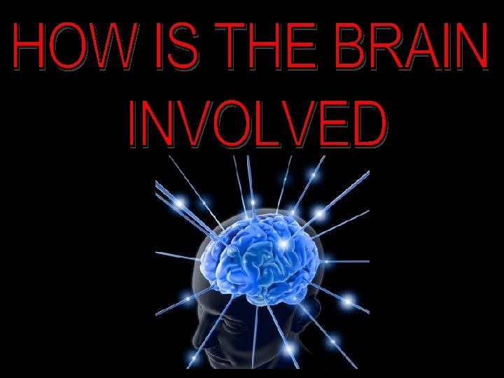
 K 1. Identify")
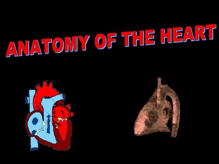
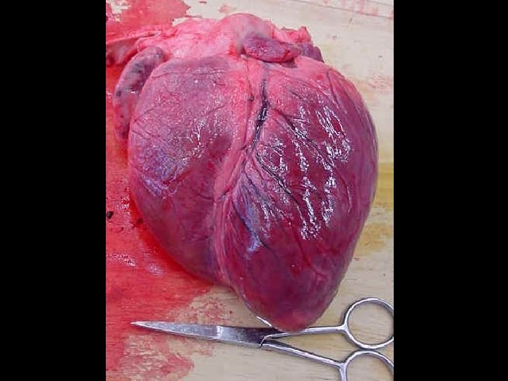
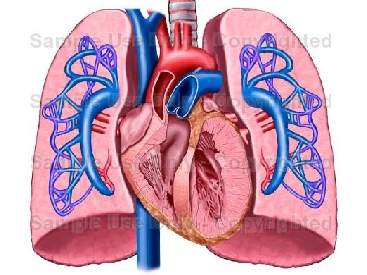
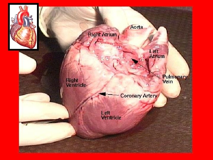
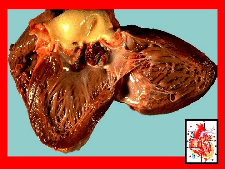
UNIT K: HEART STRUCTURE & FUNCTION (Ch. 13, pp. 228 -233) K 1. Identify & give functions for the following: • Left and right atria • Left and right ventricles • Coronary arteries and veins • Anterior and posterior vena cava • Aorta • Pulmonary arteries and veins • Pulmonary trunk • Atrioventricular valves • Chordae tendinaea • Semi-lunar valves • Septum

K 2. Describe the location and functions of the SA node, AV node, and Purkinje fibers K 3. Describe the autonomic regulation of the heartbeat by the nervous system K 4. Relate factors that affect and regulate blood pressure to hypertension and hypotension K 5. Demonstrate the measurement of blood pressure K 6. Distinguish between systolic and diastolic pressure
 valves _____ Atrium _____ Autonomic nervous")
_____ Aorta _____ Aortic valve _____ Atrioventricular (AV) valves _____ Atrium _____ Autonomic nervous system _____ AV node _____ Blood pressure _____ Brachial artery _____ Bundle of His _____ Chordae tendineae _____ Constrict _____ Coronary arteries _____ Coronary veins _____ Dilate _____ Hypertension _____ Hypothalamus _____ Inferior Vena Cava _____ Medulla oblongata _____ Pacemaker _____ Pulmonary artery _____ Pulmonary circuit _____ Pulmonary trunk _____ Pulmonary valve _____ Pulmonary vein _____ Purkinje fibres _____ SA node _____ Semi-lunar valve _____ Septum _____ Superior Vena Cava _____ Systemic circuit _____ Vagus nerve _____ Ventricle

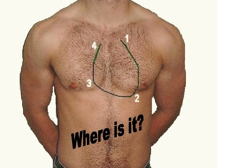
Amazing Heart Facts • Put your hand on your heart. Where is it? • Hold out your hand make a fist. size of heart: child = 1 fist; adult = 2 fists! • Your heart beats about 100, 000 times in one day and about 35 million times in a year. • During an average lifetime, the human heart will beat more than 2. 5 billion times. • Even at rest, the muscles of the heart work twice as hard as the leg muscles of a person sprinting

• In one day, the blood travels a total of 19, 000 km. That's 4 times the distance across CANADA from coast to coast. • When you are resting, it takes 35 -40 seconds for the blood to move through your body. What about when you are exercising? approximately 10 seconds • How much does your heart weigh? only 2/3 of a pound! • Give a tennis ball a good, hard squeeze. You're using about the same amount of force your heart uses to pump blood out to the body

1 • Transports oxygen from the lungs to body to be used 2 • Transports carbon dioxide and hydrogen ions to the ions lungs to be removed 3 • Transports nutrients from the small intestine to tissues 4 • Fights infections

5 • Transports water from the water digestive system to the body and lungs 6 • Carries waste products (ie: urea) to kidneys for removal in urine heat 7 • Distributes body heat from internal source to skin (to get rid of it) 8 • Seals wounds by forming blood clots

hormones 9 • Transports hormones around the body 10 • Maintains p. H in tissues p. H (acts as a buffer with HHb) levels 11 Regulates fluid levels in tissues (with Lymphatic system)

The human heart has 4 well developed chambers ØRight Atrium ØLeft Atrium ØRight Ventricle ØLeft Ventricle

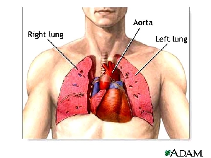
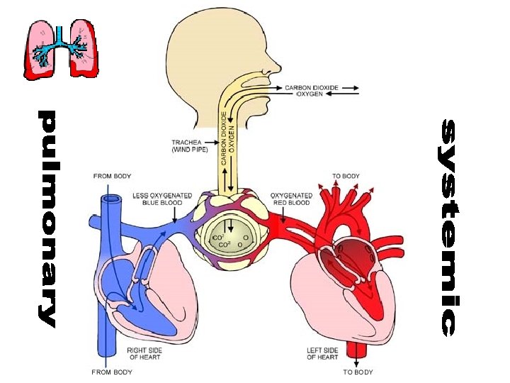
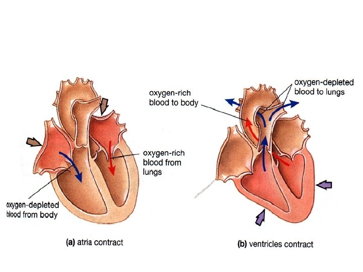
The right side of the heart pumps de. O 2 blood to the lungs. PULMONARY The left side of the heart pumps O 2 blood to the body SYSTEMIC

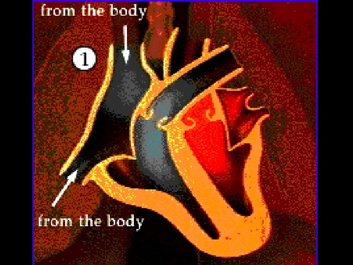
Left Atrium Right Atrium Receives deoxygenated blood from the body via the anterior & posterior vena cava. Receives oxygenated blood from the lungs via the pulmonary veins

Tricuspid Valve 1. Separate the atria from the ventricles 2. They open when the atria contract 3. They prevent the blood from going backwards when the ventricles contract. Bicuspid Valve
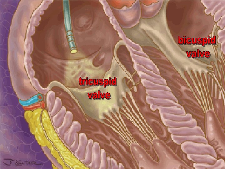
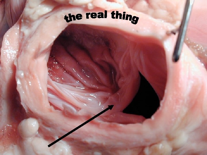

Bicuspid Valve

Tendon-like pieces of tissue They keep the AV valves from inverting when the ventricles contract

They hold & support the chordae tendinae

Open Can’t open properly Closed Normal Heart Valves Can’t close properly Heart Murmur

• When the right atrium contracts, it pushes the blood through the tricuspid valve and into the right ventricle. • When the RIGHT VENTRICLE contracts, BP forces the TRICUSPID valve to close. Right Ventricle • The BP forces the PULMONARY VALVE open & the blood moves into the PULMONARY TRUNK.

The PULMONARY ARTERIES take the de. O 2 blood to the lungs. The CO 2 is removed from the blood and is replaced with O 2. The PULMONARY VEINS take the O 2 blood to the heart.

The protein HEMOGLOBIN binds the O 2 tightly and carries it to the body cells as OXYHEMOGLOBIN! OXYHEMOGLOBIN

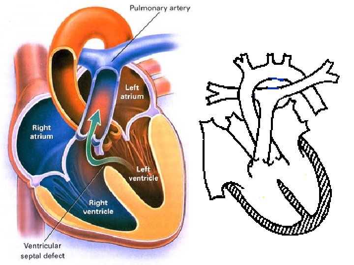
A muscular wall that separates the right side of the heart from the left side. Keeps the de. O 2 blood from mixing with the O 2 blood

Some people are born with a hole in their septum

• When the left atrium contracts, it pushes the blood through the bicuspid valve and into the left ventricle. • When the LEFT VENTRICLE contracts, the BICUSPID VALVE is forced closed. • Blood is forced through the AORTIC VALVE and enters the AORTA • The left ventricle has a THICKER muscle layer. WHY? Left Ventricle

• To the head • To the arms & chest Aortic Arch • Coronary Artery: to the heart Dorsal Aorta • To the lower body The aorta takes O 2 blood to the body.

The first branches of the aorta take the blood to the coronary arteries Takes blood into the heart muscle itself. The coronary veins return the de. O 2 blood to the vena cava right atrium.

Superior Vena Cava These are the BIGGEST VEINS! They bring the de. O 2 blood back to the heart so that it can be pumped to the lungs. Inferior Vena Cava

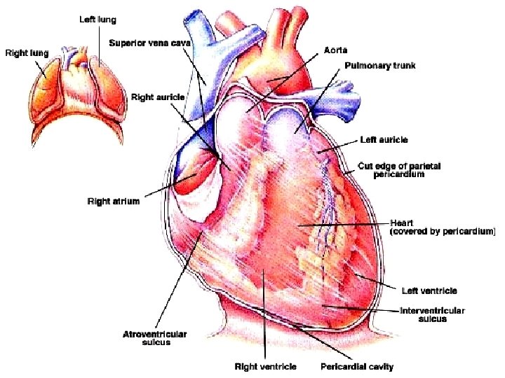
Aortic Arch Superior vena cava Pulmonary Trunk Coronary artery Right Atrium Right Ventricle Inferior Vena Cava Coronary Vein Left Ventricle
 Valve")
Aortic Arch Superior Vena Cava Right Pulmonary Artery Right Pulmonary Veins Pulmonary (semi-lunar) Valve Tricuspid (AV) Valve Inferior Vena Cava Left Pulmonary Artery Pulmonary Trunk Left Atrium Bicuspid (AV) Valve Right Atrium Left Pulmonary Veins Left Ventricle Aortic (semilunar) Valve Right Ventricle Dorsal Aorta

• Heart cells naturally beat slowly if ATP is present • If there was no coordination, coordination the heart cells would all beat randomly Beating Human Heart: http: //www. youtube. com/watch? v=Gnv 54 V 8 Jj 1 U http: //www. youtube. com/watch? v=84 Pr. Hx. Jri 9 Q

• There are two spots of specialized tissue in the heart. • Both are located in the right atrium • Nodal tissue is unique: made of specialized muscle cells combined with nerve cells • It has the ability to contract independent of other stimuli.
 SA NODE This node is found along the wall of")
The SA NODE (sino-atrial) SA NODE This node is found along the wall of the right atrium chamber. l It fires on average, every 0. 85 seconds (or 72 times per minute). l It stimulates the simultaneous contraction of the atria l It also sends a nerve impulse along a nerve trunk called the BUNDLE OF HIS to the AV NODE l

S A NODE

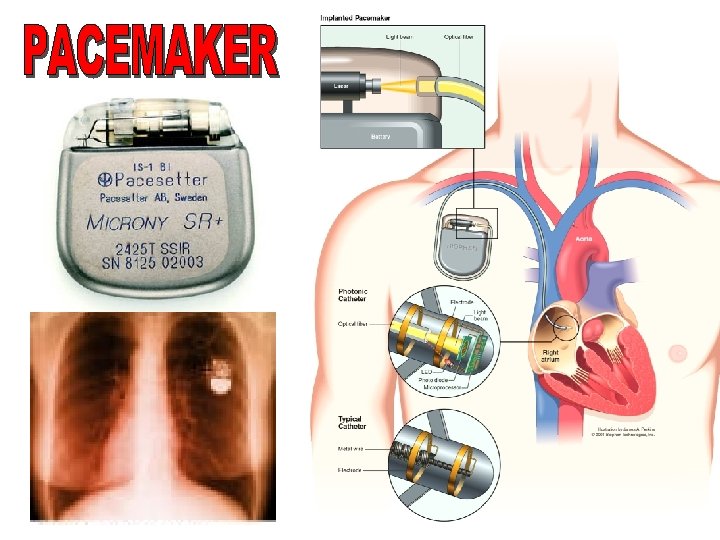
The SA node initiates the heartbeat and has been given the nickname of the “PACEMAKER” PACEMAKER a small electronic device that stimulates the SA node to fire http: //www. youtube. com/watch? v=Cx. JS 0 o. EQe. BQ People with irregular heartbeats may have to have an artificial pacemaker ‘inserted’.
 • In the right atrium close to the AV (tricuspid)")
The AV NODE (atrioventricular) • In the right atrium close to the AV (tricuspid) valve • When the AV node receives the impulse from the SA node, it fires to initiate the contraction of the LARGE ventricles A. V. NODE PURKINJE FIBRES • The AV node sends its message through the Purkinje Fibres (P. F. ), P. F. ) which cause ventricles to contract.

A V NODE & Purkinje fibres

• Atria beat from top down, then pause, and the ventricles beat from bottom up.

http: //www. youtube. com/watch? v =ew 6 Jp 74 va. N 4 http: //www. youtube. com/watch? v =v 3 b-Yh. Zm. Qu 8

FROG DISECTION http: //www. youtube. com/watch? v=f. O 7 l. BX 5 CSxw

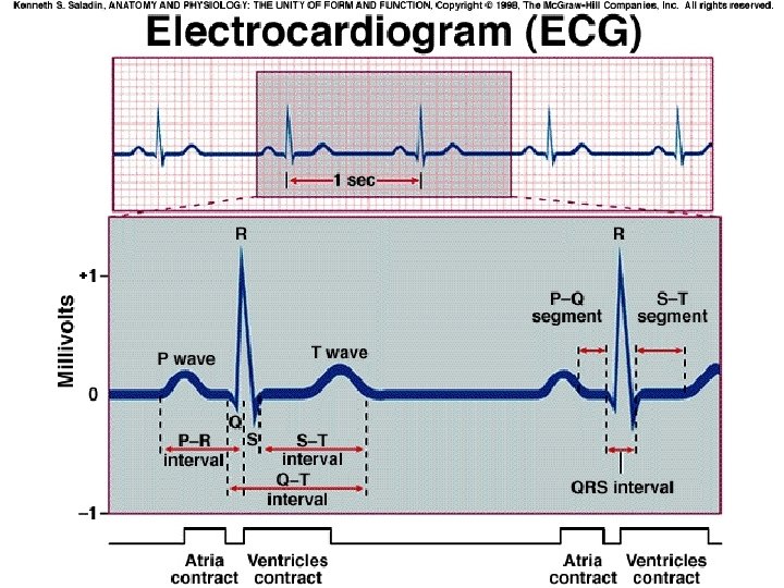
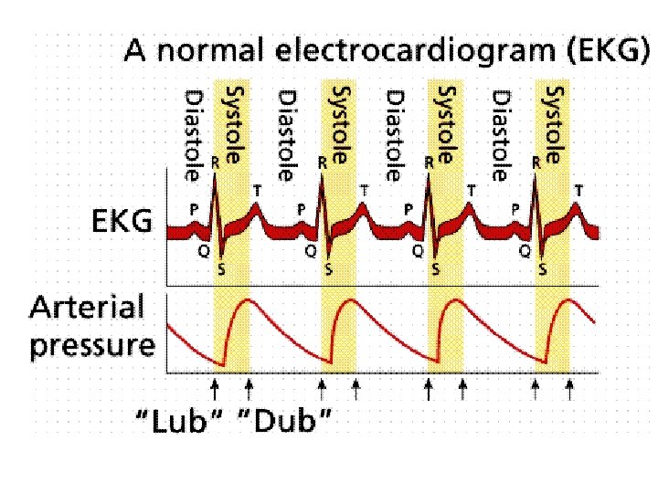
There are two parts to the contraction of the heart The heart beat is a double sound (‘lub-dub’). EKG An EKG (electrocardiogram) registers the voltage changes across the surface of the heart as it beats. The letters PQRST are the standard labels used to identify the parts of the EKG.
 QRS =")
P = the simultaneous contraction of the atria (caused by SA node) QRS = the contraction of the ventricles (caused by AV node & purkinje fibres) T = the recovery of the ventricles (preparation for next contraction)

KNOW THIS DIAGRAM! R P Q S KNOW THIS DIAGRAM!! T
 Ventricular Fibrillation")
Some Abnormal EKG’s NORMAL Tachycardia (a heart rate of over 100 beats/min) Ventricular Fibrillation (uncoordinated ventricles) Heart Block (failure to stimulate ventricles after atrial contraction)

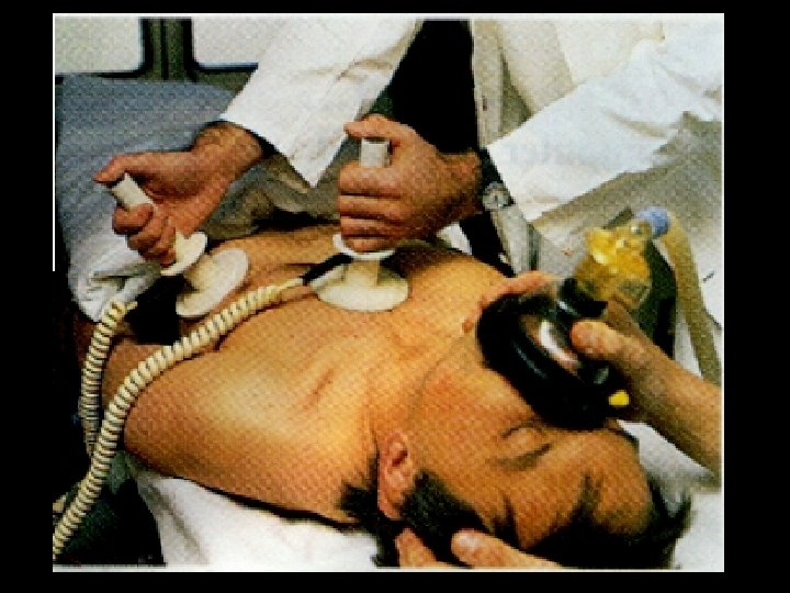
• If the system really breaks down, the heart could go into fibrillation This is uncoordinated contractions of the cardiac muscle. • When this happens, the pacemaker (SA NODE) will send a strong shock through the heart. • Hopefully the heart muscle will reset itself • This is known as defibrillation

• We also try to do this artificially when people are in cardiac arrest!

The natural average resting heart rate is 72 beats per minute The SA node is connected to the brain by the VAGUS NERVE (cranial nerve #10).
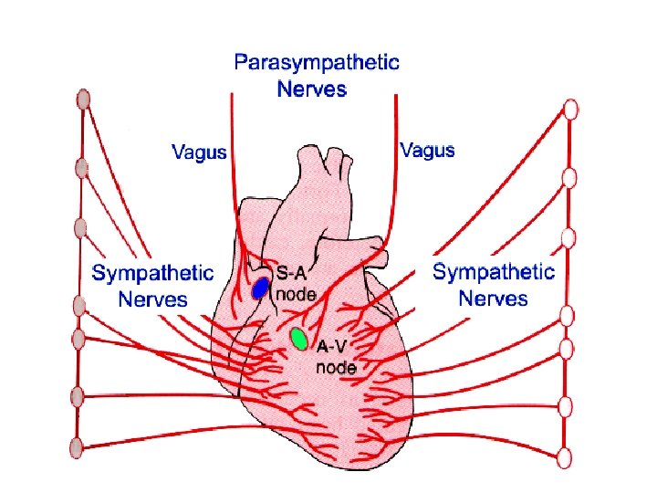

The regulation of the heartbeat is under the influence of the AUTONOMIC NERVOUS SYSTEM (not under conscious control) Sympathetic Nervous System: When the brain is not receiving blood quickly enough, the brain will signal the SA node (via the vagus nerve) to speed up its contraction. This will usually occur in circumstances of FIGHT or FLIGHT. It will also occur when the blood pressure is too low. Parasympathetic Nervous System: this system will reestablish the resting heart rate (~60 -70 beats/minute) by sending a message via the vagus nerve to slow the heart rate.

The part of the brain that governs the speed of the heart rate is called the MEDULLA OBLONGATA • It will speed up or slow down the heart rate when needed. **Under normal circumstances, the heart controls itself.

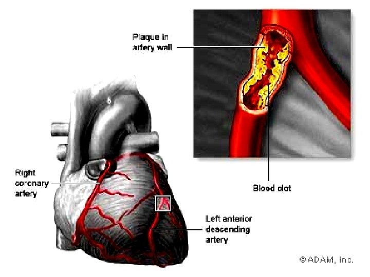
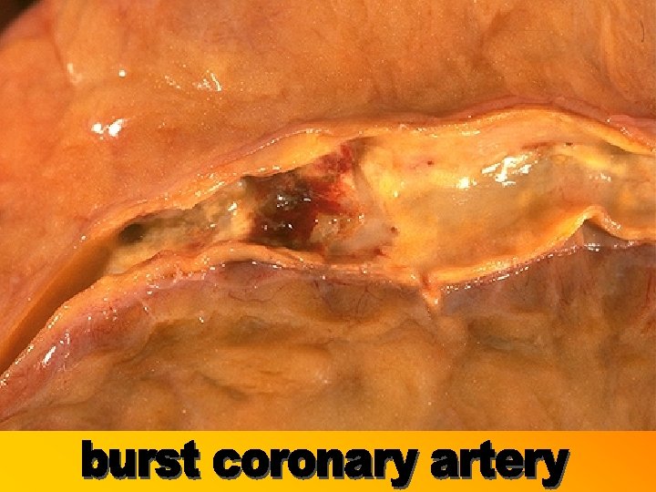
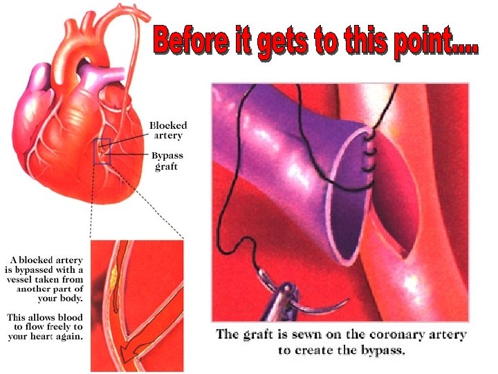
Hardening of the arteries
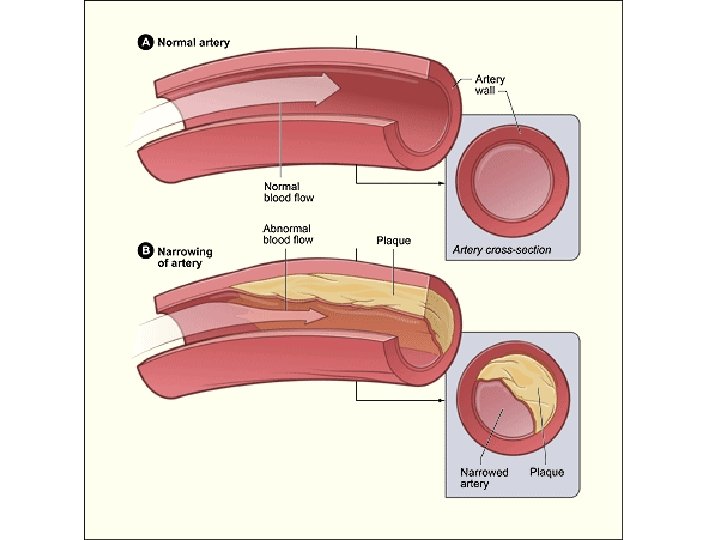

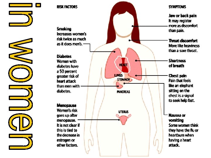
Cholesterol in the blood can become deposited on the inside walls of the arteries. This seems to happen faster in people who: • Smoke • Have high blood pressure • Eat high fat, high cholesterol foods, or, for other reasons, have high cholesterol • Are overweight • Have a lot of tension and stress • Do not exercise regularly • Have diabetes and/or family members with a history of atherosclerosis

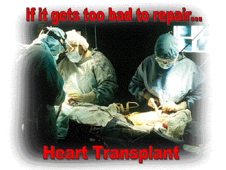

Perform your own heart transplant: http: //www. pbs. org/wgbh/nova/eheart/transplantwave. html

Made from titanium metal and a special type of plastic. A rechargeable internal battery Normally an external power pack transmits power across the skin.

• Feel your pulse by placing two fingers at pulse points on your neck or wrists. Now count the beats for 15 seconds & multiply by 4. • As a kid, your resting pulse might range from 90 to 120 beats per minute. • As an adult, your pulse rate slows to an average of 72 beats per minute. What is YOUR pulse rate at rest? After exercise?

BLOOD PRESSURE: the ventricles pump approx. 70 m. L of blood each time they contract. The pulse you feel is blood stopping and starting as it moves through your arteries
: blood pressure when the ventricles are contracting. Blood is being forced")
SYSTOLIC PRESSURE (systole): blood pressure when the ventricles are contracting. Blood is being forced through the arteries (approx. 120 mm. Hg)
: blood pressure as ventricles are refilling. This is between contractions, and")
DIASTOLIC PRESSURE (diastole): blood pressure as ventricles are refilling. This is between contractions, and the blood pressure is less (appox 80 mm. Hg).

Blood pressure is normally measured along the BRACHIAL ARTERY of the arm. A reading of 120/80 mm. Hg is normal.

When you are younger, your BP will be higher. Example: 13 -15 yrs. will be 136 -144 over 86 -92
")
HIGH BLOOD PRESSURE (ie: 150/100)

High blood pressure sometimes goes unnoticed until complications set in. Limit Alcohol Eat less salt Be Active Don’t Smoke Eat Healthy Lose Weight

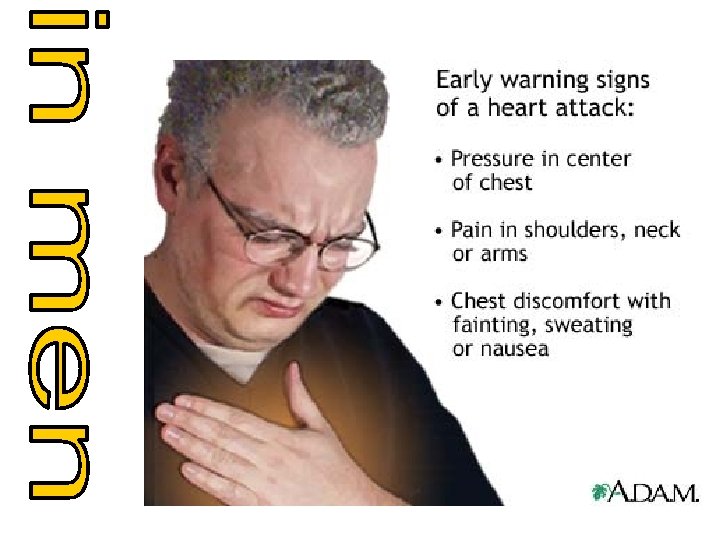
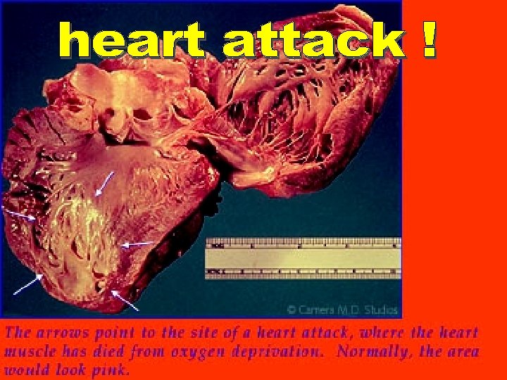
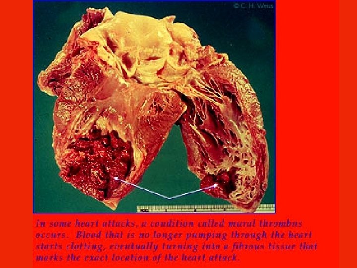
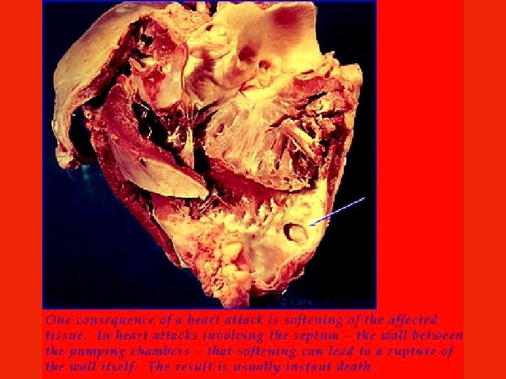
• High Blood Pressure puts constant strain on the tissues (especially the capillary beds). • May cause capillaries to burst • If this happens in the brain = a stroke. If it happens in the heart = heart attack! • The longer you have high BP, the greater the potential for tissue damage. • Sometimes high BP is normal (ie: when doing physical activity) • However, the brain should return the BP to a normal, lower level.
")
LOW BLOOD PRESSURE (ie: 100/60)

Low blood pressure is not particularly a good thing either. It can result from: • Genetics • Anemia *not enough iron* • Dehydration *not enough water* • Blood loss • Shock Proper kidney function can only be maintained if there is sufficient pressure for filtration.
 = lower BP smaller (constrict) = higher BP")
1. Vessel diameter: diameter bigger (dilate) = lower BP smaller (constrict) = higher BP Vasodilation to lower BP Vasoconstriction to increase BP
 = higher BP Thin (lots of")
2. Blood viscosity: viscosity Thick blood (little water) = higher BP Thin (lots of water) = lower BP

3. Total blood vessel length: length More fat = higher BP Thinner = lower BP More fat = more vessels = more resistance = increased BP
. elasticity Elastic vessels = lower BP")
4. Vessel elasticity: affected by plaques (fatty deposits). elasticity Elastic vessels = lower BP Hardened vessels = high BP ATHEROSCLEROSIS (hardening of arteries = decreased elasticity).

5. Blood volume: volume Sweat a lot = less volume/water = lower BP Eat lots of salt = more volume/water stays in body = higher BP

6. Cardiac output: output Heart rate increases = higher BP Heart rate decreases = lower BP

7. Age: as you get older, there is a loss of elasticity in the Age blood vessels. Young = very elastic = low BP Old = not elastic = high BP

8. Stress: constricts blood vessels which means Stress increased pressure to move the blood. Stressed = constricted vessels = Higher BP Calm = normal vessels = Lower BP

CIRCULATION SONG: http: //www. youtube. com/watch? v=q 0 s-1 MC 1 hc. E

Heart Animations and Interactives Animation: Your heart valves at work Interactive: Label the heart Interactive: Listen to the heart with a virtual stethoscope Interactive: Explore the structures of the heart and trace the pathway of blood through the heart, lungs, and body Animation: See the flow of blood to and from the exterior heart
- Slides: 111