http www personal psu edupzb 4electrophoresis swf Agarose
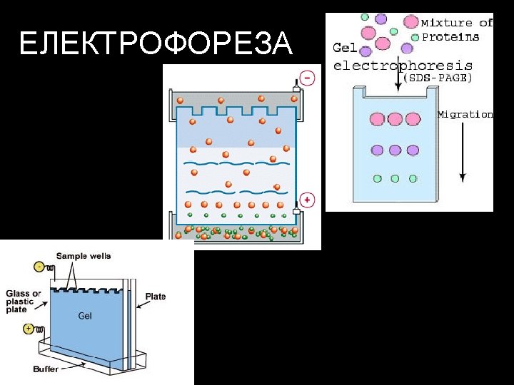

http: //www. personal. psu. edu/pzb 4/electrophoresis. swf


Agarose D-galactose 3, 6 -anhydro L-galactose • Sweetened agarose gels have been eaten in the Far East since the 17 th century. • Agarose was first used in biology when Robert Koch* used it as a culture medium for Tuberculosis bacteria in 1882 Агарозата е линеарен полимер што се екстрахира од морските алги.


Протеинските серуми се испитуваат со хоризонтална геле елктрофореза The figure was found at http: //www. mun. ca/biology/desmid/brian/BIOL 2250/Week_Three/electro 4. jpg


Serum protein electrophoresis Hydragel – agarose gel • Серумските протеини се сепарираат во 6 групи: • Albumin • α 1 - globulins • α 2 - globulins • β 1 - globulins • β 2 - globulins • γ - globulins Figure is found at http: //www. sebia-usa. com/products/protein. Beta. html#


ЦИРОЗА • Цироза –зголемена конзумација на алкохол или хепатит • ↓ albumin • ↓ α 1, α 2 и β globulins • ↑ Ig A in γ-fraction Figure is found at http: //erl. pathology. iupui. edu/LABMED/INDEX. HTM γ-globulins
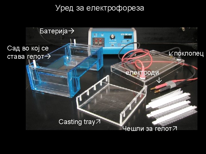

Gel casting tray & combs

Preparing the Casting Tray Seal the edges of the casting tray and put in the combs. Place the casting tray on a level surface. None of the gel combs should be touching the surface of the casting tray.

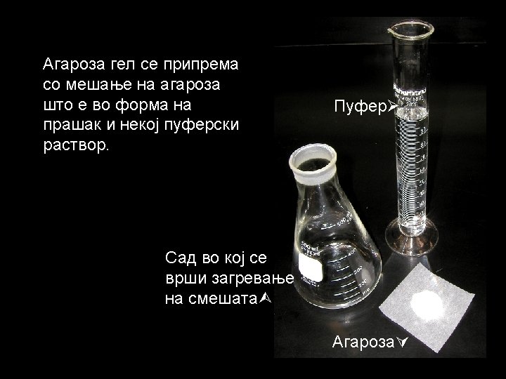
Agarose Buffer Solution Combine the agarose powder and buffer solution. Use a flask that is several times larger than the volume of buffer.
. The agarose solution is boiled")
Melting the Agarose is insoluble at room temperature (left). The agarose solution is boiled until clear (right). Gently swirl the solution periodically when heating to allow all the grains of agarose to dissolve. ***Be careful when boiling - the agarose solution may become superheated and may boil violently if it has been heated too long in a microwave oven.
 and then carefully")
Pouring the gel Allow the agarose solution to cool slightly (~60ºC) and then carefully pour the melted agarose solution into the casting tray. Avoid air bubbles.

Each of the gel combs should be submerged in the melted agarose solution.

When cooled, the agarose polymerizes, forming a flexible gel. It should appear lighter in color when completely cooled (30 -45 minutes). Carefully remove the combs and tape.

Place the gel in the electrophoresis chamber.
 Anode (positive) Add enough electrophoresis buffer to cover the")
DNA buffer wells Cathode (negative) Anode (positive) Add enough electrophoresis buffer to cover the gel to a depth of at least 1 mm. Make sure each well is filled with buffer.

Sample Preparation Mix the samples of DNA with the 6 X sample loading buffer (w/ tracking dye). This allows the samples to be seen when loading onto the gel, and increases the density of the samples, causing them to sink into the gel wells. 6 X Loading Buffer: Bromophenol Blue (for color) Glycerol (for weight)

Loading the Gel Carefully place the pipette tip over a well and gently expel the sample. The sample should sink into the well. Be careful not to puncture the gel with the pipette tip.

Running the Gel Place the cover on the electrophoresis chamber, connecting the electrical leads. Connect the electrical leads to the power supply. Be sure the leads are attached correctly - DNA migrates toward the anode (red). When the power is turned on, bubbles should form on the electrodes in the electrophoresis chamber.
 wells Bromophenol Blue DNA (-) Gel Anode (+) After the current is")
Cathode (-) wells Bromophenol Blue DNA (-) Gel Anode (+) After the current is applied, make sure the Gel is running in the correct direction. Bromophenol blue will run in the same direction as the DNA.

DNA Ladder Standard 12, 000 bp 5, 000 DNA migration Note: bromophenol blue migrates at approximately the same rate as a 300 bp DNA molecule bromophenol blue 2, 000 1, 650 1, 000 850 650 500 400 300 200 100 + Inclusion of a DNA ladder (DNAs of know sizes) on the gel makes it easy to determine the sizes of unknown DNAs.

As an alternative to purchasing costly DNA ladders, one can be created using meal worm (Tenebrio molitor) DNA and a restriction enzyme. http: //people. uis. edu/rmosh 1/DNAexercise. VIIa 02. pdf

Staining the Gel • Ethidium bromide binds to DNA and fluoresces under UV light, allowing the visualization of DNA on a Gel. • Ethidium bromide can be added to the gel and/or running buffer before the gel is run or the gel can be stained after it has run. ***CAUTION! Ethidium bromide is a powerful mutagen and is moderately toxic. Gloves should be worn at all times.

Safer alternatives to Ethidium Bromide Methylene Blue Bio. RAD - Bio-Safe DNA Stain Ward’s - QUIKView DNA Stain Carolina BLU Stain …others advantages disadvantages Inexpensive Less toxic No UV light required No hazardous waste disposal Less sensitive More DNA needed on gel Longer staining/destaining time

Staining the Gel • Place the gel in the staining tray containing warm diluted stain. • Allow the gel to stain for 25 -30 minutes. • To remove excess stain, allow the gel to destain in water. • Replace water several times for efficient destain.

Ethidium Bromide requires an ultraviolet light source to visualize
 DNA ladder 1 2 3 4 5 6 7")
Visualizing the DNA (ethidium bromide) DNA ladder 1 2 3 4 5 6 7 8 wells 5, 000 bp 2, 000 1, 650 1, 000 850 650 500 400 300 200 100 PCR Product Primer dimers + - - + + Samples # 1, 4, 6 & 7 were positive for Wolbachia DNA
 wells DNA ladder PCR Product + - -")
Visualizing the DNA (Quik. VIEW stain) wells DNA ladder PCR Product + - - + 2, 000 bp 1, 500 1, 000 750 500 250 Samples # 1, 6, 7, 10 & 12 were positive for Wolbachia DNA March 12, 2006
- Slides: 43