HOW WE SEE Eye Structure External Layer Eye


 Iris")

 ● Posterior Chamber filled with Vitreous Humour − ● Pores in")


 Retina Converts light energy to nervous impulses Rods detect shades of")
 Retina (cont. ) Blind Spot Area where optic nerve leaves eye.")


 When rhodopsin is struck by light it splits and alters the")
 Cones see color by using three types of rhodopsin; one for")
 Moving parallax (diff distances, diff speed) The size a known")

 Able to see near objects well and has difficulty seeing objects that")
 Is present, a person is able to see distant objects well and")


- Slides: 34
HOW WE SEE!
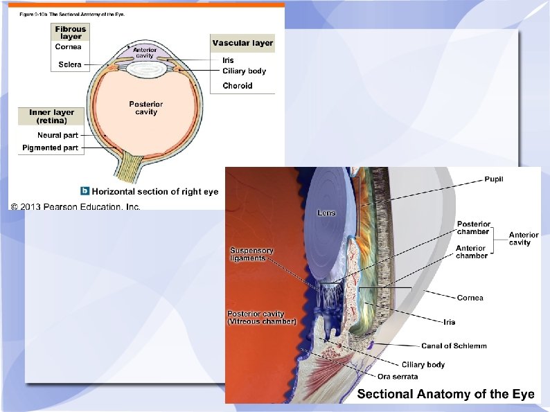
Eye Structure
External Layer
Eye Protection ● Conjunctiva − ● Fat Deposits − ● membrane lining the inside of eyelids and across the front of the eye. Prevents objects from moving behind the eye. Cushioning the eye during impacts. Sclera − Strong outer membrane. Gives shape to eye, and helps with accommodation.
Intermediate Layer ● ● Choroid – absorbs scatter light, includes blood vessels (food) Iris – regulates amount of light entering Pupil – opening of light Ciliary muscles – change shape of lens to focus
Nourishment ● Fat Deposits − ● a close energy source Choroid layer − enriched with blood vessels. Brings in nutrients and removes wastes.
Nourishment (cont. ) ● Posterior Chamber filled with Vitreous Humour − ● Pores in the ciliary muscles − ● brings nutrients to the lens. allow nutrients to diffuse from the posterior to the anterior chamber. Anterior Chamber filled with Aqueous Humour − brings nutrients to the cornea and the front of the lens.
Internal Layer ● ● ● Rods – light receptors Cones – color receptors Fovea centrailis – high density of cones, provides acute vision
Other Noteable Stuctures ● ● Lens – focuses light Humours -support eyeball with pressure of fluids Optic nerve – transmitter to brain Optic disc – blind spot, where nerve connets with eye Watch Crash Course A&P 18 https: //youtu. be/o 0 DYPu 1 r. NM? list=PL 8 d. Puua. Lj. Xt. OAKed_Mxx. WBNa. P
Objects focus on Retina The image is focused on the fovea centralis is: Smaller Upside down Reversed left and right
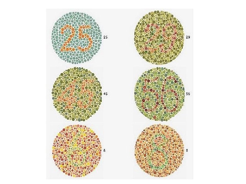
Vision (cont. ) Retina Converts light energy to nervous impulses Rods detect shades of black and white, excellent with motion, can work in low light. (peripheral) Cones • Detect colour, excellent with detail, require lots of light. (central vision) - http: //wimp. com/colorblind/ Fovea Centralis (Macula) Filled with only cones (for detail vision)
Vision (cont. ) Retina (cont. ) Blind Spot Area where optic nerve leaves eye. No room for sensory receptors.
Reflexes of Sight Light reflex bright light causes the iris to enlarge and the pupil to get smaller, while low light causes the opposite. Accommodation Reflex pupil size and lens shape changes with distance. For far objects the lens is stretched flat, while for close objects it bulges out.
Chemistry of Sight Rods and Cones contain rhodopsin pigments. Rhodopsin is made up of Vitamin A and Opsin.
Chemistry (cont. ) When rhodopsin is struck by light it splits and alters the cell chemistry, causing the neuron to fire.
Chemistry (cont. ) Cones see color by using three types of rhodopsin; one for each primary color (Red, green, blue). If you are colour blind, you have deficiency in a specific cone type.
Depth Perception Memory (brain) Moving parallax (diff distances, diff speed) The size a known object has in your brain. When you move your head from side to side, objects that are close to you move rapidly across your retina. However, objects that are far away move very little. Stereo vision (overlapping fields of view) Each eye receives a different image of an object on its retina because each eye is about 2 inches apart.
Brightness Perception The brightness of an object depends on the light reflected from itself and its background.
Nearsightedness (Myopia) Able to see near objects well and has difficulty seeing objects that are far away The image is focused in front of the retina Corrected with concave lens
Farsightedness (Hyperopia) Is present, a person is able to see distant objects well and has difficulty seeing objects that are near. Image is focused behind the retina Corrected with a convex lens.
Astigmatism Uneven curvature of the cornea and causes a distortion in vision. Corrected with a shaped lens.
Blindness Night Blindness severe vitamin A deficiency leads to a lack of rhodopsin. Cataracts Glaucoma Diabetic Retinopathy
Glaucoma increased pressure within the eyeball. aqueous humor builds up and increases pressure within the eye. damage the optic nerve directly or restrict blood flow, thus damaging the optic nerve indirectly. lead to blind spots in the visual field. can cause permanent blindness.
Glaucoma
Cataracts the lens becomes cloudy. Cataracts are caused by changes in the chemical makeup of the lens. With age, the lens becomes thicker and less clear.
Cataracts
Diabetic Retinopathy Diabetic retinopathy refers to damage to the blood vessels of the retina caused by diabetes. As new blood vessels grow on the retina, blurred vision or temporary blindness can result. Scare tissue can form and cause blindness where old blood vessles were attached to the retina.
Diabetic Retinopathy
Connectivity