
How to Request Laboratory Test in Uveitis F

How to Request Laboratory Test in Uveitis? F. Kianersi M. D 1397 / 9 / 29

Introduction Uveitis is a common and etiologically complex disease.

Introduction (History & Physical Examination) Ø A detailed History and general Physical Examination, with particular attention to potential causes of uveitis, will provide complementary information to document and characterize uveitis.
Introduction (Ophthalmic Examination) v. A complete ophthalmic examination is then performed. ü The conjunctiva, anterior segment and posterior segment findings should be documented.
Introduction (Review of Systems) Ø The review of systems is essential in developing a differential diagnosis for uveitis and utilizes an extensive review from a patient questionnaire, supplemented by direct questioning.
Introduction (Past Medical History) q. A complete past medical history is also critical and should focus on the immune status and presence of collagen vascular diseases, infectious diseases, neurologic diseases, GI disorders, and so on. q. Knowledge of underlying diabetes, hypertension, or coronary artery disease is essential for optimizing therapeutics.
Introduction Ø In patients in whom the etiology remains uncertain based upon the history and examination alone, selected laboratory testing may help to elucidate the cause.
Introduction v. Laboratory testing, however, is not a substitute for a thorough history and physical examination of any patient with ocular inflammation. v. The workup must be custom tailored for each patient depending on the History, patient’s Symptoms and Signs, and Clinical examination, and Risk factors.
Introduction (Investigations and their Purpose) 1. Identify & Confirmation of etiology, 2. Identify associated systemic disease, 3. Fitness for treatment: Liver function test before starting Methotrexate, 4. Iatrogenic complication: Blood sugar after chronic steroid treatment, 5. Sequela measurement: F/A for CNV.
Introduction ØLaboratory testing for ocular inflammatory disease is frequently a challenge for practicing ophthalmologists. ØThe myriad of tests available, the complexities of their interpretation, and the underlying concern that one may miss an important systemic disease all contribute to this challenge.
General Guidelines for Laboratory Testing Most patients presenting with unexplained uveitis should undergo laboratory workup.
General Guidelines for Laboratory Testing v. It is best for the evaluating ophthalmologist to order appropriate laboratory tests rather than referring patients to rheumatologist for workup. v. The non-ophthalmologist will not be familiar with the differential diagnosis of ocular inflammatory diseases, leading to incomplete work-up and frustration for patients and physicians.
General Guidelines for Laboratory Testing v. Only limited diagnostic testing is useful when an associated condition is not apparent despite a thorough medical History and Physical Examination. v. In either situation, the use of multiple screening tests (a “shotgun approach”) should be avoided. ü There is no “standard” uveitis workup.
General Guidelines for Laboratory Testing v. Testing need not be completed in one session. ü High-probability and high-morbidity diseases can be assessed initially. ü Less common conditions tested only if the initial workup is unrevealing. Physicians should not order tests that they cannot interpret.
General Guidelines for Laboratory Testing (Effect of Systemic condition & Medications on tests) Ø ACE levels can be depressed by systemic corticosteroids. Ø It will be near zero in patients taking ACE-inhibitor antihypertensive drugs (Captopril & Enalapril).
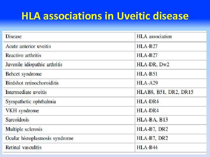
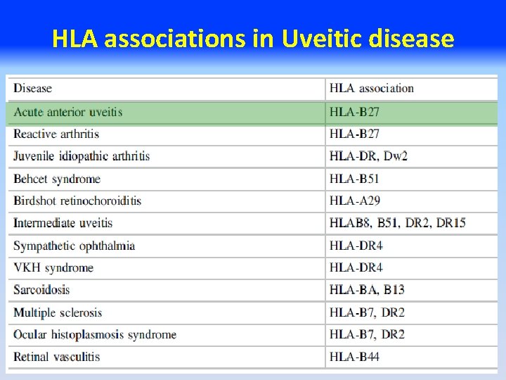
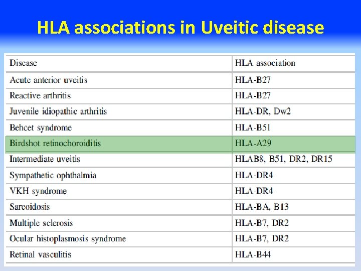
General Guidelines for Laboratory Testing (False Negative & False Positive) q. The prevalence of HLA-27 & HLA-29 in the general population is about 8%. ü So these tests have a false-positive rate of 8%.
General Guidelines for Laboratory Testing (Tests that Rule In disease versus Ruling Out disease) v. In some cases, a test being negative may be just as important as positive testing, e. g Toxoplasmosis titers. Ø A positive value does not mean a patient has toxoplasma retinochoroiditis, since nearly 30 % of the population may have been exposed to toxoplasma. Ø In contrast, a negative test is sensitive for the exclusion of toxoplasmosis.
General Guidelines for Laboratory Testing (Tests that Rule In disease versus Ruling Out disease) Ø Birdshot chorioretinitis occurs nearly exclusively in patients who are HLA‑A 29 positive. Ø Negative HLA-A 29, may be helpful in ruling out Birdshot chorioretinopathy in patients with multiple white chorioretinal lesions.
General Guidelines for Laboratory Testing (Tests that Rule In disease versus Ruling Out disease) Ø Birdshot uveitis is highly correlated with the HLAA 29 gene, with a sensitivity of 96% and a specificity of 92%. ü Thus, a negative test for HLA‑A 29 makes this diagnosis exceedingly unlikely. ü HLA‑A 29 also has an allele frequency of about 8% in the United States population, so false positives are relatively common.
General Laboratory Testing
General Laboratory Testing Ø CBC with differential can be useful in identifying systemic infection (with leukocytosis), parasitic infection (with eosinophilia), leukemia, or certain immunocompromised states.
General Laboratory Testing Ø A comprehensive metabolic panel can reveal renal or hepatic dysfunction and undiagnosed hyperglycemia. ü FBS, SGOT, SGPT, Alk Phos, Bil, BUN, Cr, U/A ………. PPD ü The results of these tests are also essential for initiating therapy with oral corticosteroids or steroid-sparing medications.
General Laboratory Testing q. Non-specific tests of inflammation, such as ESR or CRP, are rarely of utility in the diagnosis of uveitic disease.
General Laboratory Testing v. Chest x‑ray or high resolution Chest CT scans useful in the workup of sarcoidosis and tuberculosis. v. Sacroiliac CT scans useful in the workup of seronegative artheropathies.

Specific Laboratory Evaluation
Specific Laboratory Evaluation Ø The anatomical location, onset, duration, course, response to therapy, age, race and region can help guide diagnostic considerations and testing.
Specific Laboratory Evaluation (Role of Race and Region) ü TB is endemic in most parts of the world. ü In OHS & Lyme disease geography is a dominant feature. ü Behçet disease while rare in the USA and Europe, it is more common in the Middle East and Asia.

Suggested Testing Algorithm by Anatomic Classification
Suggested Testing Algorithm by Anatomic Classification Ø Grouping each uveitic patient into an anatomic class is important when deciding about what and when testing should be ordered. Ø Acute unilateral anterior uveitis suggests the possibility of a Spondyloarthropathy. Ø Bilateral chronic anterior and posterior uveitis may suggest Sarcoidosis.
Suggested Testing Algorithm by Anatomic Classification Ø Grouping each uveitic patient into an anatomic class is important when deciding about what and when testing should be ordered. ü The first episode in a patient with isolated mild to moderate acute anterior uveitis and an unremarkable history generally does not require any work-up. ü Alternatively, patients with intermediate, posterior, or panuveitis should always have testing done.
Suggested Testing Algorithm by Anatomic Classification Ø The anatomic location will also direct what tests to order or not order. ü For example, you would not send HLA-B 27 testing on a patient with posterior uveitis. ü Likewise, you would not send HLA-A 29 or Toxoplasmosis testing on a patient with isolated anterior uveitis.




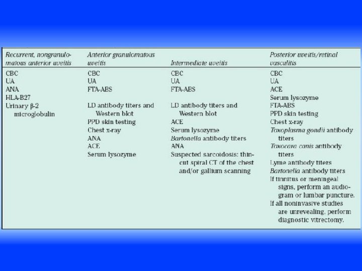
Targeted versus Screening tests • After addressing the importance of a focused or targeted laboratory work-up, it is important to acknowledge that there a few infectious uveitic diseases that cannot be defined by their clinical findings and may present in various anatomical locations.
Targeted versus Screening tests v. These are important to highlight as they are not treated with immunomodulators, and if left untreated can lead to a poor visual and in some cases systemic prognosis. ü Generally, these diseases include Lyme disease, Syphilis, and Tuberculosis.

Testing for Sarcoidosis, Syphilis, Tuberculosis, and Lyme Disease v. Nearly every presentation of ocular inflammation has potential association with Syphilis, TB, or Lyme disease, Sarcoidosis. üThese conditions must be ruled out in all uveitis cases.

Syphilis v. The mainstays of testing are direct and indirect treponemal antibody tests. v. The direct tests: FTA‑ABS (Fluorescent Treponemal antibody), TPPA and MHA‑TP, serve as seroconversion markers. v. Once a patient has been exposed to syphilis, these markers remain forever positive, even if the infection has been completely treated. v. These tests are more useful for ruling out Syphilis than establishing the diagnosis.
Syphilis v. The indirect tests: RPR (Rapid Plasmin Reagent) or VDRL (Venereal Disease Research Laboratory tests). v. The titers of these indirect antibodies vary with disease load and become undetectable in fully treated disease. ü Thus, a patient with positive FTA‑ABS and positive RPR or VDRL test results has active syphilis necessitating treatment.
Tuberculosis q The tuberculin skin test (TST), including the Mantoux test and the Purified Protein Derivative (PPD) test, is the mainstay of diagnosis worldwide. q Most positive PPD tests are not associated with active disease but will reflect exposure or latent disease. q Chest x‑ray is a useful adjunct to skin testing.
Tuberculosis q Most cases of uveitic TB miliary disease are not accompanied by pulmonary disease; thus a negative chest x‑ray in the setting of positive PPD and suspicious uveitis not rule out TB. ü In such cases, it is worth obtaining vitreous biopsy for PCR testing. ü Quanti FERON-TB Gold In Tube-Test (QFT). ü Interferon Gamma Release Assay ( IGRA).
Sarcoidosis Ø Definitive diagnosis of sarcoidosis: Histopathologic demonstration of noncaseating granulomata in biopsied tissues. ü The core diagnostic test: chest x‑ray (evidence of hilar adenopathy or interstitial fibrosis). ü High-resolution chest CT scanning with contrast.
Sarcoidosis Ø ACE is present in higher levels in patients with a high systemic granuloma load. ü The positive predictive value of ACE testing is 47%. ü Because ACE levels are not specific for sarcoidosis, a positive test never makes the diagnosis of sarcoidosis.
Lyme Disease Ø In this case, geography is a dominant feature. Ø Lyme risk varies tremendously throughout the world. ü Serologic testing, with screening ELISA followed by confirmatory Western blot screening, is the mainstay of diagnosis.
Testing for Rheumatologic Disease Ø As a group, rheumatologic diseases are commonly associated with acute anterior uveitis, scleritis, and retinal vasculitis. Ø The mainstay of testing is serology and radiographic analysis when indicated.
Testing for Rheumatologic Disease • RF (Rheumatoid factor), is useful for ruling out disease than confirming it. • c. ANCA (cytoplasmic antineutrophil cytoplasmic antibody), is positive in most patients with WG. • p. ANCA test (antimyeloperoxidase) is associated with the other systemic vasculitides.
Testing for Rheumatologic Disease Ø Approximately 5% of the population are ANA and Anti-ds. DNA positive. ü Chronic anterior uveitis in childhood is frequently associated with JIA most commonly associated with the pauci-articular form. ü The majority of these patients will have positive ANA titers, but negative RF.
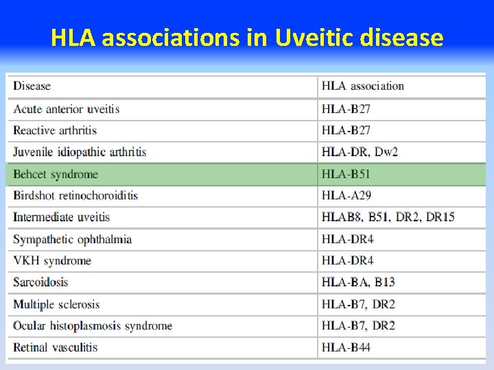
Human Leukocyte Antigen Testing Ø HLA disease associations are simply associations between a major histocompatibility complex molecule and a clinical condition. Ø Testing for HLA can provide supportive evidence for a particular diagnosis but cannot make a definitive diagnosis.
HLA associations in Uveitic disease
HLA associations in Uveitic disease
Human Leukocyte Antigen Testing • Approximately 40% to 50% of acute anterior uveitis cases are associated with HLA-B 27. • The prevalence of this allele in the general US population is about 8% (and so the test has a falsepositive rate of 8%).
HLA associations in Uveitic disease
HLA associations in Uveitic disease Ø HLA-A 29 is confirmatory rather than diagnostic, as 8% of the general population carries this haplotype, and in the absence of characteristic clinical features, an alternative diagnosis should be considered.
HLA associations in Uveitic disease
Human Leukocyte Antigen Testing v. Although HLA‑B 51 is associated with an approximately 6‑fold increased relative risk of Behçet disease, the allele is found in only 39% of Behçet patients, even in high prevalence areas, compared with 18% of control patients. ü A positive test thus does not prove disease nor does a negative test rule out disease.
Human Leukocyte Antigen Testing This type of HLA testing, however, is not universally available and predictive values of such testing are not yet clear.
Testing for Viral Disease q Serology is of little value unless negative; most patients will have been exposed to viruses at some point in their lives and will be seropositive. q PCR is capable of detection of fewer than 10 copies of viral DNA from aqueous or vitreous biopsy.
Testing for Viral Disease q. Recent studies suggest similar diagnostic sensitivity (81%) and specificity (97%) of aqueous and vitreous samples. q. Although highly sensitive, PCR is incapable of distinguishing active viral infection from latent infection.
Testing for Viral Disease q Testing for the presence of virus is rarely a first-line diagnostic investigation in the workup of uveitis. q CMV retinitis has such a typical appearance that no confirmatory testing is generally necessary. q In ARN syndrome, caused by VZV or HSV, the treatment is identical and prompt is mandatory.
Testing for Parasitic Disease (Toxoplasmosis) Ø Ig. M usually appears in the first week after infection, peaks at 1 month, and disappears after 9 months. Ø In cases of acquired Ig. M titers rise before Ig. G seroconversion. Ø Ig. G antibodies can persist at high titers for years after the acute infection. ü Negative serology effectively rules out this diagnosis.
Testing for Parasitic Disease (Toxoplasmosis) Ø For the detection of congenital toxoplasmosis, Ig. A antibodies are often used, because the Ig. M production is still weak in newborns and Ig. G antibodies can be of maternal origin. Ø If definitive diagnosis of ocular toxoplasmosis is required to initiate treatment, either Goldmann. Witmer coefficient testing or PCR of vitreous fluid can be performed.
Testing for Parasitic Disease (Toxocara ) Ø 25% of children in the U. S. have positive titers for Toxocara canis and Toxocara catis serology. Ø Negative serology does not rule out Toxocariasis. Ø PCR testing has no utility in this condition.
Testing for Masquerade Syndromes (Lymphoma ) v. Most intraocular lymphoma is due to primary CNS lymphoma. v. Typically ancillary testing including brain MRI. v. LP is useful. v. However, the mainstay of diagnosis is the vitreous biopsy.
Additional Testing in Selected Presentations Ø Pars planitis or Intermediate uveitis: ü In addition to Sarcoidosis, Syphilis, TB, and other conditions, one must have a high clinical suspicion for demyelinating disease (MS). ü If neurologic symptoms are elicited in the review of systems, MRI of the brain for demyelinating lesions is indicated.
Additional Testing in Selected Presentations Ø Neuroretinitis in one or both eyes is typical for cat scratch disease, caused by Bartonella henselae, and Bartonella quintana. ü Serologic testing is confirmatory and helps direct appropriate antibiotic therapy.
Additional Testing in Selected Presentations Ø Testing for panuveitis: ü Behçet disease, Vogt-Koyanagi-Harada disease, Sympathetic ophthalmia, and Multifocal choroiditis with panuveitis. ü As such, these diagnoses cannot be made through laboratory testing.
Additional Testing in Selected Presentations Ø The white dot syndromes: ü Including acute posterior multifocal placoid pigment epitheliopathy (APMPPE), Serpiginous Choroiditis, Punctate Inner Choroidopathy (PIC), and Multiple Evanescent White Dot Syndrome (MEWDS) do not have specific tests to establish these diagnoses. ü Fluorescein angiography is useful.
Conclusion Most patients presenting with unexplained uveitis should undergo laboratory workup.
Conclusion Laboratory workup should always be guided by, and is not a substitute for, a thorough Patient History, Review of Systems, and Clinical Examination.
Conclusion Patients should be counseled that the yield of laboratory workup is approximately 50%, and reassured that a ‘negative’ workup does not have adverse prognostic consequences for the patient’s ocular inflammation.
Conclusion There is no “standard” uveitis workup. Testing need not be completed in one session.
Conclusion High-probability and highmorbidity diseases can be assessed initially. Less common conditions tested only if the initial workup is unrevealing.
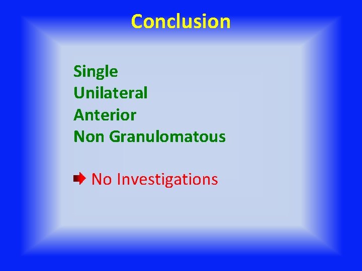
Conclusion Single Unilateral Anterior Non Granulomatous No Investigations
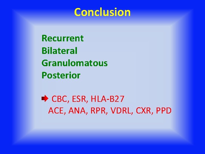
Conclusion Recurrent Bilateral Granulomatous Posterior CBC, ESR, HLA-B 27 ACE, ANA, RPR, VDRL, CXR, PPD
Conclusion Laboratory tests should be “targeted” and “shotgun approach” is not appropriate.

- Slides: 80