How to Read KUB Radiographs Presented by Dr

How to Read KUB Radiographs? Presented by ‐ Dr. Khanak Nandolia Senior Resident Moderator – Dr. Sudhir Saxena Professor and Head

Outline • Introduction • Technique • Normal anatomy • Evaluation
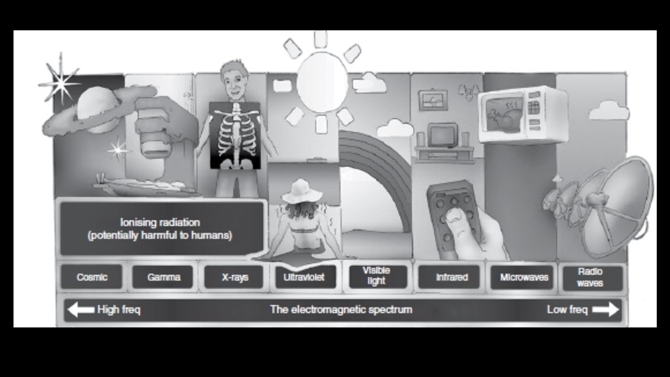

How are X-rays produced? • What is radiation? • Ionizing radiation

3 cardinal principles • Dimensional flattening • Attenuation • Contrast
 representation of a three‐dimensional (3 D) structure.")
Dimensional flattening Two‐dimensional (2 D) representation of a three‐dimensional (3 D) structure.

Attenuation Absorption of x‐rays by the material
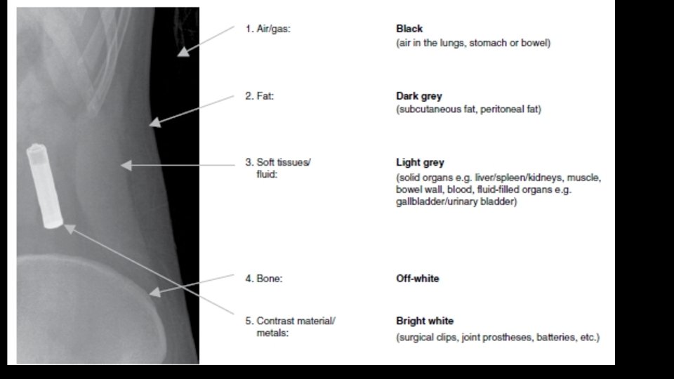

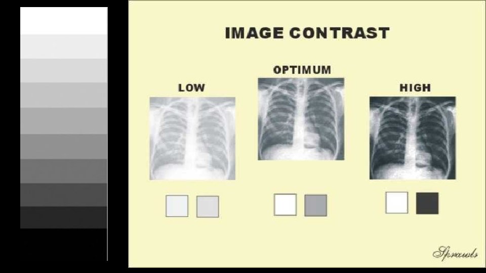
Image contrast Structures can only be seen if there is sufficient contrast with surrounding tissues (contrast is the difference in absorption between one tissue and another).

What is a filling defect?

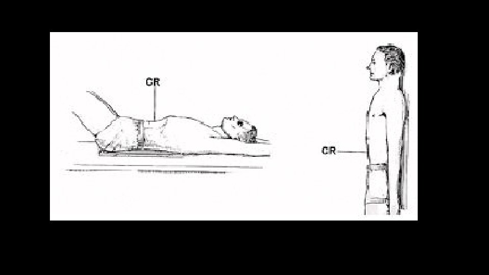
How will you read a radiograph? • Give the type of the radiograph: • Erect • Check patient details • Date of the radiograph • Quality and technical adequacy

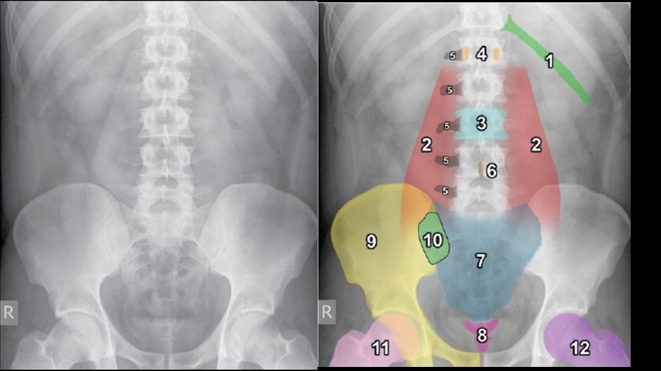
Normal Anatomy

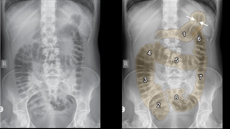
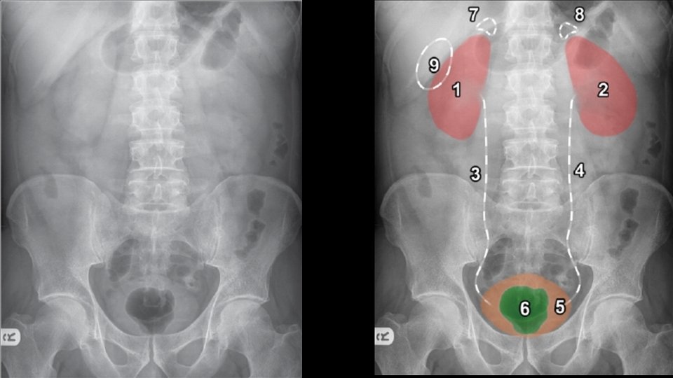
Abdominal viscera • Solid • • Liver Spleen Kidneys and adrenals Pancreas • Hollow • Stomach • Small bowel • Large bowel • Musculoskeletal parts

The most useful tip for radiographs • For a moment, forget that you are facing the examiner. • Describe as if you are describing the radiograph to a colleague, over the phone. • Speak what you are seeing, but systematically.

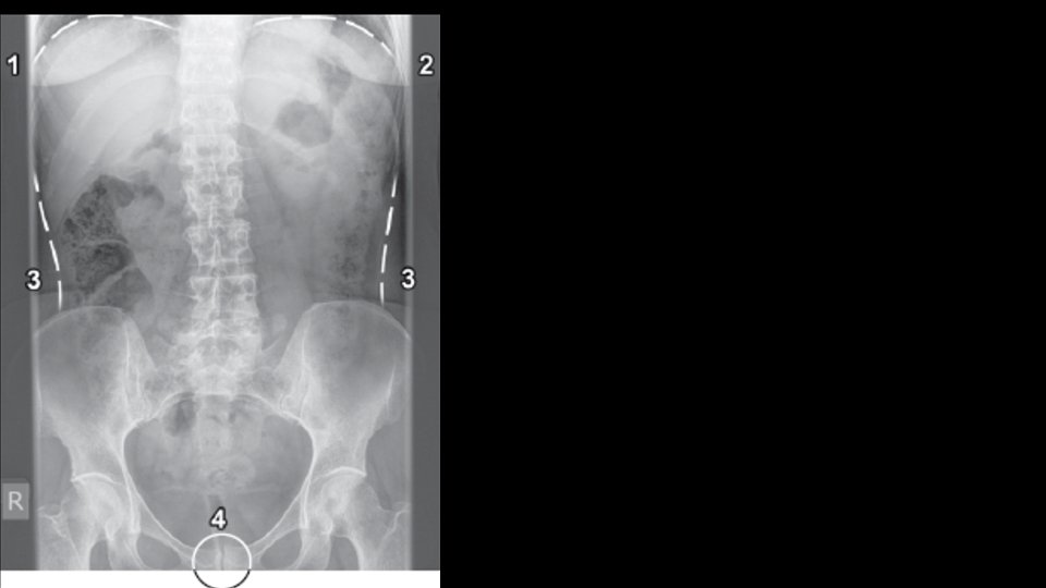
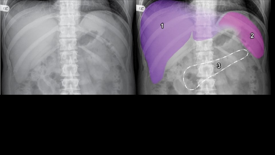
ABC of abdominal radiographs • A for Air • B for Bowel • C for Calcification

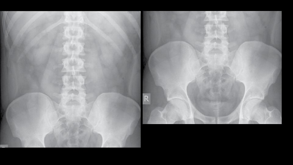
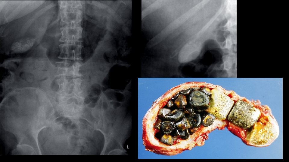
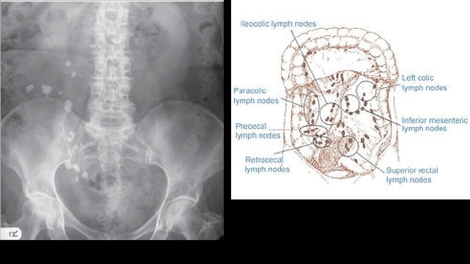
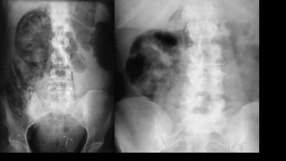
C for calcifications • Calculi – here, there and everywhere • Organ calcifications

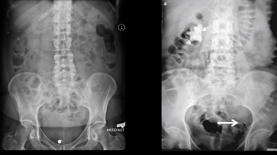
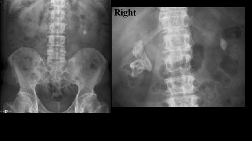
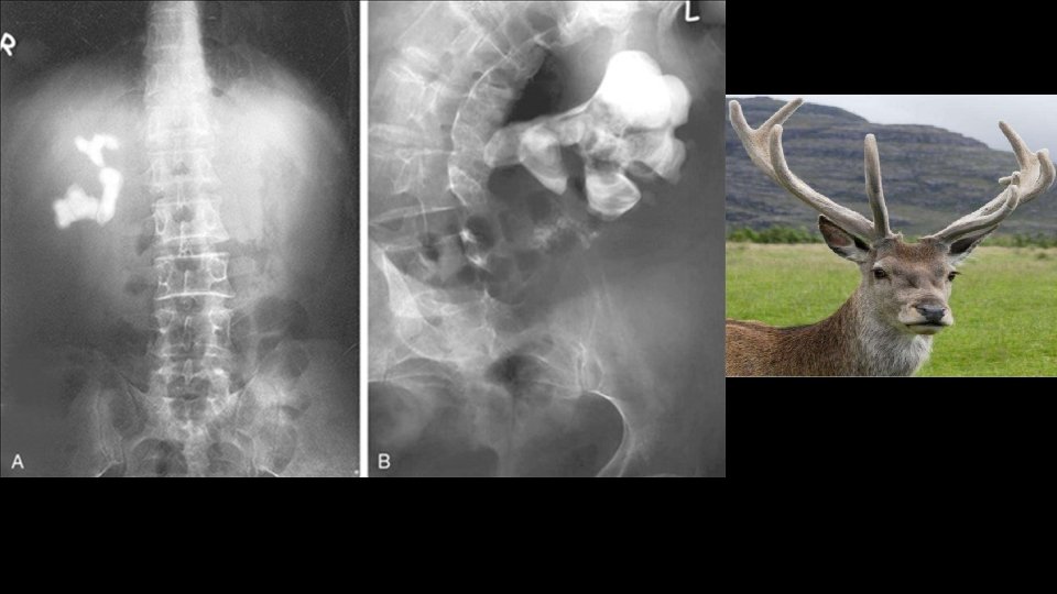
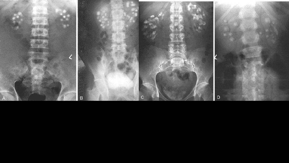
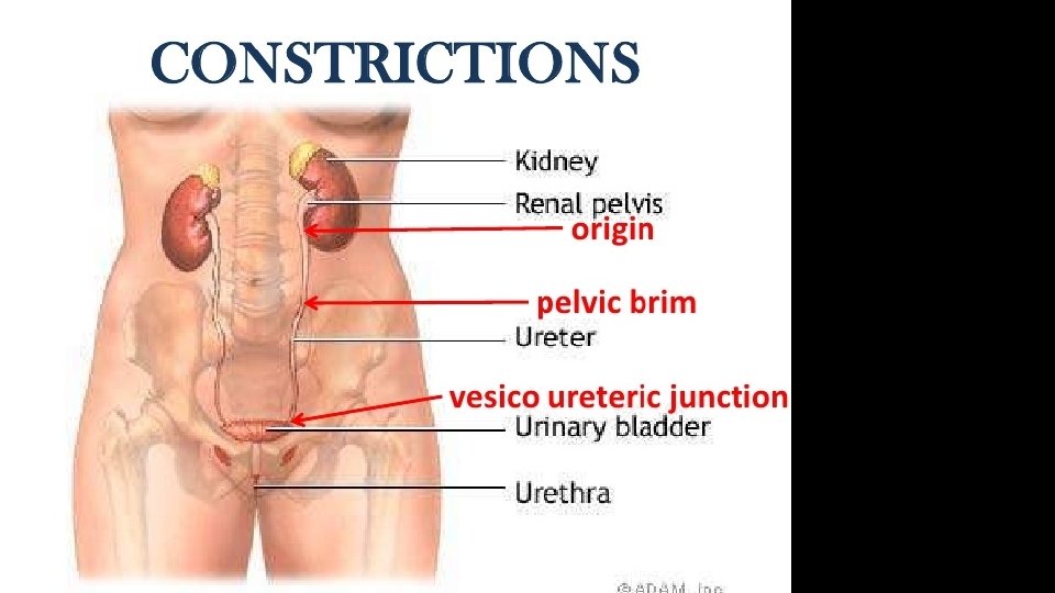
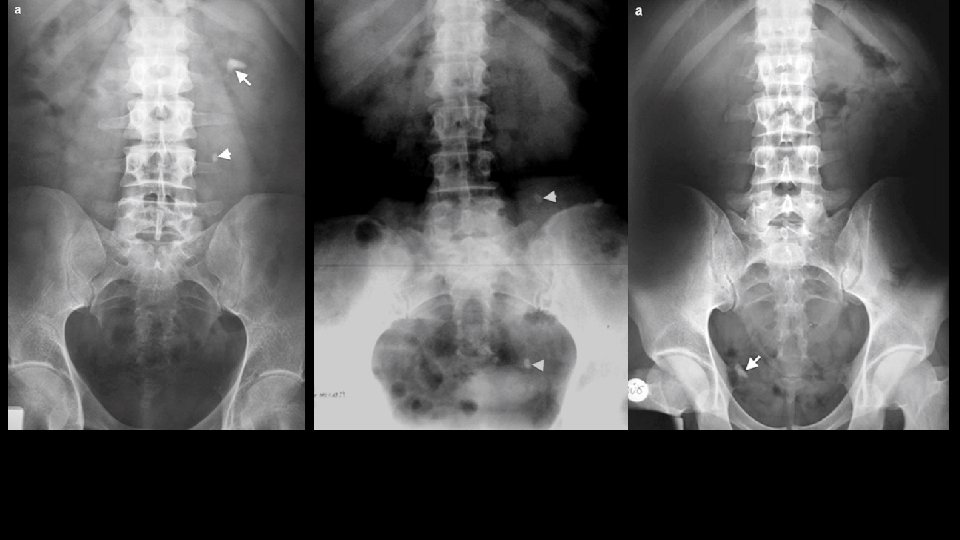
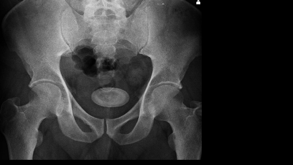
Urolithiasis Calculi along urinary tract • Anywhere • Kidney • Ureter • Bladder

Predisposing factors • • • Stagnations Hypersaturation Bacterial infections Receptor modulation Epithelial injury Nephrocalcin, uropontine

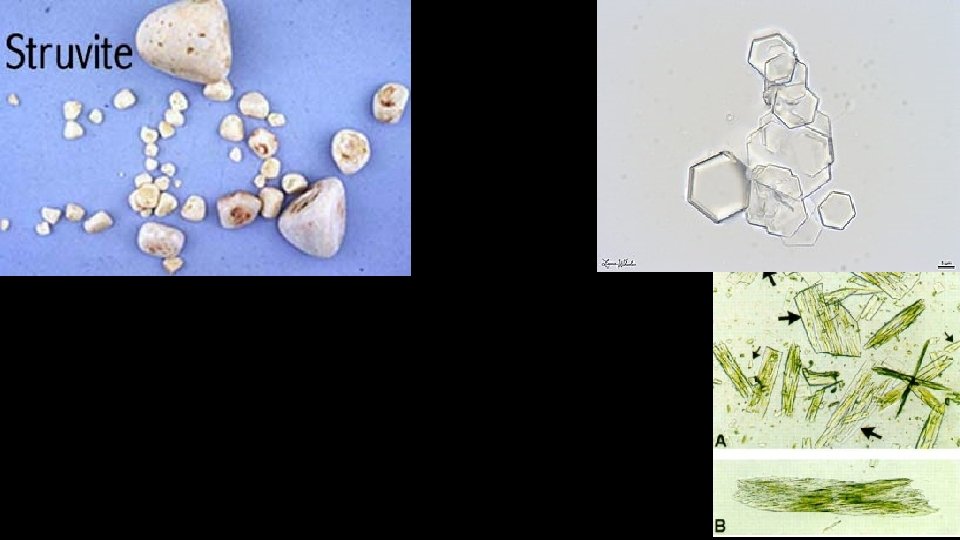
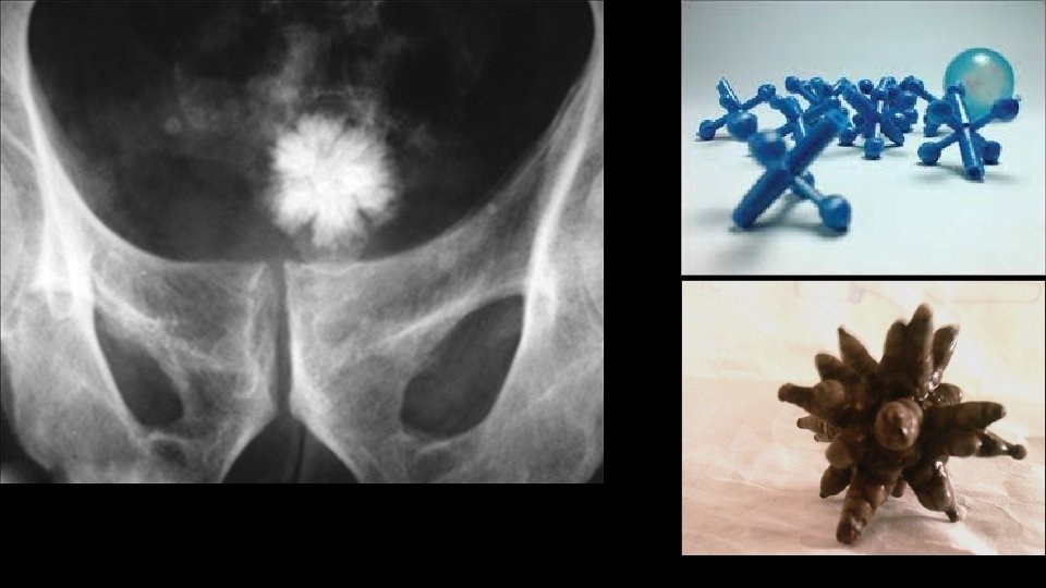
Chemical composition of calculi • Calcium containing Calcium Oxalate – metabolism derangement Struvite – UTI with urea splitting organisms • Devoid of calcium Cystine Uric acid Protease inhibitors

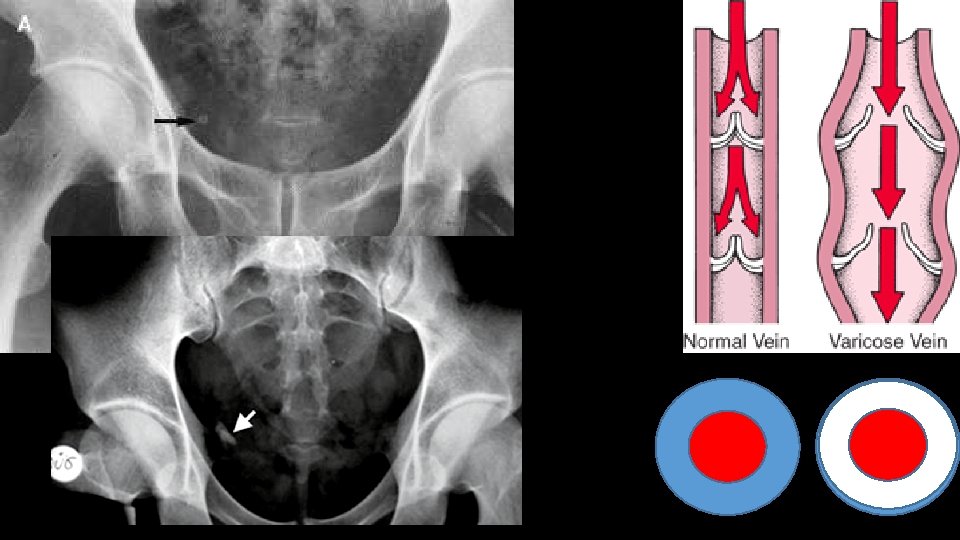
Mimics


That’s all folks Thank you
- Slides: 43