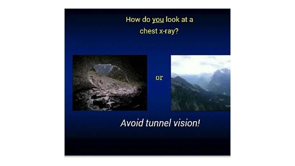
How to read a CXR Dr muna A


 fluid or other materials …it has Replacement of(air")
 • Replacement of air in one or more acini by fluid")

- Slides: 30
How to read a CXR … Dr. muna A Gh. Z
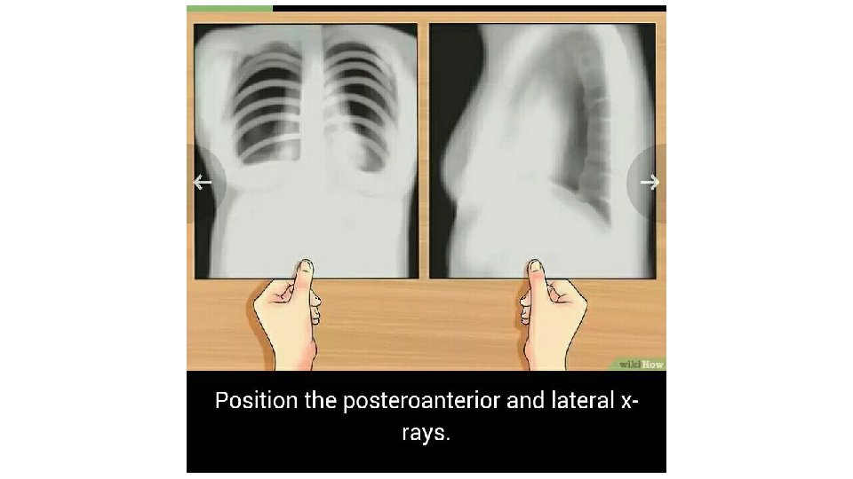
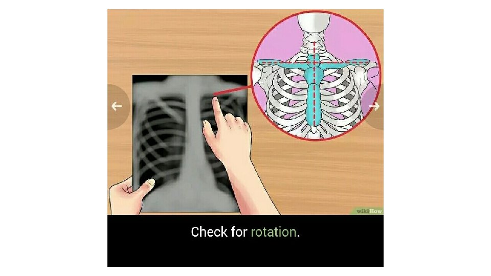
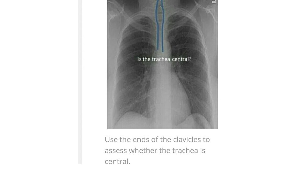
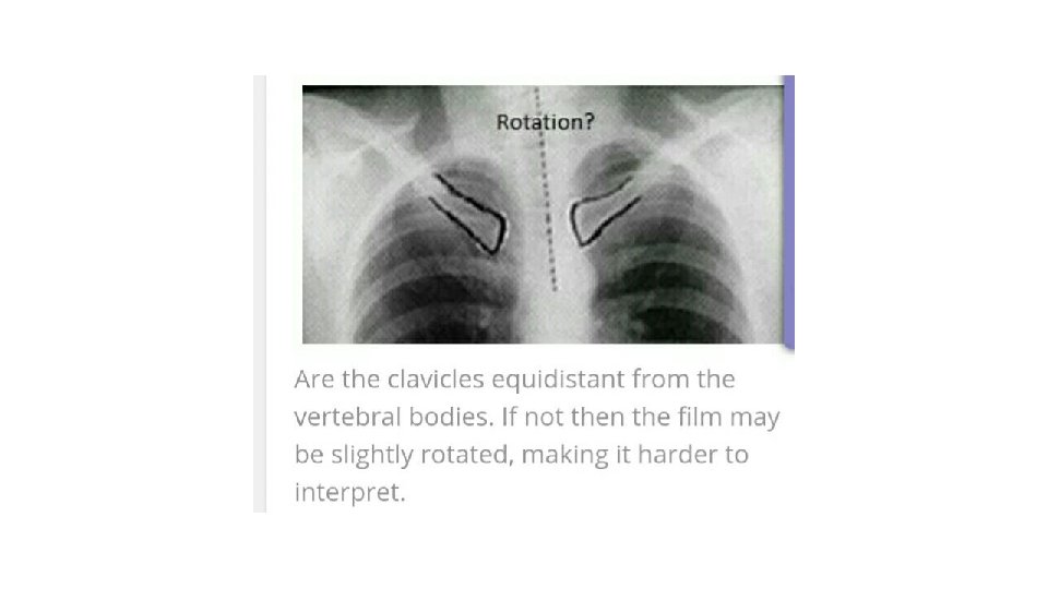
• Initial checks 1. Check the patients name 2. Look up the patients history 3. Read the date of the radiograph. 4. Look for markers 5. Position the postro -anterior and lateral view 6. Antro -posterior views taken in certain circumstances. 7. Check for any instrument insertions • film quality 1. Is it taken under full inspiration ? U should be able to see 10 posterior ribs and 6 anterior ribs. 2. Check for rotation
Check patients name
Look up the patients h. ISTORY
LOOK FOR MARKERS
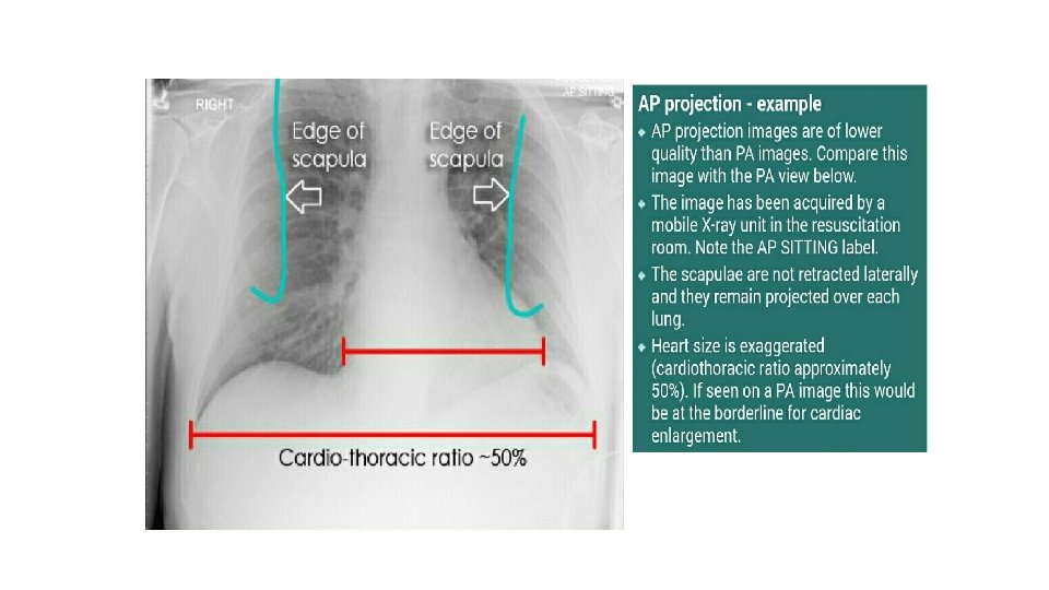
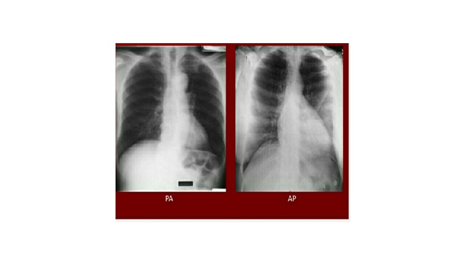
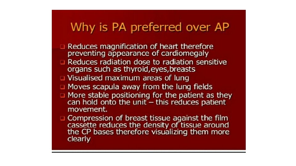
Indication of AP view 1. Severe illness 2. Pediatric s • diminish distance of x-ray beam result in more magnification of the heart
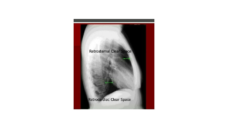
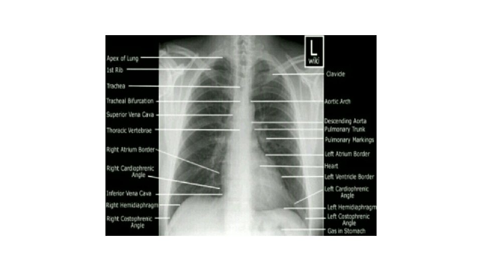
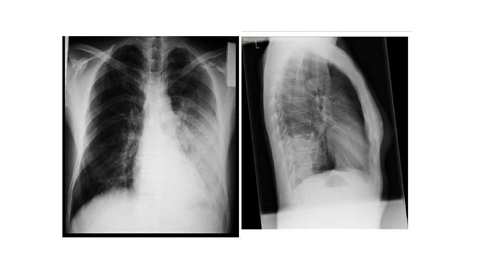
Normal chest pa and Lat. view
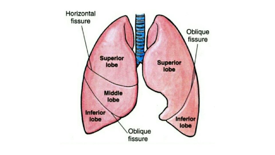
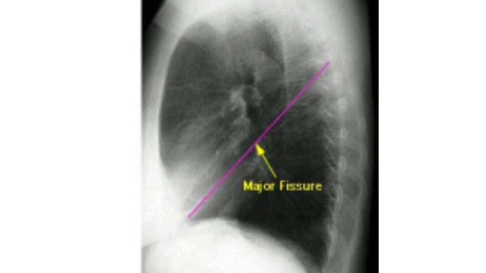
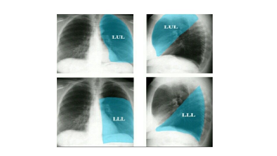
Lung zone
Inspiratory vs. expiratory films
• Check if there any instrument
2. Air space • opacification fillingby) fluid or other materials …it has Replacement of(air space in the alveoli an ill-defined border with a fissure. except when it comes in contact • Exudate …consolidation • Transudate …. pulmonary odema • Air bronchogram… air filling the normal bronchi , being made visible by the opacification of the near by alveoli. • Pulmonary collapse. (atelectasis) • Nodular , spherical opacities • Linear opacities. • Wide spread opacites • Cavitation
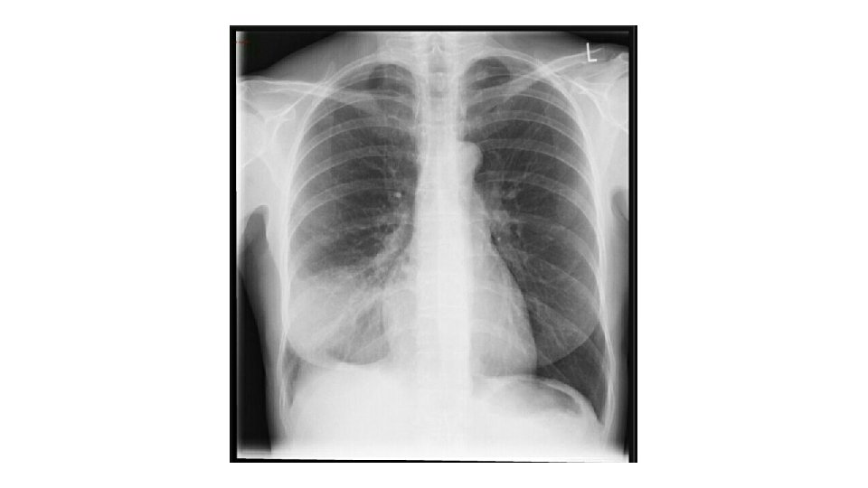
Consolidation( pneumonia ) • Replacement of air in one or more acini by fluid or solid material vol. of the lung is normal un like collapse
Middle lobe consolidation