How to look at A peripheral blood smear

How to look at A peripheral blood smear
 2 -")
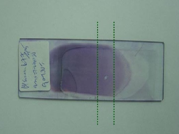
Low Power 1 - Scan the slide under low power (10 x) 2 - Find a good area 3 - Evaluate red cell distribution 4 - Evaluate the white count 1 WBC = 1000 -2000 mm 3

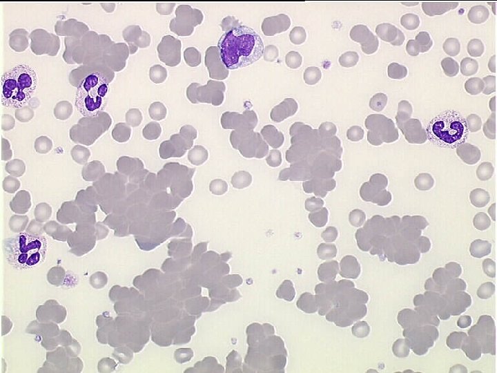
Too thick area Red cells appear as; • microcyte • hypochrome • rouleaux

Too thick area

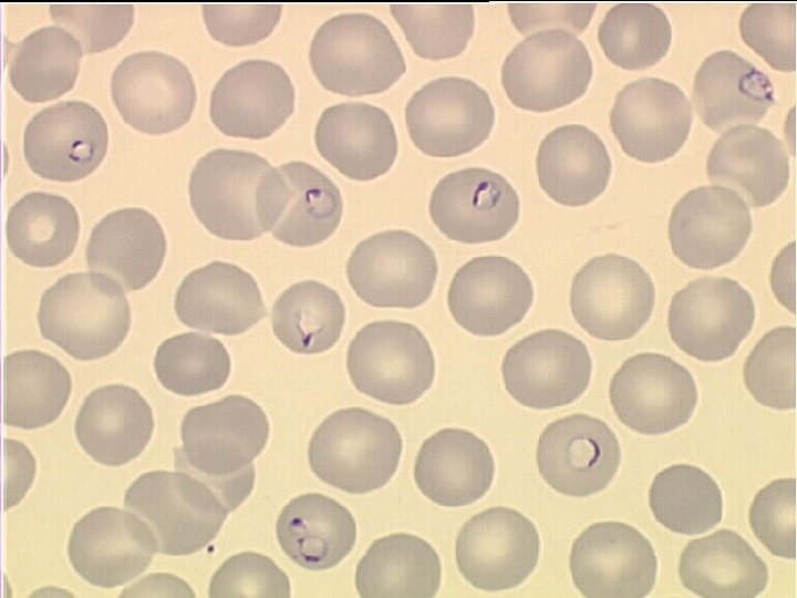
Too thin Red cells appear as • macrocyte • spherocyte • poikilocyte

Too thin area

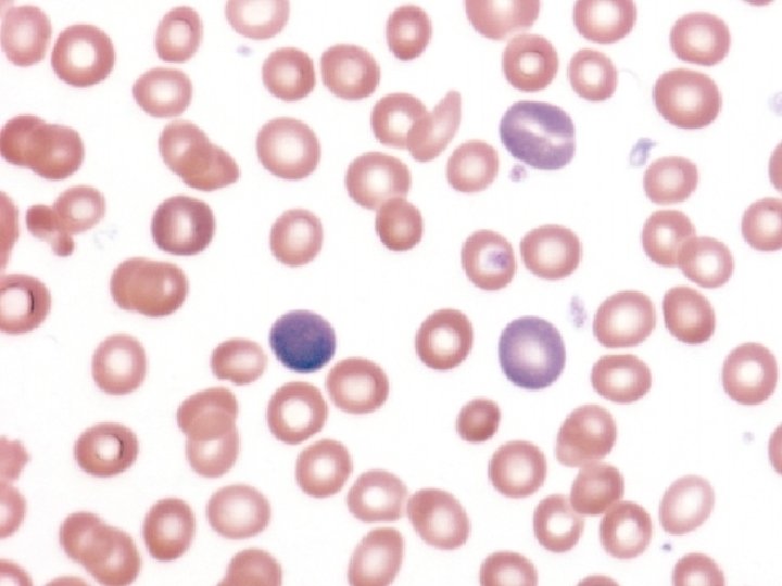
Good area
 • • Red")
What are you looking for at a peripheral smear (Oil immersion) • • Red cell morphology Platelet number and morphology WBC differential and morphology Blood parasite

RBC MORPHOLOGY

r lo Co n io ut rib st Di n o i s u l Inc Sh ap e ze i S

Evaluate red cells for size

Normocytes

Microcytes

Macrocytes

Macro-ovalocytes

Anisocytosis

Hb content & distribution

Normochrome

Hypochrome

spherocyte

Microspherocyte

Dimorphic population

Target cells

Stomatocytes

shape

Normocyte

Tear-drop cells

Acanthocytes

Echinocytes

Ovalocyte - Elliptocyte

Sickle cells
")
Keratocyte (Horn cell)

Pincher cells

Schistocytes

Distribution
")
Erythrocyte antibody rosetting (EA-rosetting)

Rouleaux formation

Inclusions
")
Hb. H(Golf ball)

Hb. C crystal

Basophilic stippling

Howell Jolly bodies

Pappenheimer bodies

Cabot ring

babesia
")
Nucleated red blood cell (NRBC)
 Water spots 2) Precipitated stain 3) Aged RBC 4) Refractiles")
Beware of artifacts! 1) Water spots 2) Precipitated stain 3) Aged RBC 4) Refractiles

Refractiles: o Humidity; under-fixation o late fixation o presence of water in alcohol used o excess buffer to stain o thick smear

Refractiles: Increased central pallor but a normal thick rim of Hb.
")
Artifact(water spots)

Precipitated stain

Reference guide for grading red blood cell morphology Normal < 5 27 -34 pg < 3 80 -100 fl 0 <6 0 <5 0 RBC type Hypochromia Polychromasia Microcytes Macrocytes Schistocytes Elliptocytes Rouleaux Spherocytes Target cells Acanthocytes Few (1+) 5 -15 22 -26 pg 3 -5 70 -79 fl 100 -11 - fl 1 -5 6 -20 1 -5 5 -10 1 -10 Moderate (2+) 16 -40 18 -21 pg 6 -20 60 -69 fl 111 -125 fl 6 -15 21 -50 11 -50 6 -20 11 -25 11 -30 Marked (3+) > 40 < 18 pg > 20 < 60 fl > 125 >15 >50 >20 >25 >30 Report if present Burr cells Bite cells Stomatocytes Teardrop cells* Agglutination Dimorphic red cells Dual population Howell-Jolly bodies Oval macrocytes Pappenheimer bodies Parasites Sickle cell >30% >4%

Follow this system regularly then you won’t miss anything
- Slides: 57