How MRI Works Magnetic resonance imaging Magnetic Resonance

, or nuclear magnetic resonance imaging (NMRI),")
 fields are used to systematically alter the alignment of")
 and the repetition time")


 The precession of the spinning proton and (B) the magnetic moment of the")
 A collection of protons at thermal equilibrium in the absence of a strong")


")

")

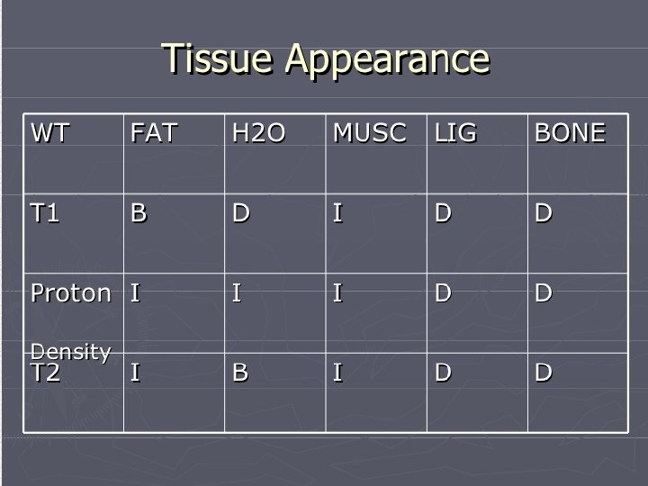
 weighted scans use a")


")
 measures signal changes in the brain that are")

 is a nuclear medicine imaging technique")


- Slides: 72
How MRI Works
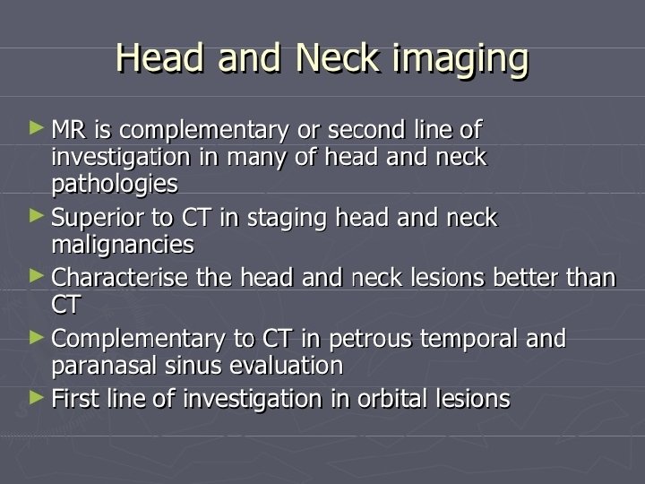
Magnetic resonance imaging • Magnetic Resonance Imaging (MRI), or nuclear magnetic resonance imaging (NMRI), is primarily a medical imaging technique most commonly used in radiology to visualize detailed internal structure and limited function of the body. MRI provides much greater contrast between the different soft tissues of the body than computed tomography (CT) does, making it especially useful in neurological (brain), musculoskeletal, cardiovascular, and oncological (cancer) imaging. Unlike CT, it uses no ionizing radiation, but uses a powerful magnetic field to align the nuclear magnetization of (usually) hydrogen atoms in water in the body.
• Radio frequency (RF) fields are used to systematically alter the alignment of this magnetization, causing the hydrogen nuclei to produce a rotating magnetic field detectable by the scanner. This signal can be manipulated by additional magnetic fields to build up enough information to construct an image of the body. • Magnetic Resonance Imaging is a relatively new technology. The first MR image was published in 1973 and the first cross-sectional image of a living mouse was published in January 1974. The first studies performed on humans were published in 1977. By comparison, the first human X-ray image was taken in 1895.
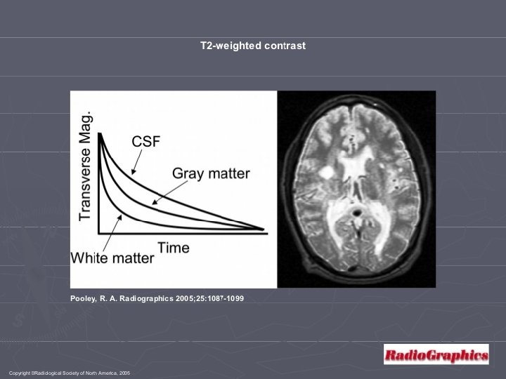
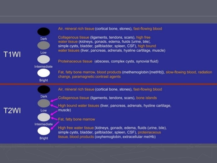
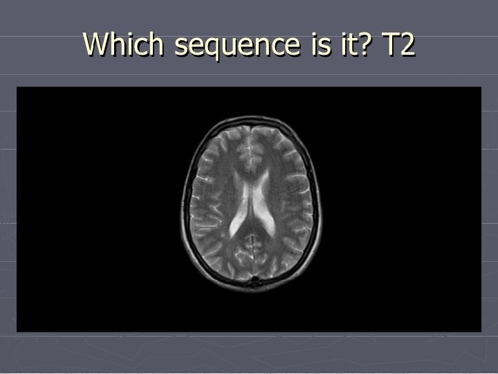
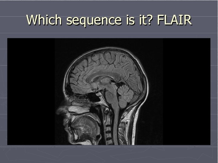
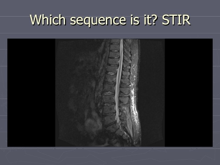
• with particular values of the echo time (TE) and the repetition time (TR), which are basic parameters of image acquisition, a sequence will take on the property of T 2 weighting. On a T 2 -weighted scan, water- and fluidcontaining tissues are bright (most modern T 2 sequences are actually fast T 2 sequences) and fat-containing tissues are dark. The reverse is true for T 1 -weighted images. Damaged tissue tends to develop edema, which makes a T 2 -weighted sequence sensitive for pathology, and generally able to distinguish pathologic tissue from normal tissue. With the addition of an additional radio frequency pulse and additional manipulation of the magnetic gradients, a T 2 -weighted sequence can be converted to a FLAIR sequence, in which free water is now dark, but edematous tissues remain bright. This sequence in particular is currently the most sensitive way to evaluate the brain for demyelinating diseases, such as multiple sclerosis.
The hydrogen atom contains a proton that spins about an arbitrary internal axis. This spinning, positive charge produces a magnetic field, called the magnetic moment. The magnetic moment of the proton has two poles (ie, north and south) like a conventional bar magnet.
A top that is spinning slightly off the vertical axis is precessing about the vertical axis.
A hydrogen atom processes about a magnetic field.
(A) The precession of the spinning proton and (B) the magnetic moment of the proton in the presence of a strong external magnetic field.
(A) A collection of protons at thermal equilibrium in the absence of a strong magnetic field. The magnetic moments produced by the spinning of the nucleus are oriented randomly. (B) The same collection of protons at thermal equilibrium in the presence of a strong magnetic field. The magnetic moments are aligned with or against the magnetic field B 0.
All of the hydrogen protons will align with the magnetic field in one direction or the other. The vast majority cancel each other out, but, as shown here, in any sample there is one or two "extra" protons
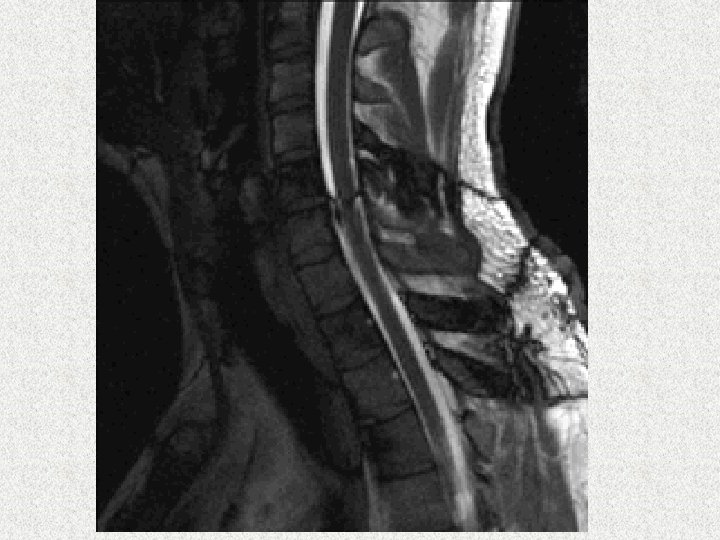
Axial, coronal and sagitall slices
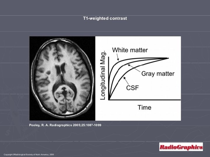
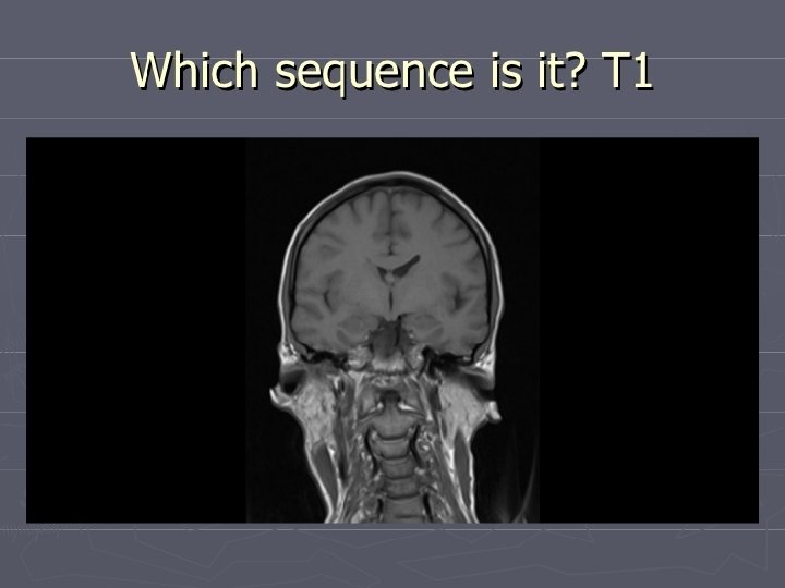
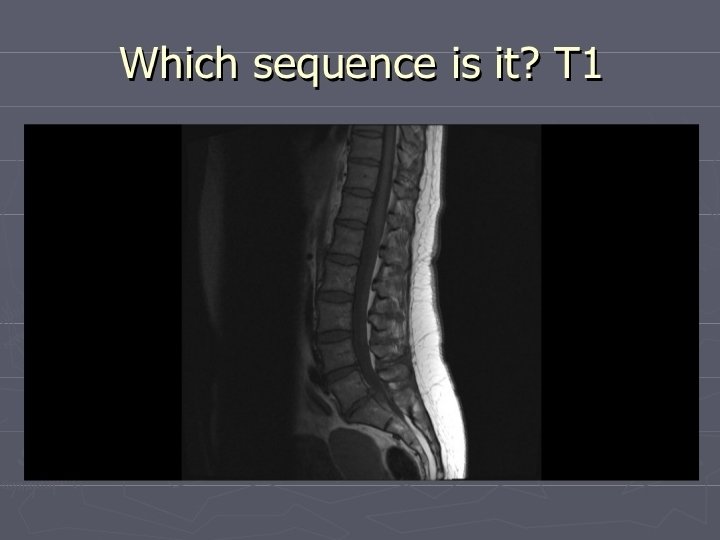
T 1 -weighted MRI • T 1 -weighted scans use a gradient echo (GRE) sequence, with short TE and short TR. This is one of the basic types of MR contrast and is a commonly run clinical scan. The T 1 weighting can be increased (improving contrast) with the use of an inversion pulse as in an MP -RAGE sequence. Due to the short repetition time (TR) this scan be run very fast allowing the collection of high resolution 3 D datasets. A T 1 reducing gadolinium contrast agent is also commonly used, with a T 1 scan being collected before and after administration of contrast agent to compare the difference. In the brain T 1 -weighted scans provide good gray matter/white matter contrast.
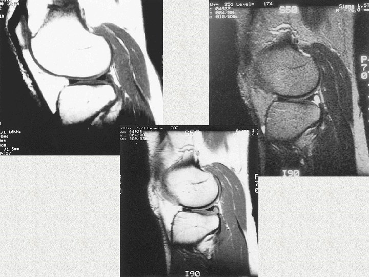
A T 1 -weighted sagittal image from the knee of a patient with a meniscal cyst. A TR of 500 msec and a TE of 15 msec have been employed using a 3 -mm slice thickness and a 16 -cm field of view with 256 points. The meniscus is very hypointense, the cyst and articular cartilage are hypointense, the muscle has moderate intensity, and the fat is hyperintense.
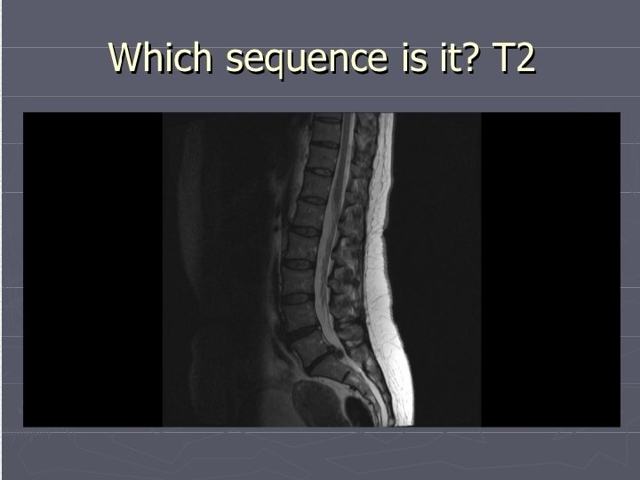
T 2 -weighted MRI • T 2 -weighted scans use a spin echo (SE) sequence, with long TE and long TR. They have long been the clinical workhorse as the spin echo sequence is less susceptible to in homogeneities in the magnetic field. They are particularly well suited to edema as they are sensitive to water content (edema is characterized by increased water content).
A T 2 -weighted sagittal image from the knee of a patient with a meniscal cyst. A TR of 2000 msec and a TE of 80 msec have been employed using a 3 -mm slice thickness and a 16 -cm field of view with 256 points. The meniscus remains hypointense, whereas the cyst is hyperintense. Fat displays moderate intensity, and muscle is hypointense. This image has a significant deficit in signal-to-noise ratio compared with that found on the T 1 -weighted and proton density weighted images.
T 2*-weighted MRI • T 2* (pronounced "T 2 star") weighted scans use a gradient echo (GRE) sequence, with long TE and long TR. The gradient echo sequence used does not have the extra refocusing pulse used in spin echo so it is subject to additional losses above the normal T 2 decay (referred to as T 2'), these taken together are called T 2*. This also makes it more prone to susceptibility losses at air/tissue boundaries, but can increase contrast for certain types of tissue, such as venous blood.
Proton Density Weighting • Images where the contrast is governed by the relative concentration of water in the tissue can be generated using long TRs (TR >2 seconds) and short TEs (TE <20 msec). These images are called proton density weighted images, and they have high SNR because the signal intensity is only slightly attenuated by T 1 or T 2 relaxation processes. Proton density weighted images provide improved anatomic detail, because the high SNR of these images can be used to visualize fine structures. They are usually obtained and are useful for interpretation of areas of high signal intensity observed on the T 2 -weighted scan in which anatomic detail is obscured. A proton density weighted image of a knee with a meniscal cyst is presented in
A proton density weighted sagittal image from the knee of a patient with a meniscal cyst. A TR of 2000 msec and a TE of 15 msec have been employed using a 3 -mm slice thickness and a 16 -cm field of view with 256 points. Less contrast among tissues is seen as compared with a T 1 weighted image.
Spin density weighted MRI • Spin density, also called proton density, weighted scans try to have no contrast from either T 2 or T 1 decay, the only signal change coming from differences in the amount of available spins (hydrogen nuclei in water). It uses a spin echo or sometimes a gradient echo sequence, with short TE and long TR.
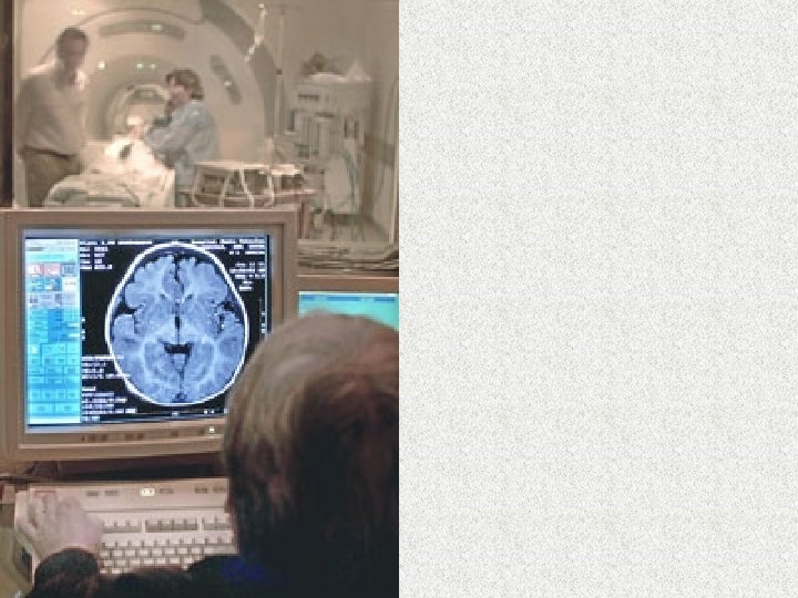
Functional magnetic resonance imaging • Functional MRI or functional Magnetic Resonance Imaging (f. MRI) is a type of specialized MRI scan. It measures the hemodynamic response (change in blood flow) related to neural activity in the brain or spinal cord of humans or other animals. It is one of the most recently developed forms of neuroimaging. Since the early 1990 s, f. MRI has come to dominate the brain mapping field due to its relatively low invasiveness, absence of radiation exposure, and relatively wide availability.
• Functional MRI (f. MRI) measures signal changes in the brain that are due to changing neural activity. The brain is scanned at low resolution but at a rapid rate (typically once every 2– 3 seconds). Increases in neural activity cause changes in the MR signal via T 2* changes; this mechanism is referred to as the BOLD (bloodoxygen-level dependent) effect. Increased neural activity causes an increased demand for oxygen, and the vascular system actually overcompensates for this, increasing the amount of oxygenated hemoglobin relative to deoxygenated hemoglobin. Because deoxygenated hemoglobin attenuates the MR signal, the vascular response leads to a signal increase that is related to the neural activity.
A f. MRI scan showing regions of activation I n orange, including the primary visual cortex
Positron emission tomography • Positron emission tomography (PET) is a nuclear medicine imaging technique which produces a three-dimensional image or picture of functional processes in the body. The system detects pairs of gamma rays emitted indirectly by a positronemitting radionuclide (tracer), which is introduced into the body on a biologically active molecule. Images of tracer concentration in 3 dimensional space within the body are then reconstructed by computer analysis.
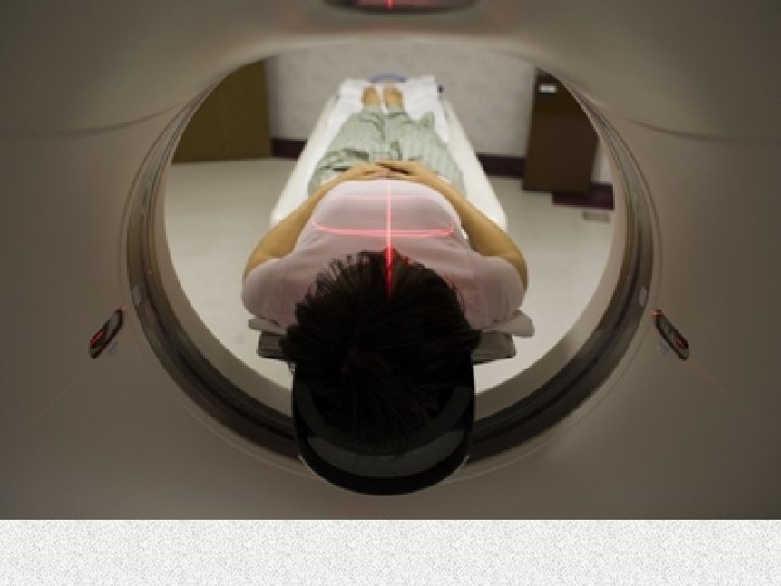
The MRI system goes through the patient's body point by point, building up a 2 -D or 3 -D map of tissue types to create images.
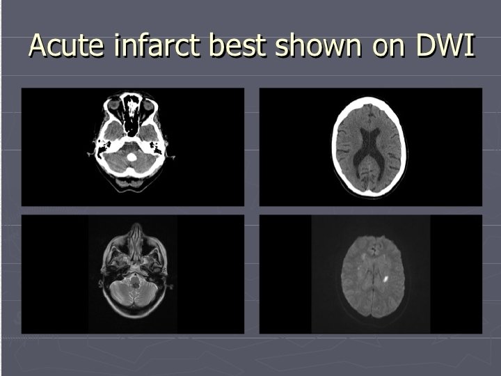
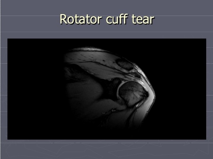
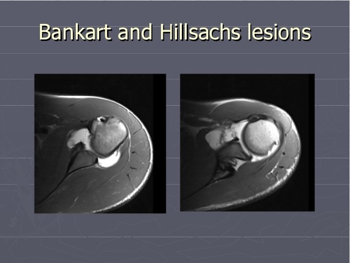
MRI is ideal for diagnosing multiple sclerosis, tumors, brain infections, torn ligaments, shoulder injuries, bone tumors, cysts or even strokes in their earliest stages.
surgical robotic system for caparoscopic surgery
A fully loaded pallet jack that has been sucked into the bore of an MRI system