How Cells Divide Chapter 10 Why do cells


 (play Thinkwell movies here)")


- Slides: 24
How Cells Divide Chapter 10
Why do cells divide? • For reproduction – asexual reproduction • one-celled organisms • For growth – from fertilized egg to multi-celled organism • For repair & renewal – replace cells that die from normal wear & tear or from injury amoeba
Bacteria divide by Bacterial binary fission. -In the single, circular bacterial chromosome replication begins at the origin of replication -chromosomes are partitioned to opposite ends a septum forms to divide the cell into 2 cells Cell Division 3
Eukaryotic Cell Cycle Cell cycle has 5 phases, first 3 make up INTERPHASE G 1 = 1 st Gap (Growth) cell doing its “everyday job” cell grows S = DNA Synthesis copies chromosomes G 2 = 2 nd Gap (Growth) prepares for division cell grows (more) produces organelles, proteins, membranes 4
Chromosome Organization ACTGGTCAGGCAATGTC DNA § DNA is organized in chromosomes double helix DNA molecule u wrapped around histone proteins u histones § like thread on spools u DNA-protein complex = chromatin § organized into long thin fiber u chromatin condensed further during mitosis duplicated mitotic chromosome 5
During the S phase Chromosomes are replicated before cell division. -Replicated chromosomes are connected to each other at their kinetochores sister chromatids: 2 copies of the chromosome within the replicated chromosome 6
Copying DNA & packaging it… Chromosomes undergo condensation coiling & mitotic folding to make a smaller package chromosome DNA chromatin Centrioles (microtubuleorganizing centers) replicate and one centriole moves to each pole.
Mitosis is divided into 5 phases: interphase prophase I. P. P. M. A. T. (pro-metaphase) cytokinesis metaphase anaphase telophase
• • • Prophase Chromatin condenses – visible chromosomes • chromatids Centrioles move to opposite poles of cell – animal cell Protein fibers cross cell to form mitotic spindle – microtubules • actin, myosin – coordinates movement of chromosomes Nucleolus disappears Nuclear membrane breaks down green = key features
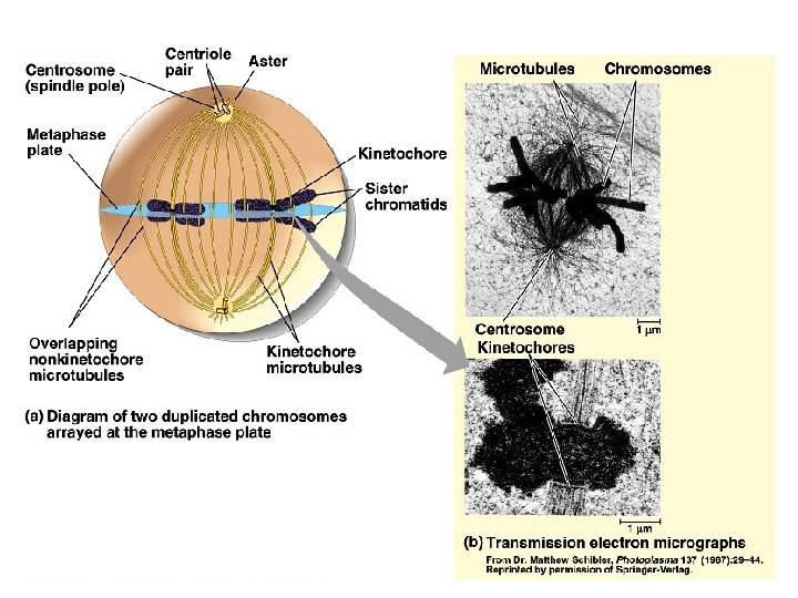
Transition to Metaphase • Prometaphase – spindle fibers attach to centromeres • creating kinetochores – microtubules attach at kinetochores • connect centromeres to centrioles – chromosomes begin moving green = key features
Metaphase • Chromosomes align along middle of cell – metaphase plate • meta = middle – spindle fibers coordinate movement – helps to ensure chromosomes separate properly • so each new nucleus receives only 1 copy of each chromosome green = key features
Anaphase • Sister chromatids separate at kinetochores – move to opposite poles – pulled at centromeres – pulled by motor proteins “walking”along microtubules • actin, myosin • increased production of ATP by mitochondria • Poles move farther apart – polar microtubules lengthen green = key features
Separation of chromatids • In anaphase, proteins holding together sister chromatids are inactivated – separate to become individual chromosomes 1 chromosome 2 chromatids double-stranded 2 chromosomes single-stranded
Chromosome movement • Kinetochores use motor proteins that “walk” chromosome along attached microtubule – microtubule shortens by dismantling at kinetochore (chromosome) end
Telophase • Chromosomes arrive at opposite poles – daughter nuclei form – nucleoli form – chromosomes disperse • no longer visible under light microscope • Spindle fibers disperse • Cytokinesis begins – cell division green = key features
Cytokinesis • Animals – constriction belt of actin microfilaments around equator of cell • cleavage furrow forms • splits cell in two • like tightening a draw string
Cytokinesis in Animals (play Cells Alive movies here) (play Thinkwell movies here)
Mitosis in whitefish blastula
Mitosis in animal cells
Cytokinesis in Plants • Plants – cell plate forms • vesicles line up at equator – derived from Golgi • vesicles fuse to form 2 cell membranes – new cell wall laid down between membranes • new cell wall fuses with existing cell wall
Cytokinesis in plant cell
Mitosis in plant cell
onion root tip