
HOSTPARASITE RELATIONSHIP By Prof Dr Zakaria Awad Mohamed
HOST-PARASITE RELATIONSHIP By Prof. Dr. Zakaria Awad Mohamed Department of Botany and Microbiology
What is the host-parasite relationship? The host-parasite relationship is the interaction between two organisms, one of which lives at the expense of the other. Parasitism is the word given to describe the action of a parasite. What is the parasite? A parasite is an organism that lives with another organism. However, a parasite only takes nutrients from its host, without host benefits. Parasites are classified based on their interactions with their hosts and on their life cycles. Those that live on its surface are called ectoparasites and those that live inside the host are called endoparasites which exist in two forms: intercellular (inhabiting spaces in the host’s body) such as bacteria or intracellular (inhabiting cells in the host’s body) such as viruses and obligate parasitic fungi.
Types of host-parasite interactions • Physical characteristics: such as the shape and texture of the host surface, the thickness of the cuticle, stomata morphology, and growth and reproduction of the pathogen. These may cause physical damage of the host, for instance by blocking vascular elements, or tearing through epidermal cell layers. • Chemical characteristics: such as enzymes which may be released or activated, altering metabolic pathways, or attacking substrates in cells. • Genetic characteristics: may change the expression of genetic information in cells by activating or repressing genes, or by introducing a new set of instructions.
Stages of host-pathogen interaction • • • Locating of pathogen on the host Attachment or adhesion to host surface Penetration and entry into host Colonization of host tissues Suppression of host defense mechanisms Reproduction of the pathogen
• The encounter between a potential pathogen and a plant host has two possible outcomes. The pathogen may successfully invade the plant, multiply, and cause disease. Alternatively, the invading pathogen may fail to establish itself in the host and disease does not develop. The whole process may be viewed as a contest between host and pathogen, with the outcome depending upon the relative balance between microbial pathogenicity factors, and host defense mechanisms (Figure 1). • The methods of entry and colonization of the host as well as the types of pathogens are summarized in Figure 2.
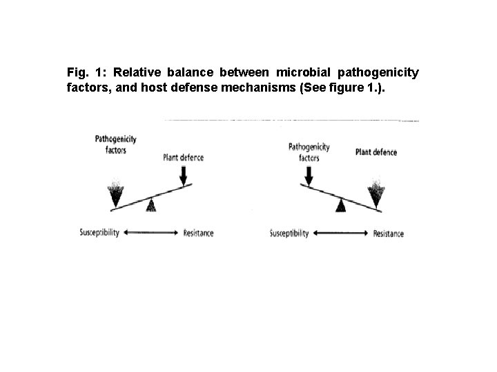
Fig. 1: Relative balance between microbial pathogenicity factors, and host defense mechanisms (See figure 1. ).

Fig. 2: Methods of entry and colonization of the host by different pathogens.
The host-pathogen interface The site of contact between a pathogen and host cells is known as the host-pathogen interface. This zone is vital in understanding the nature of different host-pathogen interactions, as it is the site at which nutrient uptake by the parasite occurs, and also where molecular communication between the two partners takes place. Three main types of interface can be distinguished. • 1. Intercellular, where the pathogen grows outside host cells; • 2. Partly intracellular, where limited penetration of cells by parasitic structures occurs; • 3. Intracellular, where growth and development takes place entirely within host cells.
Intercellular relationships are characteristic of bacteria and some fungi (e. g. Cladosporium fulvum) that grow between cell walls and through intercellular spaces. Soluble nutrients such as sugars and amino acids are scavenged from the apoplast or released from cell walls through the action of secreted hydrolytic enzymes. Hence, there is no intimate contact with living host protoplasts. Often, host cells are killed in advance of invasion, through the action of enzymes or toxins. Different host-parasite interfaces are represented in Table 1 and Figure 3.
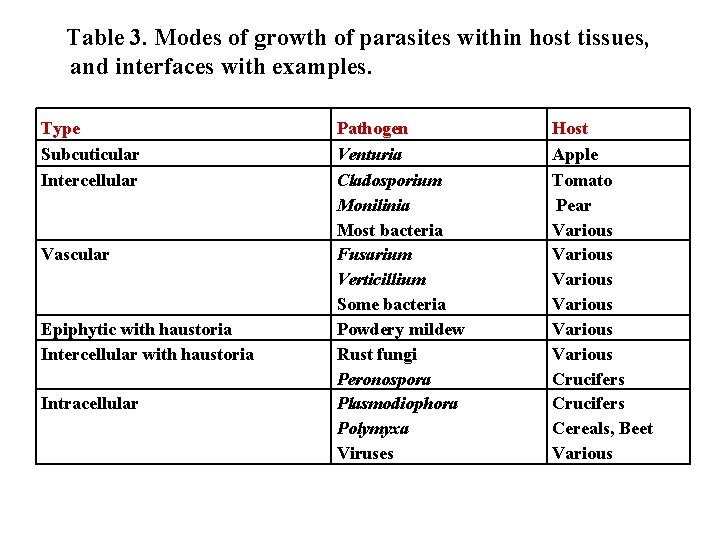
Table 3. Modes of growth of parasites within host tissues, and interfaces with examples. Type Subcuticular Intercellular Vascular Epiphytic with haustoria Intercellular with haustoria Intracellular Pathogen Venturia Cladosporium Monilinia Most bacteria Fusarium Verticillium Some bacteria Powdery mildew Rust fungi Peronospora Plasmodiophora Polymyxa Viruses Host Apple Tomato Pear Various Various Crucifers Cereals, Beet Various

Fig. 3. Some patterns of pathogenic invasion of plant tissues. 1. subcuticular; 2. epiphytic with haustoria; 3. intracellular; 4. intercellular; 5. intercellular with haustoria; 6. vasculate.

Host-Parasite Relationship of Rust Fungi (as an Example of Obligate Parasites) in Their Hosts Structure of the haustorium of rust fungi Many biotrophic fungi such as rust fungi form modified hyphae known as haustoria, which enter host cells. Haustoria typically develop from intercellular hyphae as narrow branches which penetrate the plant cell wall and then expand inside the cell. They are diverse in morphology, ranging from small, club-shaped extensions to much larger, lobed or branched structures. Although haustoria and equivalent structures are formed within plant cells, the host plasma membrane is not penetrated and remains intact as an inavgination surrounding the fungal cell.
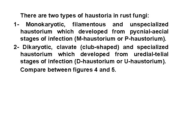
There are two types of haustoria in rust fungi: 1 - Monokaryotic, filamentous and unspecialized haustorium which developed from pycnial-aecial stages of infection (M-haustorium or P-haustorium). 2 - Dikaryotic, clavate (club-shaped) and specialized haustorium which developed from uredial-telial stages of infection (D-haustorium or U-haustorium). Compare between figures 4 and 5.

M-haustorium D-haustorium
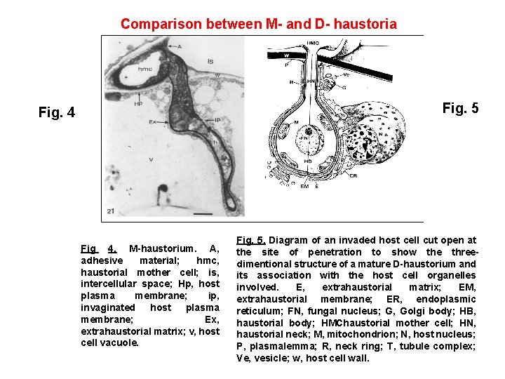
Comparison between M- and D- haustoria Fig. 5 Fig. 4 Fig 4. M-haustorium. A, adhesive material; hmc, haustorial mother cell; is, intercellular space; Hp, host plasma membrane; ip, invaginated host plasma membrane; Ex, extrahaustorial matrix; v, host cell vacuole. Fig. 5. Diagram of an invaded host cell cut open at the site of penetration to show the threedimentional structure of a mature D-haustorium and its association with the host cell organelles involved. E, extrahaustorial matrix; EM, extrahaustorial membrane; ER, endoplasmic reticulum; FN, fungal nucleus; G, Golgi body; HB, haustorial body; HMChaustorial mother cell; HN, haustorial neck; M, mitochondrion; N, host nucleus; P, plasmalemma; R, neck ring; T, tubule complex; Ve, vesicle; w, host cell wall.
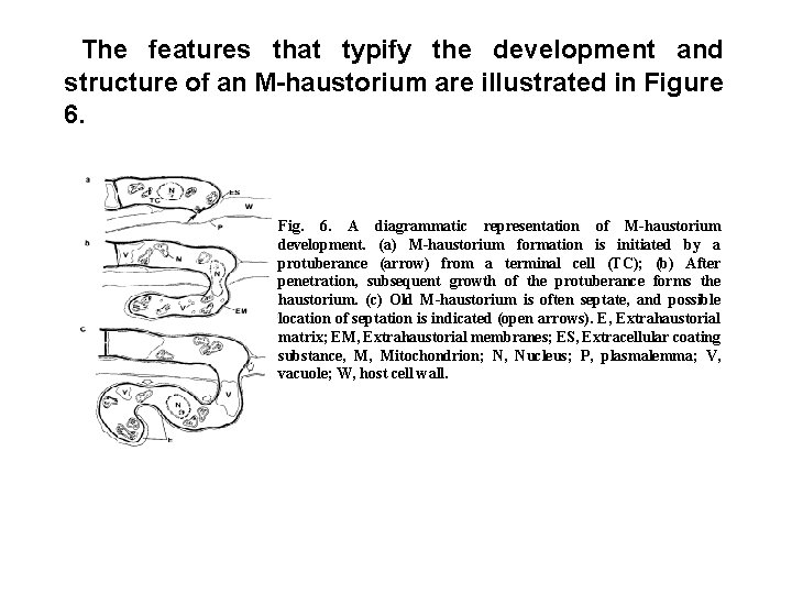
The features that typify the development and structure of an M-haustorium are illustrated in Figure 6. Fig. 6. A diagrammatic representation of M-haustorium development. (a) M-haustorium formation is initiated by a protuberance (arrow) from a terminal cell (TC); (b) After penetration, subsequent growth of the protuberance forms the haustorium. (c) Old M-haustorium is often septate, and possible location of septation is indicated (open arrows). E, Extrahaustorial matrix; EM, Extrahaustorial membranes; ES, Extracellular coating substance, M, Mitochondrion; N, Nucleus; P, plasmalemma; V, vacuole; W, host cell wall.
Characteristics of M-haustorium: 1 - The haustorium arises from a terminal intercellular hyphal cell. 2 - There is no differentiation of a specialized haustorial mother cell (HMC). 3 - There is only slight constriction of the penetration peg. 5 - The wall of the terminal intercellular hyphal cell is continuous with that of the M-haustorium and remains unmodified. 6 - The growth of the M-haustorium is filamentous, and no neck ring is formed.
7 - Centripetal septum formation may occur at any point intracellularly or at various points outside the penetration region. 8 - The host plasmalemma becomes invaginated to form an EH membrane, and an EH matrix is evident. 9 - A collar may or may not form around the neck. 10 -There is less extensive association of host ER with the M-haustorium, and the development of membranous transfer-type complexes in the host cytoplasm has not been observed.
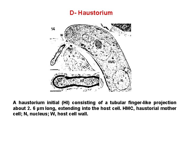
D- Haustorium A haustorium initial (HI) consisting of a tubular finger-like projection about 2. 6 µm long, extending into the host cell. HMC, haustorial mother cell; N, nucleus; W, host cell wall.
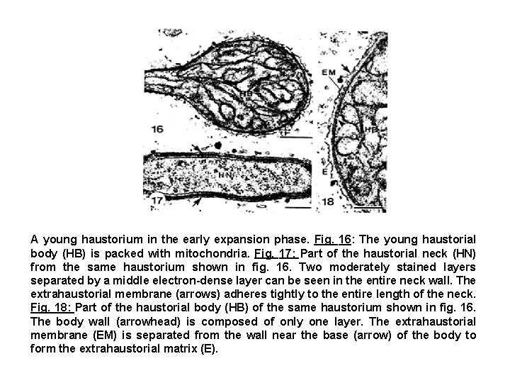
A young haustorium in the early expansion phase. Fig. 16: The young haustorial body (HB) is packed with mitochondria. Fig. 17: Part of the haustorial neck (HN) from the same haustorium shown in fig. 16. Two moderately stained layers separated by a middle electron-dense layer can be seen in the entire neck wall. The extrahaustorial membrane (arrows) adheres tightly to the entire length of the neck. Fig. 18: Part of the haustorial body (HB) of the same haustorium shown in fig. 16. The body wall (arrowhead) is composed of only one layer. The extrahaustorial membrane (EM) is separated from the wall near the base (arrow) of the body to form the extrahaustorial matrix (E).
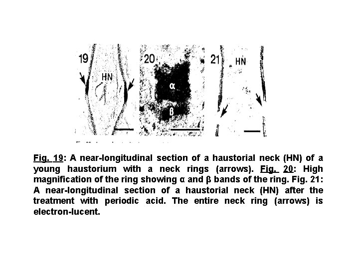
Fig. 19: A near-longitudinal section of a haustorial neck (HN) of a young haustorium with a neck rings (arrows). Fig. 20: High magnification of the ring showing α and β bands of the ring. Fig. 21: A near-longitudinal section of a haustorial neck (HN) after the treatment with periodic acid. The entire neck ring (arrows) is electron-lucent.
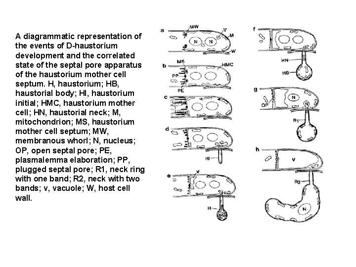
A diagrammatic representation of the events of D-haustorium development and the correlated state of the septal pore apparatus of the haustorium mother cell septum. H, haustorium; HB, haustorial body; HI, haustorium initial; HMC, haustorium mother cell; HN, haustorial neck; M, mitochondrion; MS, haustorium mother cell septum; MW, membranous whorl; N, nucleus; OP, open septal pore; PE, plasmalemma elaboration; PP, plugged septal pore; R 1, neck ring with one band; R 2, neck with two bands; v, vacuole; W, host cell wall.
• • The terms used, their definitions, and abbreviations for D-haustoria are as follows: Collar: an irregularly occurring deposition of material between the host plasmalemma and host cell wall at the penetration site. The collar may extend around the haustorial neck up to the base of the haustorial body. The collar is not a part of the haustorial apparatus. Extrahaustorial matrix (EH matrix): a region of varying dimensions and density that occurs between the haustorial body wall and the EH membrane. Extrahaustorial membrane (EH membrane): an extension of the host plasmalemma that surrounds the entire intracellular haustorium. Haustorial body: the irregularly shaped bulk of the haustorium that begins where the neck expands at its distal end.
• Haustorial body wall: the fungal cell wall enclosing the haustorial body. • Haustorial neck: the constricted portion of the haustorium originating inside the host cell wall and extending to the base of the haustorial body. • Haustorium initial: the post-penetration finger-like projection into host cell. After swelling at its distal end to form the haustorial body, it becomes the haustorial neck. • Haustorium mother cell (HMC): a slightly swollen, terminal cell of an intercellular hypha that attaches to a host cell and gives rise to the haustorium. It found only in dikaryotic infections.
• HMC septum: the septum that delimits the HMC from the penultimate hyphal cell. • Neck ring: an electron-dense portion of the neck wall that occurs approximately midway along the haustorial neck. Where the neck ring is composed of more than one ring, the composite is the neck ring and each portion is designated as a band. • Neck wall: the fungal cell wall extending along the haustorial neck. • Penetration peg: the narrowest portion of the haustorium that passes through the host cell wall.
Function of haustorium • The structure and location of haustoria, which provide an enlarged surface area of the parasite directly adjacent to nutrient sources such as chloroplasts, suggests that they play a role in nutrient uptake. Experiments with labeled sugars and amino acids have shown that solutes cross the extrahaustorial (EH) membrane, and cell cytoplasm plays an essential role in transporting assimilates into haustoria. The main compound initially moving from the host to the fungus is sucrose. Thus, the pathway of carbon flow is from the source (chloroplasts) into the sink (the fungus), via haustoria. The plant sugars are eventually converted into fungal metabolites such as mannitol and glycogen.
• What is the actual mechanism by which solutes are removed from host cells? Electron micrographs of stained or freeze-fractured haustoria suggest that the EH membrane, is altered in structure and composition by comparison with the rest of host plasma membrane. In particular, the EH membrane lacks intramembrane particles, and ATPase, an enzyme involved in the active transport of solutes. • ATPase activity can be detected in the host membrane where it lines the plant cell wall, and also in the fungal plasma membrane inside the haustorium, but not in the EH membrane. It appears therefore that both the host cell protoplast and the fungal protoplast are actively importing solutes, while the membrane enclosing the haustorium has diminished control of solute transport, and leaks nutrients into the extrahaustorial matrix, from where they are scavenged by the fungus.
• One further feature of this model is the electrondense band of impermeable material (neckband) (Fig. 12) where the EH membrane contacts the haustorial neck. This is presumed to prevent solutes diffusing along the haustorial cell wall in the neck region. • The extrahaustorial matrix and the haustorial wall are therefore a sealed compartment, and any solutes leaking across the EH membrane can only enter the fungus via active transport through the haustorial plasma membrane. These experiments confirm the importance of haustoria in nutrient uptake by at least one group of biotrophic fungi. The mechanism of nutrient uptake by rust haustoria is illustrated in figure 23.
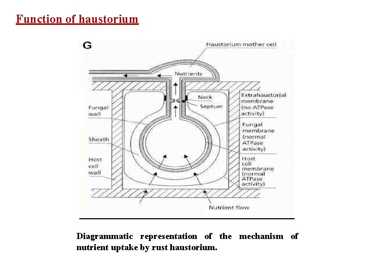
Function of haustorium Diagrammatic representation of the mechanism of nutrient uptake by rust haustorium.
Components of The Host-Parasite Interface 1 - The extrahaustorial (EH) membrane: During the growth of rust haustoria in their host cells, the host plasmalemma becomes invaginated and surrounds the entire haustorium. The invaginated plasmalemma consists of newly synthesized membrane. The part of the invaginated host plasmalemma that surrounds the haustorium, beginning at the penetration site, is referred to as the extrahaustorial (EH) membrane.
The EH membrane is closely associated with the haustorial neck wall, but around the haustorial body a matrix of material intervenes between the body wall and EH membrane. • In most conventionally processed tissue the EH membrane is undulated (Fig. 24). The conformation of the membrane may be affected by the age of the haustorium. The EH membrane in Fig. 25 represents a view of a moderately rough EH membrane. • One possible role of EH membrane is the control of the flow of metabolites into or out of the haustorium through alterations in permeability. Membrane sterol is known to play a role in membrane permeability, thus the change in sterol content may reflect such a role.
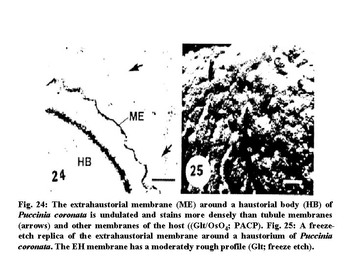
Fig. 24: The extrahaustorial membrane (ME) around a haustorial body (HB) of Puccinia coronata is undulated and stains more densely than tubule membranes (arrows) and other membranes of the host ((Glt/Os. O 4; PACP). Fig. 25: A freezeetch replica of the extrahaustorial membrane around a haustorium of Puccinia coronata. The EH membrane has a moderately rough profile (Glt; freeze etch).
2 -The Extrahaustorial (EH) Matrix: • In rust fungi, the EM matrix is of universal occurrence around the body of the haustorium but is slightly variable in appearance. The matrix ranges from a narrow, electronlucent band to a broader zone containing fibrillar or granular electron-dense substances. The variability in appearance has frequently been related to the age of the haustorium or degree of compatibility with the host. The matrix has been considered to be host origin. • Recent work has provided some information on the composition of the matrix. These EM matrices were shown to contain mixtures of lipid, larger amounts of polysaccharides, and protein. At least two types of polysaccharides were apparent: cellulose, which may be a response of the host to build a wall at this interface, and glycoprotein. The EH matrix may represent a larger wall surface area for solute transfer.
3 -Association of host endoplasmic reticulum (ER) to haustorium • Invasion of the host cell results in marked alteration of the distribution and configuration of host ER. This appears to be generalized phenomenon throughout the rusts. Endoplasmic reticulum occurs around the bodies of young haustoria of P. graminis, but the greatest association of ER is in the neck region of young developing haustoria. The extent of ER association in Puccinia graminis is seen in Fig. 26. where much of the ER radiates from the haustorial neck region into the surrounding host cytoplasm. The association of ER cisernae with young haustoria tend to be parallel to the EH membrane (Fig. 27). • The association of ER with young haustoria may play a role in nutrient transport from host cell to haustorium in this particular stage.
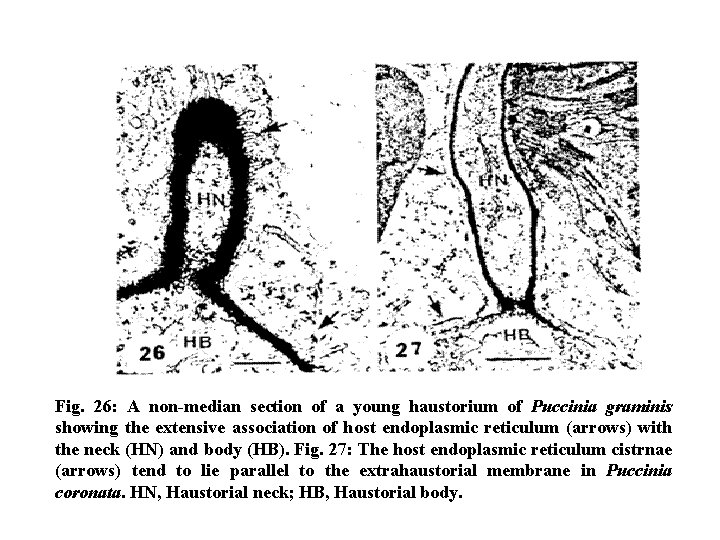
Fig. 26: A non-median section of a young haustorium of Puccinia graminis showing the extensive association of host endoplasmic reticulum (arrows) with the neck (HN) and body (HB). Fig. 27: The host endoplasmic reticulum cistrnae (arrows) tend to lie parallel to the extrahaustorial membrane in Puccinia coronata. HN, Haustorial neck; HB, Haustorial body.
4 -Association of Host Membrane Complexes to haustorium: • A number of reports describe vesicles or tubular membranous structures in the host cytoplasm near the haustoria. The vesicular configurations are probably parts of tubules as indicated in work on P. graminis and P. coronota. In P. coronata the tubules develop as an irregular network (Fig. 28), whereas in P. graminis there is a more highly organized complex of small and large tubules (Figs. 30 and 31). The latter complexes were shown to be derived from the host ER, and the same appears to be true for P. coronata. The small and large tubules in P. graminis infections are interconnected, and the entire complex may surround part of the haustorium.
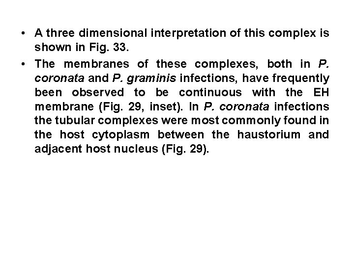
• A three dimensional interpretation of this complex is shown in Fig. 33. • The membranes of these complexes, both in P. coronata and P. graminis infections, have frequently been observed to be continuous with the EH membrane (Fig. 29, inset). In P. coronata infections the tubular complexes were most commonly found in the host cytoplasm between the haustorium and adjacent host nucleus (Fig. 29).
• The haustorium-associated membranous complexes are synthetic or secretory bodies related directly to the requirements of the fungus. These complexes are open directly to the EH matrix, and are themselves interconnected via the host ER system. This network most likely includes the nucleus, as the ER is also continuous with the outer membrane of the nuclear envelope. The “functional” interface in effect extends throughout the host cell.
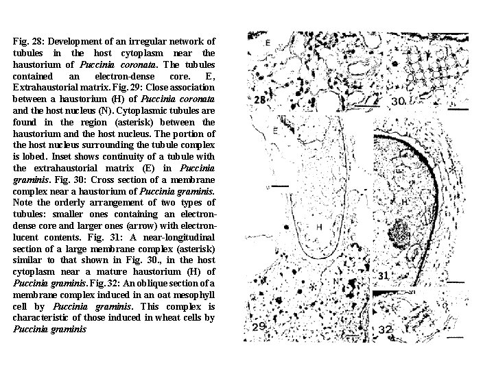
Fig. 28: Development of an irregular network of tubules in the host cytoplasm near the haustorium of Puccinia coronata. The tubules contained an electron-dense core. E, Extrahaustorial matrix. Fig. 29: Close association between a haustorium (H) of Puccinia coronata and the host nucleus (N). Cytoplasmic tubules are found in the region (asterisk) between the haustorium and the host nucleus. The portion of the host nucleus surrounding the tubule complex is lobed. Inset shows continuity of a tubule with the extrahaustorial matrix (E) in Puccinia graminis. Fig. 30: Cross section of a membrane complex near a haustorium of Puccinia graminis. Note the orderly arrangement of two types of tubules: smaller ones containing an electrondense core and larger ones (arrow) with electronlucent contents. Fig. 31: A near-longitudinal section of a large membrane complex (asterisk) similar to that shown in Fig. 30. , in the host cytoplasm near a mature haustorium (H) of Puccinia graminis. Fig. 32: An oblique section of a membrane complex induced in an oat mesophyll cell by Puccinia graminis. This complex is characteristic of those induced in wheat cells by Puccinia graminis
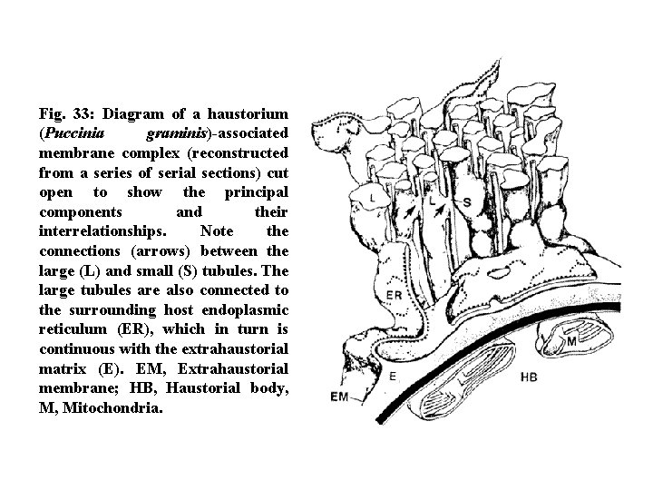
Fig. 33: Diagram of a haustorium (Puccinia graminis)-associated membrane complex (reconstructed from a series of serial sections) cut open to show the principal components and their interrelationships. Note the connections (arrows) between the large (L) and small (S) tubules. The large tubules are also connected to the surrounding host endoplasmic reticulum (ER), which in turn is continuous with the extrahaustorial matrix (E). EM, Extrahaustorial membrane; HB, Haustorial body, M, Mitochondria.
Responses of Host Tissues to The Infection by Rust Fungi I-Infection of vascular tissues by rust fungi A- Invasion of host vascular tissue by M-haustoria and not by D-haustoria: Most studies on rust infection showed that the infection of vascular tissues to be by monokaryon rather than dikaryon. More recent studies by light and electron microscopes of the haustoria of pycnial-aecial stage (monokaryon) of Puccinia poarum and P. lagenophorae showed dense growth in the vascular tissue of Tussilago farfara and Senecio vulgaris, respectively, especially in the phloem region, xylem parenchyma and bundle sheath cells.
On the other hand, the dikaryotic haustoria invade only the bundle sheath cells of Poa pratensis and Senecio vulgaris. B-Physiological significance of vascular infection: • In the specialized D–haustoria of the rust dikaryon, the invaginated plasmalemma adjacent to the fungal wall has been shown to be modified in a manner to facilitate movement of solutes from host cell to fungus. This type of extrahaustorial membrane does not show the ATP-ase activity and other cytochemical reactions, characteristic of the rest of the plasmalemma lining the host cell wall, from which it is separated by the osmiophilic neckband.
Comparative investigations of the distribution of ATP-ase activity on the invaginated host plasmalemma around the unspecialized filamentous haustoria (monokaryon) and that of specialized, clavate haustoria (dikaryon) have demonstrated a different situation. The EH membrane enclosing M-haustoria display levels of ATP-ase, contrasting with the membrane surrounding D-haustoria which lacks ATP-ase activity. This may give an indication that M-haustoria have a short functional period compared with that of D-haustoria. However, by gaining access to nutrients in the host vascular system, M- haustorium may compensate for this inefficiency of nutrient uptake.
As far as can be determined from ultrastructural and cytochemical studies, the filamentous haustoria are basically similar to intercellular hyphae. Direct uptake of water and nutrients from the conducting system of the host, is indicated by autoradiographic evidence from Puccinia poarum showing transfer of 3 H via intercellular hyphae to spores when tritium-labeled glycerol was supplied to rust infected regions of leaves through the vascular tissue.
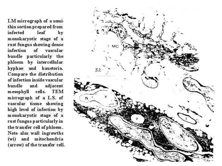
LM micrograph of a semithin section prepared from infected leaf by monokaryotic stage of a rust fungus showing dense infection of vascular bundle particularly the phloem by intercellular hyphae and haustoria. Compare the distribution of infection inside vascular bundle and adjacent mesophyll cells. TEM micrograph of a L. S. of vascular tissue showing high level of infection by monokaryotic stage of a rust fungus particularly in the transfer cell of phloem. Note also wall ingrowths (wi) and mitochondria (arrow) of the transfer cell.
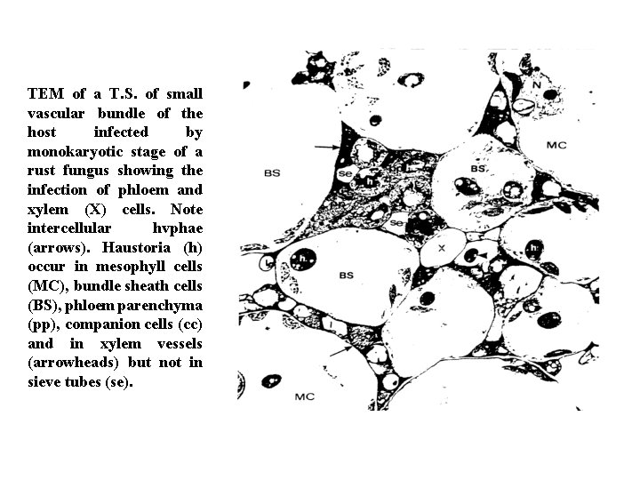
TEM of a T. S. of small vascular bundle of the host infected by monokaryotic stage of a rust fungus showing the infection of phloem and xylem (X) cells. Note intercellular hvphae (arrows). Haustoria (h) occur in mesophyll cells (MC), bundle sheath cells (BS), phloem parenchyma (pp), companion cells (cc) and in xylem vessels (arrowheads) but not in sieve tubes (se).
II- Structural changes in rusted host tissues A-Collars A common response of the host cell to the invasion of the rust fungi is the deposition of a collar of material around the fungus in the region of host cell penetration. Collars are not an integral part of the haustorial apparatus. Collars are not formed in every invaded cell, and their formation is frequently linked to the degree of host-rust compatibility. Collar formation is seen as a nonspecific response by the host to wall off the fungus, and in some cases of incompatibility, the entire invading haustorium may be enclosed by the collar.
In genotypically compatible interactions a collar may form at the point of penetration. Collars are more frequently observed in older infections in a variety of host-rust combinations, which is probably related to a general decrease in the degree of host-rust fungal compatibility in older infections. The mode of collar formation is interpreted in the drawing in Fig. 34. These collars are formed after fungal penetration. Although these collars surround the haustorial neck, a zone of host material normally intervenes between the collar and the neck. In one version of collar formation, small membrane-bound vesicles, some is containing electron-opaque material, aggregate near the base of the neck. These vesicles appear to be derived from Golgi bodies that have aggregated at this site.
The vesicles then apparently coalesce to form the bulk of the collar. Collars frequently have long projections radiating into host cytoplasm. Host ER and Golgi bodies are associated with these projections (Fig. 35). The collars in most rust infections are of variable electron density after conventional processing. Calloselike compounds have been suggested to be a major component of collars, although other carbohydrate substances may also present. From Fig. 36. It is clear that the collar is integral with the inner layer of the host wall. Collars in P. coronata (Fig. 36) or P. graminis (Fig. 37) show intense staining with PA-TCH-SP (specific procedure for detection of polysaccharides), particularly for P. coronata.
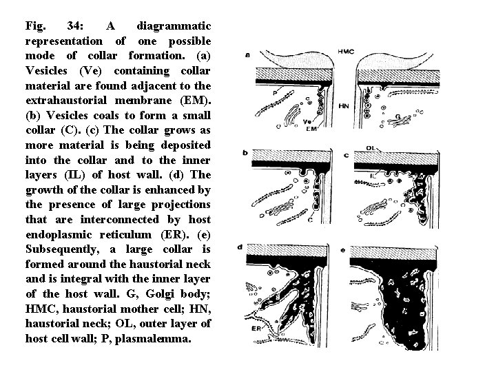
Fig. 34: A diagrammatic representation of one possible mode of collar formation. (a) Vesicles (Ve) containing collar material are found adjacent to the extrahaustorial membrane (EM). (b) Vesicles coals to form a small collar (C). (c) The collar grows as more material is being deposited into the collar and to the inner layers (IL) of host wall. (d) The growth of the collar is enhanced by the presence of large projections that are interconnected by host endoplasmic reticulum (ER). (e) Subsequently, a large collar is formed around the haustorial neck and is integral with the inner layer of the host wall. G, Golgi body; HMC, haustorial mother cell; HN, haustorial neck; OL, outer layer of host cell wall; P, plasmalemma.
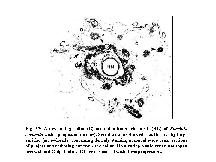
Fig. 35: A developing collar (C) around a haustorial neck (HN) of Puccinia coronata with a projection (arrow). Serial sections showed that the nearby large vesicles (arrowheads) containing densely staining material were cross sections of projections radiating out from the collar. Host endoplasmic reticulum (open arrows) and Golgi bodies (G) are associated with these projections.
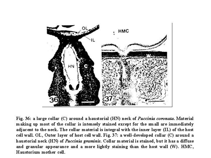
Fig. 36: a large collar (C) around a haustorial (HN) neck of Puccinia coronata. Material making up most of the collar is intensely stained except for the small are immediately adjacent to the neck. The collar material is integral with the inner layer (IL) of the host cell wall. OL, Outer layer of host cell wall. Fig. 37: a well-developed collar (C) around a haustorial neck (HN) of Puccinia graminis. Collar material is stained, but it has a diffuse and granular appearance and a more lightly staining than the host wall (W). HMC, Haustorium mother cell.
B-The juvenile host response For the first 4 -6 days after inoculation, colonized host tissues at each infection site are maintained in a juvenile condition in which normal leaf senescence is retarded. This is directly visible if senescence is accelerated by detaching or shading infected leaves. As non-colonized tissues become yellow, the colonized tissue remains green, producing a “green island” at each infection site (Fig. 38). As the fungus starts to sporulate, the central tissues, in contrast, may become chlorotic, leaving a green ring.
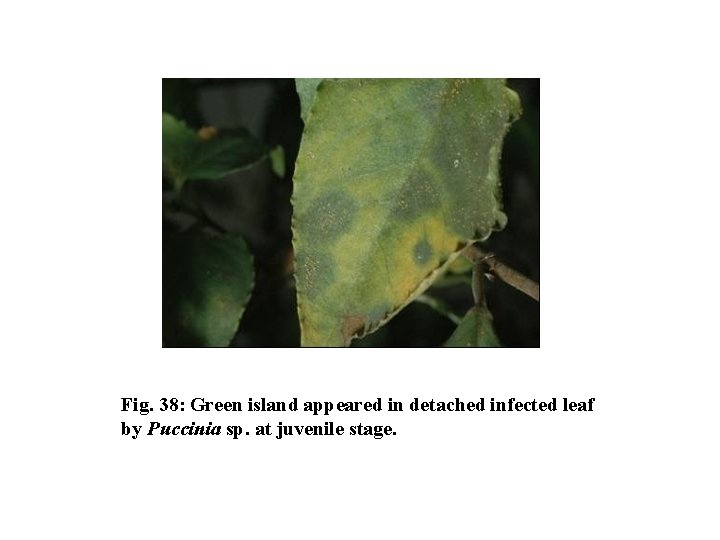
Fig. 38: Green island appeared in detached infected leaf by Puccinia sp. at juvenile stage.
• Host cell cytoplasm in the juvenile response generally resembles cytoplasm of young healthy leaves. This is the most evident in the vicinity of haustoria within infected cells. An extensive network of endoplasmic reticulum (ER) develops, which sometimes touches the extrahaustorial membrane, and unique tubular complexes, thought to be synthetic or secretory structures related to requirements of the fungus, may be present in host cytoplasm. Golgi bodies increase in number, and the volume of cytoplasm seems to be increased. • The rust haustorium is universally found in contact with the host nucleus in cereal or grass hosts. Rust infection of cereal cells generally stimulates a marked increase in volume of the host nucleus.
• This increase begins in the juvenile host response stage, but extends into the beginnings of autolytic, chlorotic stage. The increase in nuclear volume is accompanied by an increase in volume of the host nucleolus and by a shift from several nucleoli per nucleus to a single nucleolus. • The period in which the host nucleus and nucleolus usually enlarge coincides with increased synthesis of nucleolar and extranucleolar RNA, and of cytoplasmic protein in host cells. Apparently, the enlarged nucleoli and nuclei produce increased amounts of ribosomes and messenger RNA (m. RNA) that are used for protein synthesis in the cytoplasm. As noted, these changes are not strictly associated with the juvenile host response and instead may provide enzymes active in the autolytic stage to follow.
C- The autolytic host response • By 10 days after infection, the cytoplasmic organelles of host cells at pustule centers begin to degenerate and eventually disappear seem to be a slow autolytic digestion process. The ER becomes less abundant, breaking into vesicles, and mitochondria lose their cristae, swell, and become vesiculate. Mitochondria may gain small electron-dense inclusions. • Host chloroplasts sometimes appear swollen. Volume of chloroplast stroma can increase. The chloroplast lamellae may become less compacted as grana structure becomes disorganized. The outer chloroplast membrane can become vesiculate and eventually ruptured.
• As part of the autolytic stage, the host nucleus shrinks rapidly. With wheat stem rust, decrease in nuclear width and volume occurred by 14 days after inoculation. With wheat leaf rust, a reduction in host cell nuclear volume occurred as a result of chromatin within the nucleus condensed. • As autolysis progresses, most host cells remain alive and turgid for as long as 3 to 4 weeks after inoculation. Vacuoles can form residues of degenerated organelles, membranes, and electron-dense bodies. In advanced stages of autolysis, virtually the only host cell contents remaining are the host nucleus and the fungal haustorium, as the cytoplasmic layer lining the cell wall becomes highly attenuated.
• The autolytic host response resembles normal senescence of plants. The sporulating fungus seemingly accelerates the senescence of the host tissues; the result is digestion of host constituents, which are then probably utilized largely by the sporulating fungus.
D- Changes in host membranes During the juvenile response, host cells probably retain a full capacity for active uptake of metabolites. Later as disease progresses into the autolytic stage, host tissues become leaky and readily lose ions, sugars, amino acids, and probably other substances if the tissues are immersed in water. Such leakiness is a characteristic of uninfected, naturally senescing tissues as enzymes located on membranes lose activity. The host plasmalemma after infection has increased permeability to nonelectrolytes, which now thought to enter through the phospholipid portions of the membrane. Some evidence for changes of phospholipid components of host membranes in bean rust was obtained.
Responses of host cell organelles after infection by rust fungi as observed by microscopy 1 - Host chloroplast • Infection with plant pathogens may result in marked changes in a decrease in the number and the structure of chloroplasts in infected cells compared with uninfected cells. • The examination of semi-thin section using the light microscope indicates that the number of chloroplasts of infected host cells of Cirsium arvense during late infection of pycnial stage of the rust fungus Puccinia punctiformis is lower than in adjacent uninfected cells (See the Table). Compared with uninfected cells, infected mesophyll cells show a 53. 7 % decrease in chloroplast number at this stage.
• Similar but more drastic reduction in chloroplasts numbers (76. 1%) has been found in infected mesophyll cells during the uredial stage of infection, although the number of chloroplasts increased with age. • The loss in number of chloroplasts in infected hosts may be due to the decrease of chloroplast reproduction after infection or due to their degeneration.
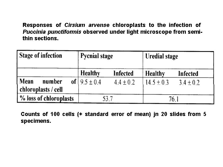
Responses of Cirsium arvense chloroplasts to the infection of Puccinia punctiformis observed under light microscope from semithin sections. Counts of 100 cells (+ standard error of mean) jn 20 slides from 5 specimens.
• Many reports describe ultrastructural changes of chloroplasts after infection by pathogens. In this connection, it is of interest that some workers have demonstrated that the ultrastructural changes in the chloroplasts in rust infected tissue are generally similar to those observed after infection by viruses or bacteria or during the processes of fruit ripening or natural senescence or after chemical treatment. • Such changes are similar to the changes during leaf senescence or chromoplast development. Increased of a peripheral reticulum after rust infection has also been reported. Prolarmellar bodies like carotenoid crystalloids occurring in the chloroplasts near the sporulating regions of Uromyces phaseoli also indicated.
Following infection by rust fungi, degradation of starch grains in chloroplasts has been observed. The chloroplasts of infected host cells are usually closely associated with uredial haustoria. Moreover, a decrease in size of chloroplasts in infected zones has been reported. The chloroplasts show disorganization of the membrane structure and/or a reduction in the number of photosynthetic lamellae. The increase in number and size of plastoglobuli after infection has also been reported.
• Electron microscopical examination revealed that the chloroplast from uninfected cells of Cirsium arvense show well-developed membrane structure with very dense stacks of grana, intergranal lamellae and chloroplast-bounding membrane with starch grains (See the figure). Chloroplasts of infected mesophyll cells during the pycnial stage of infection by Puccinia puncitiformis show a more spherical shape and the breakdown of grana, intergranal lamellae, chloroplast envelope and the appearance of peripheral reticulum, and the chloroplasts become more closely associated to the haustoria (See the figures).
• In contrast, at the uredial stage of infection, chloroplasts appear to be spherical, show degradation of grana and disappearance of the chloroplast envelope. An increase in the number of plastoglobuli and the appearance of marked peripheral reticulum was also seen. Starch grains become globoid or reduced in size (See the figure). • Many reports describe ultrastructural changes of chloroplasts after infection by rusts, viruses, or bacteria, and during the processes of fruit ripening, senescence or chemical treatments. In addition, most stages of infection show an increase in number and size of plastoglobuli of infected chloroplasts. Such increases are also recorded during leaf senescence, chromoplast development and thylakoid breakdown.
• The increase of plastoglobuli in chloroplasts, however, contributes to the increase of lipids or it may derive from the breakdown of chloroplast membranes. • The changes of chloroplast ultrastructure by rust infection may correlate also with changes in the rate of photosynthesis. Starch grains in chloroplasts show degradation during the infection. This supports theory that during the first stages of infection, the fungus stimulates starch synthesis of chloroplast of infected cells but later results in degradation and death. • The table summarizes the effect of pycnial and uredial stages of infection on the ultrastructure of chloroplast.
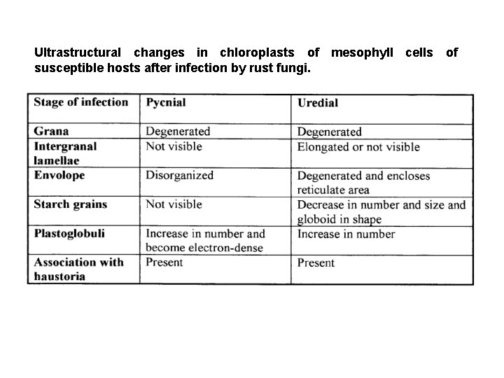
Ultrastructural changes in chloroplasts of mesophyll cells of susceptible hosts after infection by rust fungi.
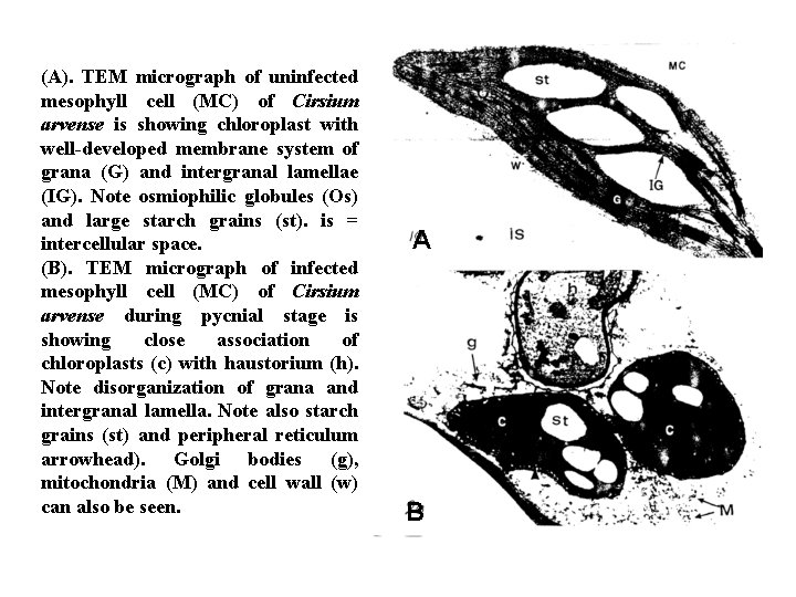
(A). TEM micrograph of uninfected mesophyll cell (MC) of Cirsium arvense is showing chloroplast with well-developed membrane system of grana (G) and intergranal lamellae (IG). Note osmiophilic globules (Os) and large starch grains (st). is = intercellular space. (B). TEM micrograph of infected mesophyll cell (MC) of Cirsium arvense during pycnial stage is showing close association of chloroplasts (c) with haustorium (h). Note disorganization of grana and intergranal lamella. Note also starch grains (st) and peripheral reticulum arrowhead). Golgi bodies (g), mitochondria (M) and cell wall (w) can also be seen. A B
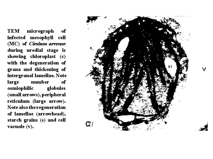
TEM micrograph of infected mesophyll cell (MC) of Cirsium arvense during uredial stage is showing chloroplast (c) with the degeneration of grana and thickening of intergranal lamellae. Note large number of osmiophilic globules (small arrows), peripheral reticulum (large arrow). Note also the regoneration of lamellae (arrowhead), starch grains (s) and cell vacuole (v). C
2 - Host nucleus • The nuclei in host cells in rust infection were altered in size and appearance and are closely associated with the uredial and pycnial haustoria. • When the semi-thin sections are examined using the light microscope, the nuclei in infected cell of Cirsium arvense by pycnial stage of Puccinia punctiforms show an increase of volume up to 10 % greater than of uninfected nuclei (See the table). The nuclei of infected mesophyll cells during the uredial stage of the same pathogen shows a further significant increase in volume (about 27. 7%). • The enlargement of nuclei after infection may be correlated with the increase of protein synthesis and amount of RNA as reported by many authors.
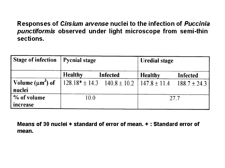
Responses of Cirsium arvense nuclei to the infection of Puccinia punctiformis observed under light microscope from semi-thin sections. Means of 30 nuclei + standard of error of mean. + : Standard error of mean.
• Uninfected mesophyll cells of Cirsium arvense show an elongated nucleus bounded by a well-defined double membrane and obvious differentiation between heterochromatin and euchromatin. The nucleus contains a nucleolus, generally lying close to the nuclear envelope. The heterochromatin is less in amount than the euchromatin and concentrated adjacent to the nuclear membrane (See the figure). At the pycnial stage of infection the nucleus enlarges, takes an irregular-shape and the heterochromatin is dispersed within the nuclear material. The nucleolus is present in the nucleus during this stage of infection and sometimes contains an electron-dense body appear to originate from it. As in the case of chloroplasts, the infected nuclei are closely associated with haustoria (See the figure).
• At uredial stage, the nuclei are always associated with haustoria. They become elongated and strongly curved around the haustorium. The nucleoli are always absent at this stage and small amount of heterochromatin appears as irregular clumps at the periphery of the nuclei (See the figure). During the infection, the host nuclei show a decrease in heterochromatin aggregation, an increase in the electron-density of euchromatin. This is may be due to the increase of RNA contents and losses of DNA. It is known that the heterochromatin regions as repressed DNA while, euchromatin regions are active in RNA synthesis. The table summarizes the effect of pycnial and uredial stages of infection on the ultrastructure of nucleus.
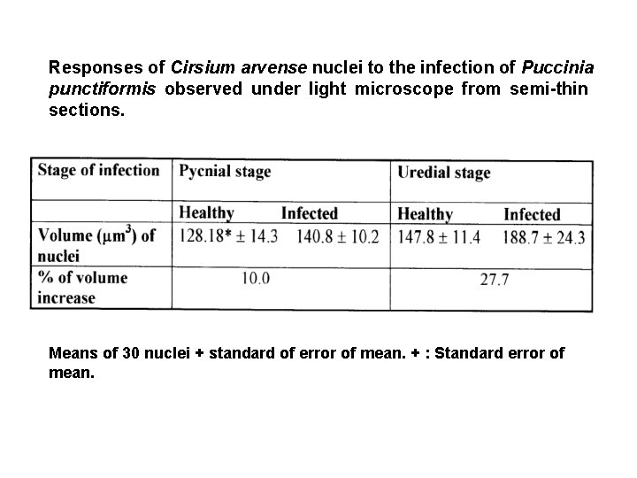
Responses of Cirsium arvense nuclei to the infection of Puccinia punctiformis observed under light microscope from semi-thin sections. Means of 30 nuclei + standard of error of mean. + : Standard error of mean.
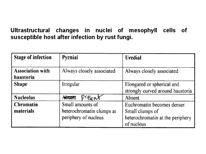
Ultrastructural changes in nuclei of mesophyll susceptible host after infection by rust fungi. cells of
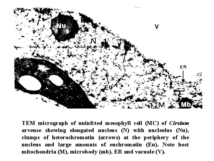
TEM micrograph of uninfcted mesophyll cell (MC) of Cirsium arvense showing elongated nucleus (N) with nucleolus (Nu), clumps of heterochromatin (arrows) at the periphery of the nucleus and large amounts of euchromatin (Eu). Note host mitochondria (M), microbody (mb), ER and vacuole (V).
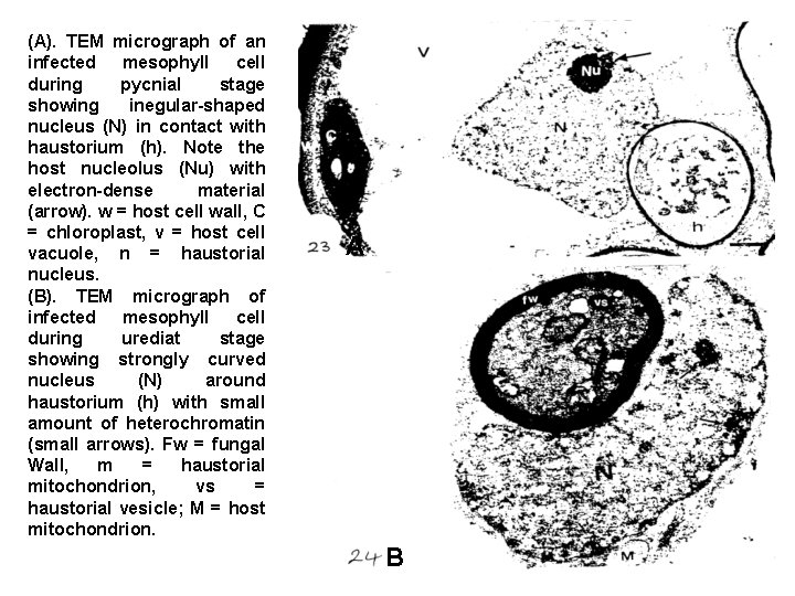
(A). TEM micrograph of an infected mesophyll cell during pycnial stage showing inegular-shaped nucleus (N) in contact with haustorium (h). Note the host nucleolus (Nu) with electron-dense material (arrow). w = host cell wall, C = chloroplast, v = host cell vacuole, n = haustorial nucleus. (B). TEM micrograph of infected mesophyll cell during urediat stage showing strongly curved nucleus (N) around haustorium (h) with small amount of heterochromatin (small arrows). Fw = fungal Wall, m = haustorial mitochondrion, vs = haustorial vesicle; M = host mitochondrion. A B
3 - Host microbodies • Microbodies occur in both infected and healthy mesophyll cells. A single membrane bound the microbodies in healthy mesophyll cells. Microbodies of healthy cells always lie in cytoplasm near to the chloroplasts and host cell wall (See the figure). During pycnial stage of infection, the microbodies tend to be closely associated with Golgi bodies (See the figure) and there is a decrease in the number of both crystalcontaining microbodies and non crystal-containing microbodies. During the uredial stage of infection, both types of microbodies decrease in number and are observed close to mitochondria, chloroplast and haustoria (See the figure).

• The crystal-containing microbodies, which have been seen in close proximity to the haustoria, may play a role in host resistance. Crystal containing microbodies were observed in the resistance zone around tobacco mosaic virus lesions. When leaf tissue was made resistant to virus infection artificially by a brief heat treatment, the resistant tissue contained crystal-containing microbodies. A number of workers have also reported that peroxidase activity increased after rust infection. The frequency of microbodies and the presence or absence of their crystals in hosts after infection by parasites may be correlated with the production of enzymes present in microbodies, which may be affected by the presence of the parasites.
4 - Host mitochondria, endoplasmic reticulum (ER) and Golgi bodies • Mitochondria of uninfected mesophyll cells of C. arvense lie in the cytoplasm close to chloroplasts, microbodies, and host cell wall (See the figure). During pycnial stage of infection, mitochondria are associated with chloroplasts and nucleus (Fig 26). However, at the uredial stage, mitochondria of infected cells, together with the microbodies, chloroplasts, nucleus and Golgi bodies are found clustered around the haustorium (See the figure). At this stage, mitochondria of infected cells contain small osmiophilic bodies, which are not observed in mitochondria of uninfected cells.
• The endoplasmic reticulum in uninfected mesophyll cells of C. arvense is frequently observed close to nucleus, mitochondria and microbodies (See the figure). At both stages of infection, the ER is more closely associated with the haustoria (See the figure) • Golgi bodies in uninfected cells are normally found adjacent to the host cell wall. During the pycnial stage of infection, Golgi bodies together closely associated with different organelles and haustoria (Fig. 26). Similarly, at the uredial stage, Golgi bodies lie close to haustoria (See the figure) and are rarely observed close to the host cell wall. Figure (26) shows degeneration of Golgi bodies, their vesicles appear separated away from their cisternae possibly migrating to the extrahaustorial plasma membrane.
• Mitochondria in infected mesophyll cells increase in numbers. This increase may be correlated with the increased rate of respiration after infection. Further change observed in infected mesophyll cells, was an increase in Golgi bodies and in the amounts of ER compared with uninfected cells. Many authors reported that Golgi bodies and ER remains still active following infection. Golgi bodies are frequently closely associated with the haustoria of the parasites and also with the extrahaustorial membrane.
• Golgi bodies and their vesicles have been noted in the cytoplasm around haustoria in many hostparasite interactions. The vesicles may also be continuous with extrahaustorial membrane. The frequency of such observations strongly suggests a functional significance. They may play a role in transporting of materials of extrahaustorial matrix. • Table 10 summarizes the quantitative changes in host microbodies, mitochondria and Golgi bodies in mesophyll cells of C. arvense infected by both stages of infection of P. punctifbrmis.
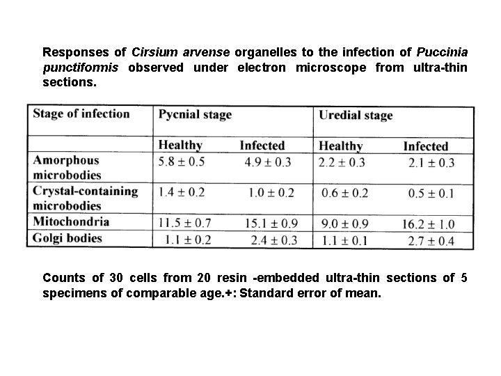
Responses of Cirsium arvense organelles to the infection of Puccinia punctiformis observed under electron microscope from ultra-thin sections. Counts of 30 cells from 20 resin -embedded ultra-thin sections of 5 specimens of comparable age. +: Standard error of mean.
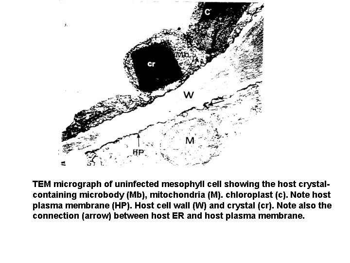
TEM micrograph of uninfected mesophyll cell showing the host crystalcontaining microbody (Mb), mitochondria (M). chloroplast (c). Note host plasma membrane (HP). Host cell wall (W) and crystal (cr). Note also the connection (arrow) between host ER and host plasma membrane.
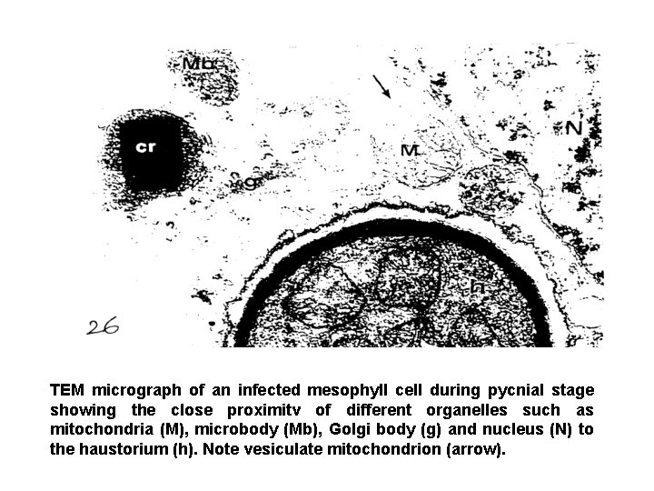
TEM micrograph of an infected mesophyll cell during pycnial stage showing the close proximitv of different organelles such as mitochondria (M), microbody (Mb), Golgi body (g) and nucleus (N) to the haustorium (h). Note vesiculate mitochondrion (arrow).
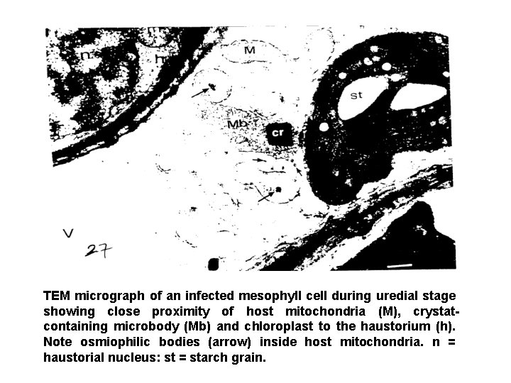
TEM micrograph of an infected mesophyll cell during uredial stage showing close proximity of host mitochondria (M), crystatcontaining microbody (Mb) and chloroplast to the haustorium (h). Note osmiophilic bodies (arrow) inside host mitochondria. n = haustorial nucleus: st = starch grain.
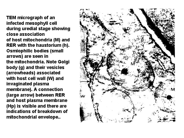
TEM micrograph of an infected mesophyll cell during uredial stage showing close association of host mitochondria (M) and RER with the haustorium (h). Osmiophilic bodies (small arrows) are seen in the mitochondria. Note Golgi body (g) and their vesicles (arrowheads) associated with host cell wall (W) and invaginated plasma membrane). A connection (large arrow) between RER and host plasma membrane (Hp) is visible and there are indications of breakdown of mitochondrial envelope. .
5 - Host wall encasement • A wide matrix (encasement) containing membranous materials always encloses necrotic haustoria. This matrix represents a type of wall apposition around haustoria and the membranous materials are radiate from an electron-dense layer between the fungal wall and matrix (Fig. 31). In this case, the haustorium is highly vacuolated and the host plasma membrane is completely enclosed this matrix.
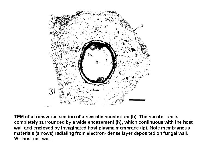
TEM of a transverse section of a necrotic haustorium (h). The haustorium is completely surrounded by a wide encasement (K), which continuous with the host wall and enclosed by invaginated host plasma membrane (ip). Note membranous materials (arrows) radiating from electron- dense layer deposited on fungal wall. W= host cell wall.
The physiology of diseased host The invasion of the host by a foreign organism leads, sooner or later, to changes in host physiology. If the organism is a pathogen, these changes will eventually prove deleterious to the host. Alternatively, where the pathogen fails to establish itself, these changes may be important in preventing the pathogen from gaining a foothold. Plants infected by quite different types of pathogens often exhibit very similar physiological symptoms. These similarities at first sight suggest that plants employ a common pathway of response to infection.
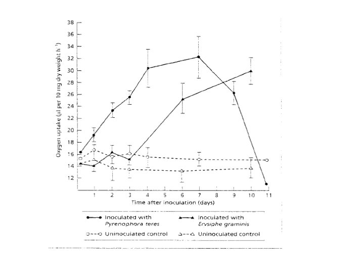
Post-infectional changes in host physiology 1 - Respiration As parasitism involves a nutritional relationship much of the work on the physiology of diseased plants has been concerned with energy metabolism. One of the most prominent changes, which occur following infection, is a substantial increase in respiration rate. This is equally true for diseases involving fungi, bacteria and viruses, although most of the available information has obtained from plants infected by biotrophic fungi. The figure shows the rate of oxygen uptake in barley leaves infected by two different fungi: one, Pyronophora teres, is a nectrotroph; the other, Erysiphe graminis, is a biotroph.
Powdery mildew fungi are only in intimate contact with their host where the haustoria enter epidermal cells. It is possible to peel off the epiphytic mycelium of the pathogen and measure the respiration rate of the host leaf with only an insignificant portion of the fungus (namely, the haustoria) remaining. Experiments like this have shown that the increase in respiration is maintained even after removal of the pathogen. All in all, the rise in respiration rates following infection would seem mainly to represent a response by host tissues. This response bears similarities to the transitory increase in respiration observed in plants subjected to mechanical injury.
2 - Photosynthesis is the most distinctive physiological activity of green plants. The capture of solar energy by chlorophyll and its subsequent utilization to fix carbon dioxide into organic compounds is the basis of life on this planet. However, in spite of its fundamental importance, comparatively little is known about the effects of pathogens on photosynthesis. Any pathogen, which attacks green aerial tissues, is likely to affect crop yield. In many cases the harmful effects of a pathogen can directly attributed to the destruction of photosynthetic tissues.
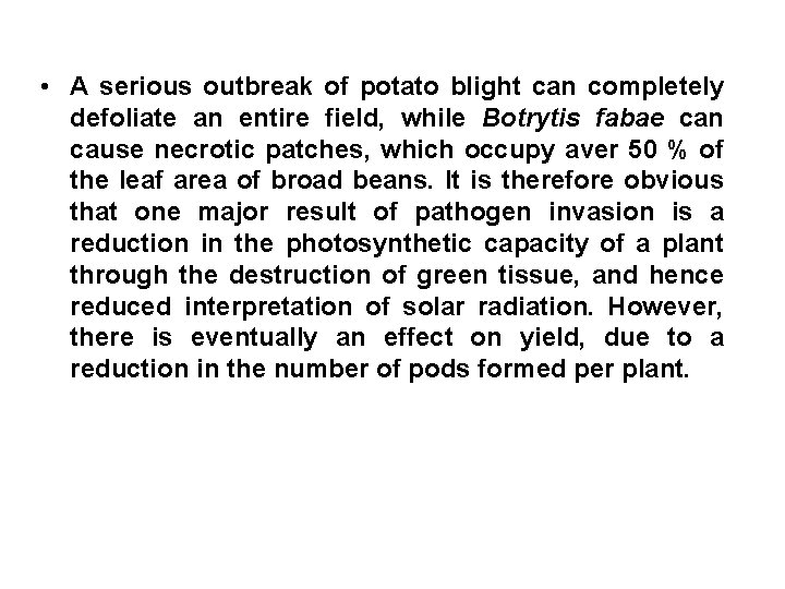
• A serious outbreak of potato blight can completely defoliate an entire field, while Botrytis fabae can cause necrotic patches, which occupy aver 50 % of the leaf area of broad beans. It is therefore obvious that one major result of pathogen invasion is a reduction in the photosynthetic capacity of a plant through the destruction of green tissue, and hence reduced interpretation of solar radiation. However, there is eventually an effect on yield, due to a reduction in the number of pods formed per plant.
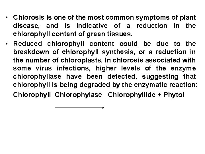
• Chlorosis is one of the most common symptoms of plant disease, and is indicative of a reduction in the chlorophyll content of green tissues. • Reduced chlorophyll content could be due to the breakdown of chlorophyll synthesis, or a reduction in the number of chloroplasts. In chlorosis associated with some virus infections, higher levels of the enzyme chlorophyllase have been detected, suggesting that chlorophyll is being degraded by the enzymatic reaction: Chlorophyll Chlorophylase Chlorophyllide + Phytol
• Symptoms of virus infection in leaves often include characteristic mosaics of green and yellow areas in which chloroplasts are reduced in number, or show ultrastructural abnormalities such as swelling and fewer lamellae, which are the sites of photochemical reactions. The chlorotic areas are often rich in starch, suggesting that virus infection affects both photosynthetic capacity and carbon partitioning within diseased leaves. • In leaves infected by biotrophic fungi there is a progressive loss in overall photosynthetic activity, although this usually only noticeable in the later stages of infection, when premature senescence of the leaf may set in.
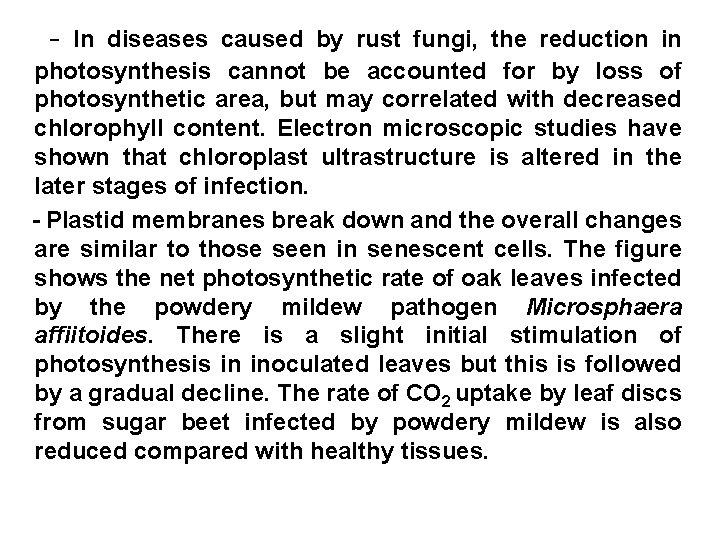
- In diseases caused by rust fungi, the reduction in photosynthesis cannot be accounted for by loss of photosynthetic area, but may correlated with decreased chlorophyll content. Electron microscopic studies have shown that chloroplast ultrastructure is altered in the later stages of infection. - Plastid membranes break down and the overall changes are similar to those seen in senescent cells. The figure shows the net photosynthetic rate of oak leaves infected by the powdery mildew pathogen Microsphaera affiitoides. There is a slight initial stimulation of photosynthesis in inoculated leaves but this is followed by a gradual decline. The rate of CO 2 uptake by leaf discs from sugar beet infected by powdery mildew is also reduced compared with healthy tissues.
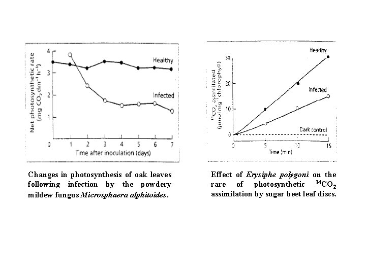
Changes in photosynthesis of oak leaves following infection by the powdery mildew fungus Microsphaera alphitoides. Effect of Erysiphe polygoni on the rare of photosynthetic 14 CO 2 assimilation by sugar beet leaf discs.
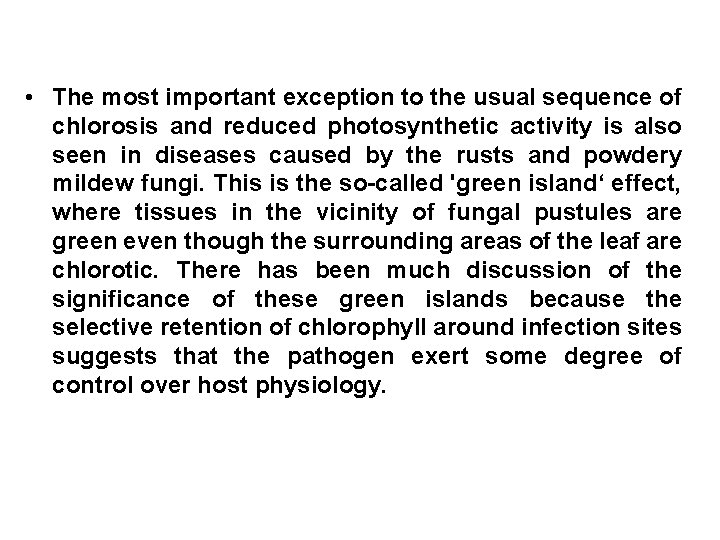
• The most important exception to the usual sequence of chlorosis and reduced photosynthetic activity is also seen in diseases caused by the rusts and powdery mildew fungi. This is the so-called 'green island‘ effect, where tissues in the vicinity of fungal pustules are green even though the surrounding areas of the leaf are chlorotic. There has been much discussion of the significance of these green islands because the selective retention of chlorophyll around infection sites suggests that the pathogen exert some degree of control over host physiology.
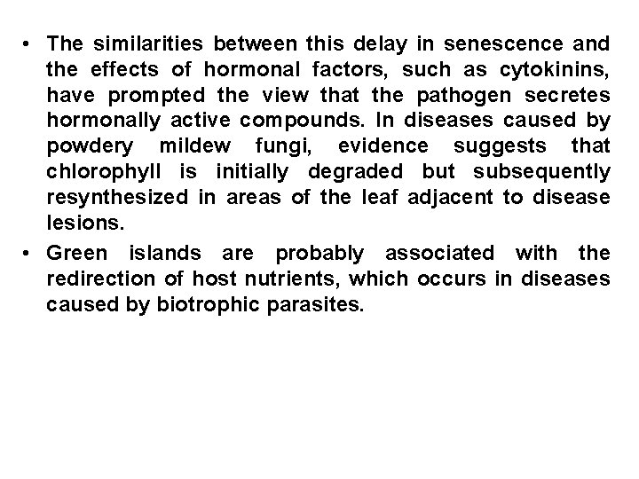
• The similarities between this delay in senescence and the effects of hormonal factors, such as cytokinins, have prompted the view that the pathogen secretes hormonally active compounds. In diseases caused by powdery mildew fungi, evidence suggests that chlorophyll is initially degraded but subsequently resynthesized in areas of the leaf adjacent to disease lesions. • Green islands are probably associated with the redirection of host nutrients, which occurs in diseases caused by biotrophic parasites.
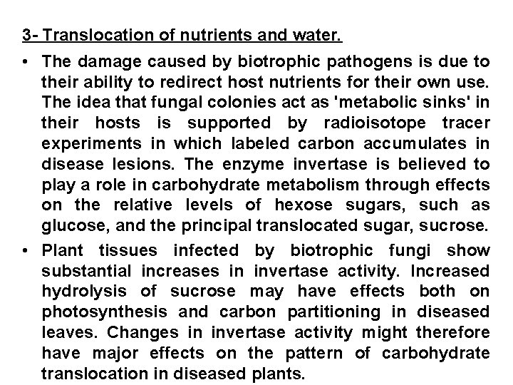
3 - Translocation of nutrients and water. • The damage caused by biotrophic pathogens is due to their ability to redirect host nutrients for their own use. The idea that fungal colonies act as 'metabolic sinks' in their hosts is supported by radioisotope tracer experiments in which labeled carbon accumulates in disease lesions. The enzyme invertase is believed to play a role in carbohydrate metabolism through effects on the relative levels of hexose sugars, such as glucose, and the principal translocated sugar, sucrose. • Plant tissues infected by biotrophic fungi show substantial increases in invertase activity. Increased hydrolysis of sucrose may have effects both on photosynthesis and carbon partitioning in diseased leaves. Changes in invertase activity might therefore have major effects on the pattern of carbohydrate translocation in diseased plants.
• The nutrient stress imposed by a redirection of host nutrients to satisfy the energy and biosynthetic needs of the pathogen is very different from that caused by pathogens which actually colonize the transporting tissues of the plant. Vascular wilt pathogens impair the flow of water and mineral salts through the xylem. Translocation of sugars through phloem tissues is also disrupted by some pathogens; a number of virus infections cause necrosis of phloem elements, resulting in nutrient imbalances in the host.
4 - The wilt syndrome The most pronounced effect of vascular wilt pathogens is one the water economy of the host. ln tomatoes infected by Fusarium oxysporum f. sp. Lycopersici, the resistance to water flow through the xylem is substantially increased compared to the resistance of uninfected stems. This effect can be partially explained on the basis of physical obstruction of the vessels by hyphae, but the vascular wilt syndrome is complex and involves host responses to infection as well as direct effects of the pathogen and its products (Fig. 14). Its secretion of polysaccharides and pectolytic enzymes compounds, blockages caused by the growth of the pathogen; in turn the host responds by producing gums and mucilages and by forming tyloses in the vessels. The end result in that water flow may be reduced to less than 59% of that in healthy plants.
• A further factor interfering with xylem function may be gas bubbles breaking the water column, this has been observed in sapwood colonized by the Dutch elm pathogen, Ophiosloma nova-ulmi. As well as causing severe water stress, infection by wilt pathogens also reduces the passage of essential mineral ions to the leaves. The overall consequences of vascular blockage are, however, difficult to assess, as these pathogens also secrete toxins which have physiological effects throughout the plant. Many plant pathogenic bacteria can enter the vascular system through wounds, and spread and multiply in xylem vessels, while secretion of extracellular slime or other high-molecular-weight materials further reduces flow by plugging pit membranes.
5 - Transpiration Wilting is one of the most common disease symptoms in plants, but the physiological basis of the symptom is not the same in all cases. The water economy can be disrupted through reduced water uptake, reduced flow rate or increased water loss through transpiration. Root rot pathogens destroy root tissues, and therefore reduce the surface area available for uptake and disrupt the transport of water through the root system. The vascular wilt fungi reduce flow and at the same time reduce the transpiration rate. Many other pathogens increase the transpiration rate (Fig. 15). The few studies, which have been made of the water relations of virus-infected plants indicate that the transpiration rate is reduced, and the total water content of the host is lower. This is especially true in severely diseased plants.
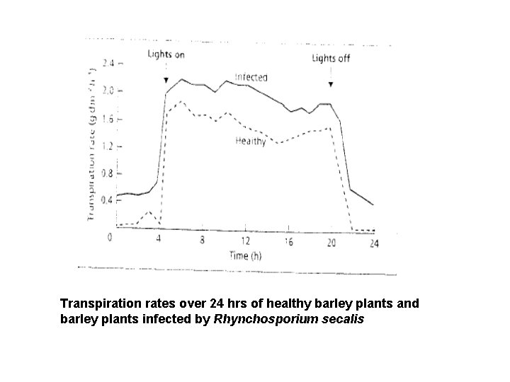
Transpiration rates over 24 hrs of healthy barley plants and barley plants infected by Rhynchosporium secalis
6 - Growth regulation • All plant pathogens affect the growth and development of their hosts to a greater or lesser extent. The diversion of nutrients orthe destruction of host tissues will inevitably lead to reduce performance, and in some cases may severely stunt the plant. These effects on plant growth are, however, essentially indirect and therefore different from the specific growth abnormalities induced y a variety of pathogens (Table 4). symptoms such as galls and tumors, excessive branching, leaf epinastry, abnormal induction of adventitious roots and premature leaf abscission are all associated with changes in the control of plant growth and differentiation. Such deranged growth is characteristic of many diseases involving microbial pathogens (Fig. 16).
Although plant morphogenesis is influenced by environmental conditions, hormonal compounds such as indoleacetic acid, gibberellins, abscisic acid and ethylene mediate control of the basic processes of cell division and differentiation. Changes in the concentration or distribution of these hormones have widespread effects on the physiology of plants. Alterations in growth regulation can often be attributed to the production of hormonally active compounds by the pathogen.
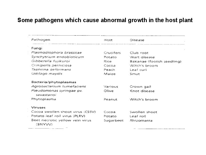
Some pathogens which cause abnormal growth in the host plant
7 - Nucleic acids Here we focus on compatible host-parasite combinations and the series of changes that occur within the host at disease progresses. 1. RNA As part of the juvenile host response before the fungus sporulates, host cells should maintain or possibly increase their ability to synthesize protein. They should maintain the machinery needed for DNA-dependent transcription of m. RNA and production of ribosomes. Protein synthesis occurs in cytoplasm, synthesis of ribosomes occurs in nucleoli, and synthesis of m. RNA occurs in the extranucleolar protein of nuclei.
Enhanced metabolic activity in both the nucleolar and extranucleolar portions of host nuclei has been demonstrated cytologically for wheat rust. As the volumes of host cell nuclei and nucleoli increase, the amount of both nucleolar and extranucleolar RNA doubles. Incorporation of radioactively labeled uridine and cytidine (precursors of RNA) into nuclei was doubled, as was incorporation of leucine into nuclear protein. Furthermore, the diffuse interchromatin network of the nucleus, where transcription occurs, increased in electron density. Finally, the amount of histone within the nucleus decreased, and apparently also the incorporation of amino acids into histone. Histones thought to repress transcription non-specifically.
Together, these cytological studies indicate that transcriptional activities increase in host nuclei as a consequence of rust. 2. RNase In line with the probable enhancement of RNA degradation, the activity of RNase increases in rusted tissues. With wheat stem rust, RNase activity doubles at 1 to 4 days after inoculation at levels two to five times those of uninfected leaves. Apparently, the new RNase is the type found in uninfected leaves and not a fungal type. Furthermore, the RNase in rusted flax was of the type that degrades RNA and of the type involved in posttranscriptional processing of RNA. The large amount of RNase activity in cereal rusts is probably involved in rapid RNA turnover.
3. DNA Because cereal host cells do not enlarge or divide in rusted tissues, no increase in the amount of host DNA is expected; indeed, the amount of DNA in host nuclei as measured microspectrophotometrically does not change with wheat stem rust until 9 days after inoculation when a slow decline begins. Total DNA of host and parasite combined tends to remain constant, because it has usually expressed on a dry-weight basis, and because the amount of nuclear host DNA eventually declines as new fungal DNA is produced.
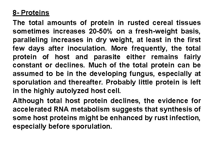
8 - Proteins The total amounts of protein in rusted cereal tissues sometimes increases 20 -50% on a fresh-weight basis, paralleling increases in dry weight, at least in the first few days after inoculation. More frequently, the total protein of host and parasite either remains fairly constant or declines. Much of the total protein can be assumed to be in the developing fungus, especially at sporulation and thereafter. Probably little protein is left in the highly autolyzed host cell. Although total host protein declines, the evidence for accelerated RNA metabolism suggests that synthesis of some host proteins might be enhanced by rust infection, especially before sporulation.
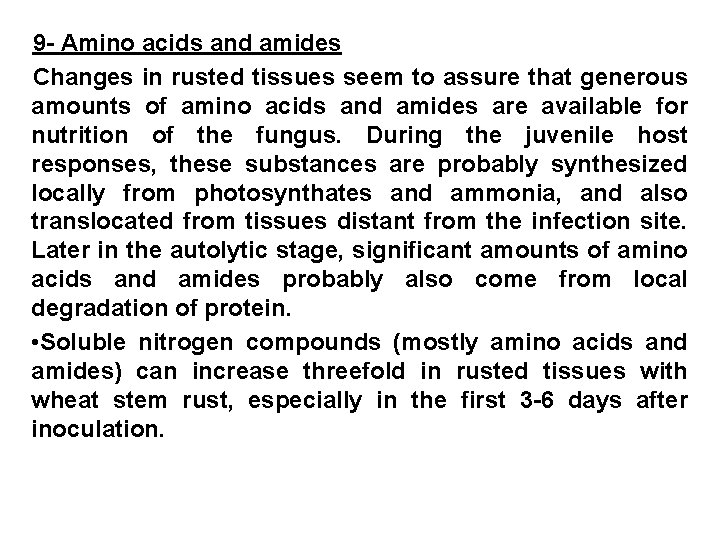
9 - Amino acids and amides Changes in rusted tissues seem to assure that generous amounts of amino acids and amides are available for nutrition of the fungus. During the juvenile host responses, these substances are probably synthesized locally from photosynthates and ammonia, and also translocated from tissues distant from the infection site. Later in the autolytic stage, significant amounts of amino acids and amides probably also come from local degradation of protein. • Soluble nitrogen compounds (mostly amino acids and amides) can increase threefold in rusted tissues with wheat stem rust, especially in the first 3 -6 days after inoculation.
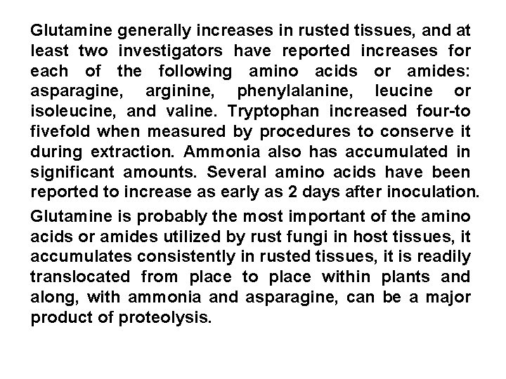
Glutamine generally increases in rusted tissues, and at least two investigators have reported increases for each of the following amino acids or amides: asparagine, arginine, phenylalanine, leucine or isoleucine, and valine. Tryptophan increased four-to fivefold when measured by procedures to conserve it during extraction. Ammonia also has accumulated in significant amounts. Several amino acids have been reported to increase as early as 2 days after inoculation. Glutamine is probably the most important of the amino acids or amides utilized by rust fungi in host tissues, it accumulates consistently in rusted tissues, it is readily translocated from place to place within plants and along, with ammonia and asparagine, can be a major product of proteolysis.
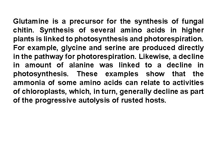
Glutamine is a precursor for the synthesis of fungal chitin. Synthesis of several amino acids in higher plants is linked to photosynthesis and photorespiration. For example, glycine and serine are produced directly in the pathway for photorespiration. Likewise, a decline in amount of alanine was linked to a decline in photosynthesis. These examples show that the ammonia of some amino acids can relate to activities of chloroplasts, which, in turn, generally decline as part of the progressive autolysis of rusted hosts.
- Slides: 121