
HOST RESPONSE BASIC CONCEPTS DR RIYAS KARIM CONTENTS

HOST RESPONSE: BASIC CONCEPTS DR RIYAS KARIM
CONTENTS: - INTRODUCTION - HISTORICAL PERSPECTIVE - ROLE OF SALIVA IN HOST DEFENSE - HOST DEFENSE PROCESSES - CELLS OF THE IMMUNE SYSTEM - IMMUNOGLOBULIN - ROLE OF ANTIBODIES IN INFLAMMATORY DISEASE - COMPLEMENT SYSTEM - IMMUNE MECHANISMS - CYTOKINES - PERIODONTAL DISEASE ACTIVITY - INFLUENCE OF HOST RESPONSE IN PERIODONTAL DISEASE - PREREQUISTES FOR PERIODONTAL DISEASE INITIATION & PROGRESSION - CONCLUSION - REFERENCES
INTRODUCTION: Microbial colonization by both pathogenic and commensal organisms invariably evokes a host response. � The balance between host and commensal flora represents a relative harmonious co-existence that can be disturbed by changes in the host defense or bacterial composition with the resultant expression of clinical disease. � Health is not a static condition. It is a dynamic state in which the living and functioning organism or tissue remains in balance with a constantly changing environment. This constant process of readjustment to maintain a functional integrity is known as homeostasis. �
The tissues of the periodontium are exposed to various environmental factors in the oral cavity. � Over 300 species of bacteria have been isolated in the oral cavity. � The periodontal tissues remain in a state of partnership with most of the bacteria and only under certain circumstances do we suffer from their attack because , host defence system strikes a balance between the two. � The host responds to the attack of bacteria and its toxins at various levels. �
HISTORICAL PERSPECTIVE: � � � During the past two decades, extraordinary advances have been made in understanding the etiologic mechanisms and pathogenesis of infectious diseases. A result of the role of the immunoinflammatory response in periodontal diseases has become clearer. The tissue destruction found in periodontal disease results, for the most part from the action of immune system and related effect or mechanisms. Microorganism especially bacteria in dental plaque cause periodontal disease. Although these microorganism produce enzyme and other factors that can directly cause degradation of periodontal tissues, the progression and severity of tissue destruction are mostly caused by host immune response to infecting bacteria. This concept was not always thought to be the case. The development of this tenet has been a dramatic albeit slow, evolution in the understanding of the pathogenesis of periodontal diseases.
ROLE OF SALIVA IN THE HOST DEFENSE: � A vehicle for swallowing bacteria � Inhibition of attachment of bacteria � Bacterial action by the perioxidase system � Bacterial action by lysozyme , lactoferrin and other factors.
Peroxidase : It is synthesized by salivary gland acini and secreted into the saliva, where it bound to bacteria and thiocyanate is secreted into saliva by the ductal cells. � Lactoferrin: It is secreted by serous salivary gland, which binds iron, an important growth factor or requirement for many microorganisms. This action is bacteriostatic rather than bactericidal. � Lysozyme: It is an antimicrobial enzyme in the saliva secreted mainly by mucous salivary glands and it degrades mucopeptides in the cell wall of gram-positive bacteria, weakening the wall and causing lysis. �
HOST DEFENSE PROCESSES: Host-parasite reactions can be divided into innate(non specific) and adaptive(specific)responses. � Innate reactions include the inflammatory response and do not involve immunological mechanisms. � Adaptive reactions that include immunological responses tend to be very effective as the host response is specifically tailored to the offending pathogens. �
Important aspects of host defense processes: Inflammatory processes: � The host has an extensive repertoire of defensive responses to ward off invasion by pathogens. Such responses may either result in a rapid resolution of the lesion. � An ineffective response may result in a chronic lesion that does not resolve or if excessively deployed , in a lesion in which the responses contribute significantly to tissue destruction. �
In the classical description of inflammation, a tissue is presented that appears macroscopically red, swollen, hot and painful, and with possible loss of function in specific sites. � Redness and heat are due to vasodilatation and increased blood flow. � Swelling is a result of increased vascular permeability and leakage of plasma proteins that create an osmotic potential that draws fluid into the inflammed tissues. � Related to the vascular changes there is an accumulation of inflammatory cells infiltrating the lesion. �
Molecules, cells and processes: � Proteinases(proteases) � Periodontal disease results in tissue degradation, and thus proteases, derived both from the host and from bacteria, are central to the disease processes. � Proteinases cleave proteins by hydrolyzing peptide bonds and may be classified into two major classes, endopeptidases and exopeptidases, depending on the location of activity of the enzyme on its substrate. � Efforts have been made to assess endopeptidase activity in gingival tissues and in GCF. �
Matrix metalloproteinases: � The periodontium is structurally comprised of fibrous elements, including collagen, elastin and glycoproteins, minerals, lipids, water, and tissuebound growth factors. � In addition there exists a large variety of extracellular matrix components, including tropocolagen, proteoglycans and other proteins. � Matrix mettaloproteinases are responsible for remodelling and degradation of the matrix components. �
One of the MMPs that has received much attention is the neutrophil(PMN) collagenase that is found in higher concentrations in inflammed gingival specimens than in clinically healthy gingiva. � The increased presence of these MMP enzymes in diseased over healthy sites, their increase during experimental gingivitis, and decreased after periodontal treatment suggest that MMPs from PMNs are involved in periodontal tissue breakdown. � An important feature of connective tissues in general and the periodontal ligament in particular, is the process of constant renewal of the extracellular matrix components involving MMP. �
Prostaglandins: � Prostaglandins are derivatives of arachidonic acid and are important mediators of inflammation. � Macrophages in particular, but also several other cells produce prostaglandins, particularly PGE 2 which is a potent vasodilator and inducer of cytokine production by various cells. � PGE 2, acts on fibroblasts and osteoclasts to induce production of MMPs, which are of importance for tissue turnover and tissue destruction in gingivitis and periodontitis. �
� Polymorphonuclear leukocytes: � The PMN is the predominant lekocyte within the gingival crevice/pocket in both health and disease. � PMNs are attracted from the circulation to the affected area via chemotactic stimuli elicited from, for example , microorganism in the biofilm, and host derived chemokines.
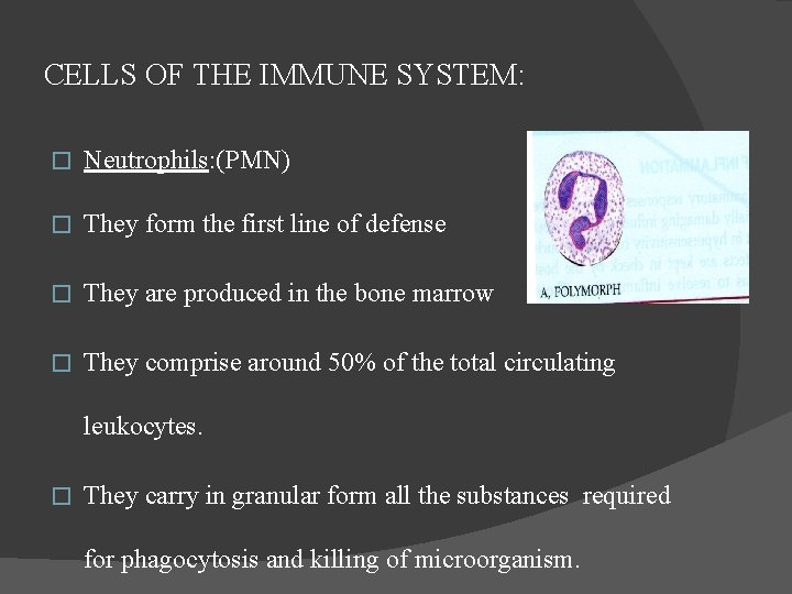
CELLS OF THE IMMUNE SYSTEM: � Neutrophils: (PMN) � They form the first line of defense � They are produced in the bone marrow � They comprise around 50% of the total circulating leukocytes. � They carry in granular form all the substances required for phagocytosis and killing of microorganism.
Functions of neutrophils: Emigration: Leukocytes normally travel along the center of the lumen of the blood vessel, but in inflamed tissues the blood flow is slowed by fluid exudation and they adhere more readily to endothelial cells, the mechanism is called rolling and margination. When the neutrophils migrate across the endothelium it is called diapedesis and interendothelial transmigration. � Chemotaxis: It is the directed movement of a cell along a chemical gradient. The neutrophils are attracted by chemical signals from multiple sources. �
The agents acting as potent chemotactic substances for different leucocytes called chemokines are as follows: 1. Leukotriene B 4 2. Platelet factor 4 3. Components of complement system 4. Cytokines 5. Soluble bacterial products 6. Monocyte chemoattractant protein 7. Chemotactic factor for CD 4+T cells 8. Eotaxin chemotactic for eosinophils �
� In addition , chemotactic agents also induce leucocyte activation that includes: The production of arachidonic acid metabolites, degranulation and secretion of lysosomal enzymes, generation of oxygen metabolites , increased intracellular calcium and increase in leucocyte surface adhesion molecules.
Mechanism of action of neutrophils: � Protective mechanisms 1. Pavementing/margination 2. Chemotaxis 3. Phagocytosis: Engulfing and destroying noxious substances such as immune complexes and damage tissues, apart from various microorganisms and antigenic substrates within phagolysosomes.
The process of phagocytosis is similar for both polymorphs and macrophages and involves the following four steps: 1. Recognition and attachment stage: (Opsonisation) The phagocytic cells are recognised and attracted to bacteria by chemotactic factors released by bacterial products as well as by tissue proteins. 2. Engulfment stage: The opsonised particle bound to the surface of phagocyte is ready to be engulfed. This is accomplished by formation of cytoplasmic pseudopods around the particle due to activation of actin filaments beneath the cell wall, enveloping it in a phagocytic vacuole. �
3. Degranulation stage: � During this process, the preformed granulestored products of PMNs are discharged or secreted into the phagosome and the extracellular environment. � In particular the specific or secondary granules of PMNs are discharged while azurophilic granules are fused with phagosomes. � Besides the discharged of performed granules, mononuclear phagocytes synthesise and secrete certain enzymes and oxygen metabolites. �
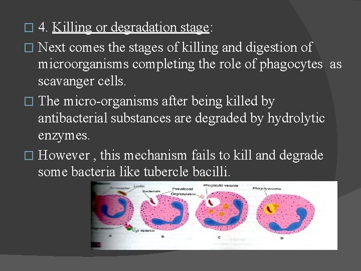
4. Killing or degradation stage: � Next comes the stages of killing and digestion of microorganisms completing the role of phagocytes as scavanger cells. � The micro-organisms after being killed by antibacterial substances are degraded by hydrolytic enzymes. � However , this mechanism fails to kill and degrade some bacteria like tubercle bacilli. �
Destructive mechanisms: � PMNs carry potent substances such as acid hydrolases, collagenase, prostaglandins etc. � They are responsible for destroying collagen and other connective tissue substances and inducing bone resorption. � These substances are released during cell death. Besides, enzymes escape from the cell to the interstitial area due to incomplete closure of the phagocytic vacuoles during phagocytosis. � These substances activate complement components via the alternate pathway. � They activate kinin producing systems. � They magnify inflammation. �
Neutrophil defects: � � - Defects in the PMN defense system are catastrophic to the periodontium. Neutrophil disorders associated with periodontal diseases: Diabetes mellitus Papillon-Lefevre syndrome Down syndrome Chediak-Higashi syndrome Drug-induced agranulocytosis Cyclic neutropenia
� Periodontal disease with neutrophil disorders: � Acute ulcerative necrotizing gingivitis � Localized juvenile periodontitis � Pre-pubertal periodontitis � Refractory periodontitis
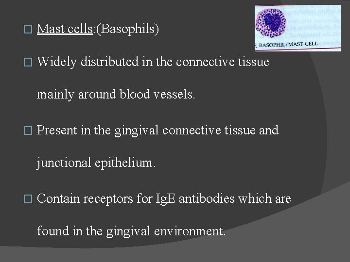
� Mast cells: (Basophils) � Widely distributed in the connective tissue mainly around blood vessels. � Present in the gingival connective tissue and junctional epithelium. � Contain receptors for Ig. E antibodies which are found in the gingival environment.
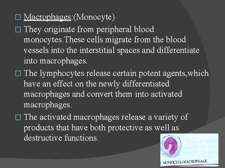
Macrophages: (Monocyte) � They originate from peripheral blood monocytes. These cells migrate from the blood vessels into the interstitial spaces and differentiate into macrophages. � The lymphocytes release certain potent agents, which have an effect on the newly differentiated macrophages and convert them into activated macrophages. � The activated macrophages release a variety of products that have both protective as well as destructive functions. �
Protective functions: � Phagocytosis and microbial killing. � Destroys damaged tissues. � Stimulate stem cell growth in the bone marrow. � Processes and presents antigen to lymphocytes. � Synthesizes and secretes: - Interferon - Complement components - Pyrogen - Pmn chemotactic factors �
� Destructive functions: � Secretes prostaglandins and cyclic nucleotides � Cytotoxic factors: cytokines � Acid hydrolases � Neutral proteases including collagenase, elastase and plasminogen activators.
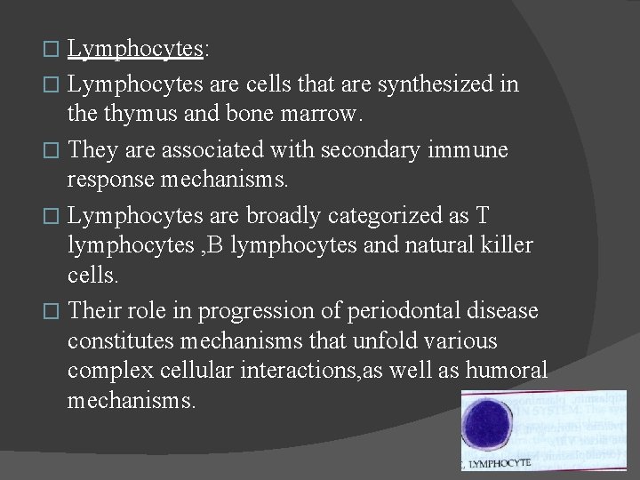
Lymphocytes: � Lymphocytes are cells that are synthesized in the thymus and bone marrow. � They are associated with secondary immune response mechanisms. � Lymphocytes are broadly categorized as T lymphocytes , B lymphocytes and natural killer cells. � Their role in progression of periodontal disease constitutes mechanisms that unfold various complex cellular interactions, as well as humoral mechanisms. �
T lymphocytes: � T lymphocytes or T cells which are derived from the thymus and play a role in cell mediated immunity. � T cells are recognized to be composed of several subsets that modulate the humoral response. These include helper T cells which aid in the cellular response of B cells to differentiate into plasma cells and produce antibodies and suppressor cytotoxic T cells, which stimulate cytotoxic and microbial activity of immune cells. �
B lymphocytes: � The B lymphocytes or B cells , which are derived from liver , spleen and bone marrow, are the precursors for plasma cells, and play a role in humoral immunity. � The B cells are identifiable by their cell surface immunoglobulin, usually Ig. M. � However, some B cells express Ig. G, Ig. A, or Ig. E. � These surface immunoglobulins serve as receptors for antigen. �
� NK cells: � The NK cells are recognized by their absence of T -cell receptors and surface immunoglobulin. � The interaction between antigens and macrophages known as antigen processing, leads to the activation of NK cells.
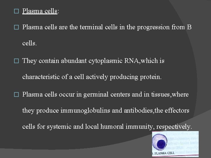
� Plasma cells: � Plasma cells are the terminal cells in the progression from B cells. � They contain abundant cytoplasmic RNA, which is characteristic of a cell actively producing protein. � Plasma cells occur in germinal centers and in tissues, where they produce immunoglobulins and antibodies, the effectors cells for systemic and local humoral immunity, respectively.
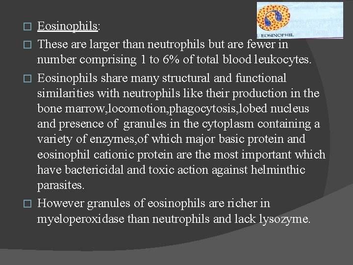
Eosinophils: � These are larger than neutrophils but are fewer in number comprising 1 to 6% of total blood leukocytes. � Eosinophils share many structural and functional similarities with neutrophils like their production in the bone marrow, locomotion, phagocytosis, lobed nucleus and presence of granules in the cytoplasm containing a variety of enzymes, of which major basic protein and eosinophil cationic protein are the most important which have bactericidal and toxic action against helminthic parasites. � However granules of eosinophils are richer in myeloperoxidase than neutrophils and lack lysozyme. �
IMMUNOGLOBULINS: Immunoglobulins are gamma globulins produced by plasma cells in response to antigens with which they can react in a specific way either in vivo or vitro. Structure of immunoglobulins: - Ig molecules are composed of two light chains (Ƙ/ƛ) and one of the five types of heavy chains(δ, µ, ε, ϒ, α). - The heavy chain denotes the class of the Ig molecules. - Basic structure of the Ig molecules resembles a “Y. ” �
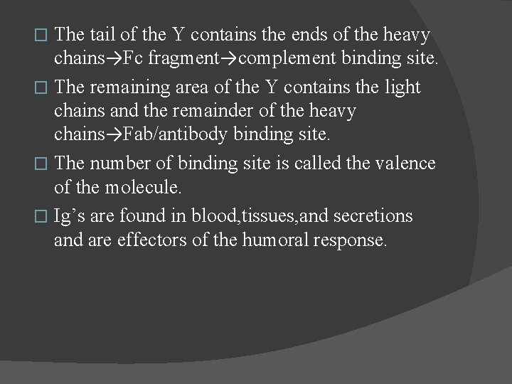
The tail of the Y contains the ends of the heavy chains→Fc fragment→complement binding site. � The remaining area of the Y contains the light chains and the remainder of the heavy chains→Fab/antibody binding site. � The number of binding site is called the valence of the molecule. � Ig’s are found in blood, tissues, and secretions and are effectors of the humoral response. �
Functions of immunoglobulins: - Ig. G: � Complement fixation � Delayed antibody response � Opsonization � Cross-placental barrier � Increased concentration in GCF
� Ig. A: � Serum Ig. A complement fixation by alternate pathway � Secretory Ig. A protects mucosal surfaces � Increased concentration in saliva � Prevents adhesion of bacteria to tissue surfaces especially in the early stages of periodontal diseases
Ig. M: � Early antibody response � Complement fixation � Ig. E: � Causes immediate hypersensitivity reactions � Ig. D: � Ig. D is found at extremely low levels in serum � unknown �
ROLE OF ANTIBODIES IN INFLAMMATORY DISEASES: � � � Complement activation by antigen-antibody complexes→protective. Phagocytosis of antigen-antibody complex→release of cytokines →protective and destructive. Blocking of lymphocytes by free antibody/antigenantibody complex. Suppression of cell mediated immunity. Neutralization of bacterial allergens/reactions toxin/histolytic enzymes→protective. Enhanced opsonization/bacteriolysis of plaque bacteria→protective.
COMPLEMENT SYSTEM: � It is an amplifier of the immune system. � It is made up of at least 22 proteins. � Upon activation its components interact in a cascade fashion, generating a variety of biologically active substances, which terminates in the lysis of antibody tagged cells.
Complement system can be broadly classified into two pathways: � CLASSICAL PATHWAY � ALTERNATE PATHWAY � CLASSICAL PATHWAY: � The classical pathway is activated by a reaction of antigen with Ig. G or Ig. M antibodies and by aggregated immunoglobulins. � The sequence is C 1, C 4, C 2, C 3, C 5. C 6, C 7, C 8 and C 9. �
� ALTERNATIVE PATHWAY: � It is activated by antibodies of immunoglobulins and also by endotoxins. � They can initiate third component of the complement without starting from the beginning of the cascade. � The sequence is C 3, C 5, C 6, C 7, C 8, C 9.
Bacterial antigens such as endotoxins and polysaccharides such as dextran are also activators of the alternative pathway. � Dental plaque and pure cultures of bacteria can also activate complement by the alternative pathway in the absence of antibody. � On activation of complement by endotoxin, biologically active fragmentation products are released. � Complement components that are present in the gingival sulcular fluid have been shown to decrease and therefore assumed to be activated by some plaque bacterial proteases. �
� Biological properties of activated complement factors: � Anaphylaxis→Histamine release from mast cells and permeability of capillaries→C 3 a+C 5 a. � Activation of B lymphocytes→C 3 b. � Chemotaxis→C 5 a, C 5 b, C 6, C 7. � Immune adherence→opsonization→C 3 b+C 5 b. � Lectin pathway.
� Membrane damage→C 8+C 9. � Enhancement of blood clotting→C 8. � Lysosomal enzyme release→C 5 a. � Promotion of phagocytosis→C 3+C 5. � Inactivation of bacterial LPS→C 5+C 6.
IMMUNE MECHANISMS: Immune mechanisms are usually protective responses by the host to the presence of foreign substances such as bacteria and viruses. � They may at the same time cause local tissue destruction by triggering several types of overreaction or hypersensitivity. � Tissue damage may occur in a sensitized host with subsequent exposure to the sensitizing antigen. �
� Four types of hypersensitivity reactions have been described: � Type 1(Anaphylaxis or immediate hypersensitivity) � Type 2(Cytotoxic reactions) � Type 3(Immune complex or Arthus reactions) � Type 4(Cell-mediated immunity or Delayed hypersensitivity)
� � � Type 1(Anaphylaxis or immediate hypersensitivity): Two variations in anaphylactic hypersensitivity occur, depending on the route of administration of the antigen. If the antigen is injected locally into the skin, the reaction is called cutaneous anaphylaxis. If the antigen is injected intravenously, it is called systemic or generalized anaphylaxis. The basic mechanisms in both types of immediate hypersensitivity are the same. Although both Ig. E and Ig. G antibodies are involved in anaphylaxis, only Ig. E plays a direct role in its pathogenesis through its ability to sensitize the skin.
� Ig. E antibodies involved in anaphylactic reactions attach strongly at the Fc portion of the antibody to receptors found on mast cells and basophilic leukocytes, primarily in the skin and other connective tissues such as gingiva. Mechanisms of Anaphylactic hypersensitivity: � Anaphylaxis occurs when two Ig. E antibodies that are fixed to a mast cell or basophil react with the antigen through the Fab portion of the antibodies. This antibody-antigen reaction causes the release of the pharmacologicallyactive substances from the sensitized-cells. �
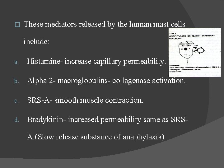
� These mediators released by the human mast cells include: a. Histamine- increase capillary permeability. b. Alpha 2 - macroglobulins- collagenase activation. c. SRS-A- smooth muscle contraction. d. Bradykinin- increased permeability same as SRSA. (Slow release substance of anaphylaxis).
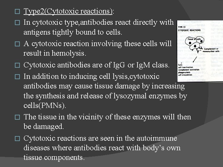
� � � � Type 2(Cytotoxic reactions): In cytotoxic type, antibodies react directly with antigens tightly bound to cells. A cytotoxic reaction involving these cells will result in hemolysis. Cytotoxic antibodies are of Ig. G or Ig. M class. In addition to inducing cell lysis, cytotoxic antibodies may cause tissue damage by increasing the synthesis and release of lysozymal enzymes by cells(PMNs). The tissue in the vicinity of these enzymes will then be damaged. Cytotoxic reactions are seen in the autoimmune diseases where antibodies react with body’s own tissue components.
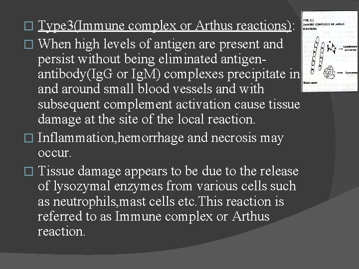
Type 3(Immune complex or Arthus reactions): � When high levels of antigen are present and persist without being eliminated antigenantibody(Ig. G or Ig. M) complexes precipitate in and around small blood vessels and with subsequent complement activation cause tissue damage at the site of the local reaction. � Inflammation, hemorrhage and necrosis may occur. � Tissue damage appears to be due to the release of lysozymal enzymes from various cells such as neutrophils, mast cells etc. This reaction is referred to as Immune complex or Arthus reaction. �
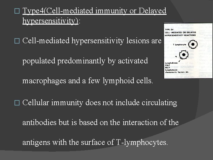
� Type 4(Cell-mediated immunity or Delayed hypersensitivity): � Cell-mediated hypersensitivity lesions are populated predominantly by activated macrophages and a few lymphoid cells. � Cellular immunity does not include circulating antibodies but is based on the interaction of the antigens with the surface of T-lymphocytes.
CYTOKINES: The complex interactions among lymphocytes, inflammatory cells, and other cellular elements in connective tissues are mediated by a series of low-molecular-weight proteins called cytokines. � Cytokines assist in the regulation and development of immune effector cells, cell to cell communication, and direct effector functions. � Some cytokines exhibit autocrine function, binding to the cell that produced them. �
Cytokines may be pleiotrophic, eliciting different biologic activities fromdifferent cells. � In addition, different cytokines may exhibit similar responses. � The accumulation of plasma cells and lymphocytes in the periodontal tissues suggests that cytokines participate in periodontal pathologic changes. � More recently the majority of cytokines have been renamed as interleukins, referring to their role in communication between leukocytes. � Presently 10 interleukins have been identified. �
� � � � Classification of interleukins: IL-1 is a pleiotrophic cytokine with a variety of activities. It includes osteoclast-activating factor because of its stimulation of osteoclasts and lymphocyteactivating factor because of its ability to stimulate proliferation of phytohemagglutination-treated T cells. It is secreted by monocytes, macrophages, B cells, fibroblasts, neutrophils, epithelial cells, and many other stimulated cell types. IL-1 occurs in gingival tissues and crevicular fluid and decreases after periodontal treatment. It also increases fibroblast procollagen, PGE 2, and bone resorption activity.
IL-2: � IL-2 was originally called T-cell growth factor because of its effect on mitogen or antigen – activating T cells and is known to play a general role in immune responses. � IL-2 also stimulates macrophage functional activity, modulates NK function, and induces NK proliferation. � It is secreted by helper- inducer T cells and NK cells and is increased in periodontal tissues in periodontitis. �
IL-3: � IL-3 supports the growth and differentiation of hematopoietic cells, including stimulation of mast cell growth and histamine secretion. � IL-4: � IL-4 was originally called T-cell-derived B-cell growth factor because of its activation of B cells. � It is known to also play a role in the activation, proliferation, and differentiation of B cells. �
IL-5: � IL-5 induces B-cell proliferation and enhances Ig. A production. � IL-6: � IL-6 stimulates plasma cell production of immunoglobulins and with IL-1, activates helperinducer Tcells production. � IL-7: � IL-7 induces T-cell proliferation by expressing IL -2 and IL-2 receptors. It is secreted by bone marrow stromal cells. �
IL-8: � IL-8 is chemotactic for neutrophils and increases their adherence to endothelial cells. It is secreted by macrophages. � IL-9: � IL-9 secreted by some helper- inducer T cells, induces proliferation of helper –inducer T cells in the absence of antigen or antigen presenting cells and promotes growh of mast cells. � IL-10: � IL-10 secreted by helper-inducer T cells. IL-10 inhibits the antigen – presenting capacity of monocytes. �
PERIODONTAL DISEASE ACTIVITY: The term periodontal disease activity refers specifically to the stage of the disease, which is characterized by loss of attachment and alveolar bone. � Models of periodontal disease activity: � Continuous paradigm: - Continuous paradigm implies slow, continuous, and progressive destruction of periodontal attachment until tooth loss eventually results. �
� Random burst theory: � Progression of periodontal disease occurs as short periods of destruction followed by periods of non destruction occurring randomly with respect to time and site within an individual. � Synchronous multiple burst theory: � Tissue destruction occurs at a definite period of time in one’s life and then pass on to a state of remission.
� Clinical indications of disease activity: � Bleeding on probing � Exudation � Assessment of GCF for presence of products of tissue destruction � Micobiological increase in gram-negative anaerobic species � Increased sulcular temperature
INFLUENCE OF HOST RESPONSE ON PERIODONTAL DISEASE: Disease aspect : � Bacterial colonization – Subgingivally, antibody complement in GCF inhibits adherence and coaggregation of bacteria→potentially reduces their number by lysis. � Bacterial invasion- Antibody-complement mediated lysis→decreases bacterial counts. Neutrophils(chemotaxis, phagocytosis+lysis)→bact erial counts.
� Tissue destruction- Antibody-mediated hypersensitivity, cell mediated immune response, Activation of tissue factors→collagenase. � Healing and fibrosis- Lymphocyte and macrophage produced chemotactic factors for fiboblastsfibroblast activating factors.
PREREQUISITES FOR PERIODONTAL DISEASE INITIATION AND PROGRESSION: Factors effecting virulence of periodontal pathogens: Local environment: - Beneficial species - Reverse antagonism - Temperature - Osmotic pressure - Concentration of iron, magnesium, calcium - Pocket depth �
� Host susceptibility: - Debilitating systemic disease: AIDS - Defects in PMN function - A poorly regulated immune response - Smoking, diet - Various systemic diseases: Diabetes mellitus.
CONCLUSION: Depletion of the host defense mechanisms can have catastrophic effects on the progression of periodontal disease. � Various types of periodontal diseases have been associated with a defective host defense mechanism. � It is therefore necessary to have a substantially toned defense system to maintain general homeostasis and well being of an individual. �
REFERENCES: - Clinical periodontology- Newman Carranza 8 th and 10 th edition. Text book of clinical periodontology-Jan Linde/Munksgaard 2 nd edition. Text book of clinical periodontology and implant dentistry-Jan Linde 5 th edition. Essential pathology-Harsh Mohan 3 rd edition. Text book of periodontology & oral implantology. Dilip nayak, Mahesh CP. Essential of clinical periodontology and periodontics -3 rd edition.

- Slides: 73