Hospital Acquired Infections HAI DR MAZIN BARRY MD
 DR. MAZIN BARRY, MD, FRCPC, FACP, DTM&H Infectious Disease Consultant")
Hospital Acquired Infections (HAI) DR. MAZIN BARRY, MD, FRCPC, FACP, DTM&H Infectious Disease Consultant Assistant Professor of Medicine

Objectives • Know different types of HAI and how to prevent them • Highlight the crucial importance of Hand Hygiene • Understand different types of Isolation Precautions and how to comply with them
 • Between 5% and 10% of patients admitted to hospitals")
Hospital Acquired Infections (HAI) • Between 5% and 10% of patients admitted to hospitals acquire one or more HAI • Causes more serious illness • Prolong hospital stay • Long-term disability • High personal burden on patients and their families • High additional financial burden • Deaths

Estimated rates of HAI worldwide • In the developed world: 5– 10% of patients acquire one or more infections • In developing countries : HAI can exceed 25% • In intensive care units: HAI affects about 30% of patients and the attributable mortality may reach 44%
![Source of infection • HAIs are caused by infectious agents from: 1] Endogenous sources](http://slidetodoc.com/presentation_image_h2/d41dddf4f31977b6a4263ffc55e17edc/image-5.jpg "Source of infection • HAIs are caused by infectious agents from: 1] Endogenous sources")
Source of infection • HAIs are caused by infectious agents from: 1] Endogenous sources such as the skin, nose, mouth, GI tract, or vagina that are normally inhabited by microorganisms (normal flora) 2] Exogenous sources external to the patient such as health care workers (HCW), visitors, patient care equipment, medical devices, or the health care environment
 Catheter-Associated Bloodstream Infections (CLABSI) Ventilator-Associated Pneumonia")
Types of HAI Catheter-Associated Urinary Tract Infections (CAUTI) Catheter-Associated Bloodstream Infections (CLABSI) Ventilator-Associated Pneumonia (VAP) Surgical site infections (SSI)
 • Indwelling urinary catheter • Urinary invasive procedures •")
Catheter-Associated Urinary Tract Infections (CAUTI) • Indwelling urinary catheter • Urinary invasive procedures • Risk Factors: – – – Advanced age Severe underlying disease Urolithiasis Pregnancy DM

CAUTI • Most common type of HAI: > 30% – Estimated > 500, 000 of hospital UTIs annually • Increased morbidity & mortality – Estimated 13, 000 attributable deaths annually – Leading cause of secondary blood stream infection with ~10% mortality • Excess length of stay: 2 -4 days

Indwelling Urinary Catheters • 15 -25% of hospitalized patients • Often placed for inappropriate indications • Physicians frequently unaware: • > 50% did not monitor which patients catheterized • 75% did not monitor duration and/or discontinuation
")
Pathogenesis of CAUTI • Source of microorganisms: – Endogenous (meatal, rectal, or vaginal ) – Exogenous, usually via contaminated hands of HCW during catheter insertion or manipulation of the collecting system

Pathogenesis of CAUTI • Formation of biofilms by urinary pathogens is common on the surfaces of catheters and collecting systems • Bacteria within biofilms are resistant to antimicrobials and host defenses • Must remove catheter for cure

CAUTI • Symptomatic UTI must meet at least 1 of the following criteria – Fever (380 C or above), urgency, frequency, dysuria, or suprapubic tenderness – Positive urine culture, that is more than 105 CFU per ml, with no more than 2 species of microorganisms • A positive culture of a urinary catheter tip is not an acceptable laboratory test to diagnose UTI

Prevention: CAUTI Bundle • Insert catheters only for appropriate indications • Leave catheters in place only as long as needed • Ensure that only properly trained persons insert and maintain catheters • Insert catheters using aseptic technique and sterile equipment (acute care setting) • Following aseptic insertion, maintain a closed drainage system • Maintain unobstructed urine flow • Daily revision of need of catheterization • Hand hygiene

Prevention: CAUTI Bundle • Minimize use in all patients, particularly those at higher risk of CAUTI and mortality : • Women, elderly, impaired immunity • Avoid its use for management of incontinence • Use catheters in operative patients only as necessary • Remove catheters ASAP postoperatively, preferably within 24 hours, unless there appropriate indications for continued use
 • • Inadequate antibiotic prophylaxis Incorrect surgical skin preparation Inappropriate")
Surgical Site Infection (SSI) • • Inadequate antibiotic prophylaxis Incorrect surgical skin preparation Inappropriate wound care Risk Factors: – – – – – Surgery duration Type of surgery: clean, clean-contaminated, dirty Type of wound Improper surgical aseptic preparation Poor glucose control malnutrition Immunodeficiency hypothermia Lack of training and supervision

SURGICAL WOUND CLASSIFICATIONS • • I. Clean: Uninfected, no inflammation Resp, GI, GU tracts not entered Closed primarily Examples: Ex lap, mastectomy, neck dissection, thyroid, vascular, hernia, splenectomy • II. Clean-contaminated: • Resp, GI, GU tracts entered, controlled • No unusual contamination Examples: Chole, SBR, Whipple, liver txp, gastric surgery, bronch, colon surgery

SURGICAL WOUND CLASSIFICATIONS • • • III: Contaminated: Open, fresh, accidental wounds Major break in sterile technique Gross Spillage from GI tract Acute nonpurulent inflammation – Examples: Inflamed appendix, bile spillage in chole, diverticulitis, Rectal surgery, penetrating wounds • • IV: Dirty: Old traumatic wounds, devitalized tissue Existing infection or perforation Organisms present BEFORE procedure – Examples: Abscess I&D, perforated bowel, peritonitis, wound debridement, positive cultures pre-op

SSI Burden • 17% of all HAI; second to UTI • 2%-5% of patients undergoing inpatient surgery Mortality • 3 % mortality • 2 -11 times higher risk of death • 75% of deaths among patients with SSI are directly attributable to SSI Morbidity • long-term disabilities

Superficial SSI • Infection occurs within 30 days after the operative procedure and involves only skin and subcutaneous tissue of the incision • Purulent drainage from the superficial incision • Organisms isolated from an aseptically obtained culture of fluid or tissue from the superficial incision • Often Clinical diagnosis: pain or tenderness, localized swelling, redness, or heat, lack of systemic symptoms (e. g. fever) • A negative culture does not rule it out

Deep SSI • Infection occurs within 30 days after the operative procedure if no implant is left in place or within 1 year if implant is in place and the infection appears to be related to the operative procedure • involves deep soft tissues (eg, fascial and muscle layers) of the incision • Clinically may have abscess, fever

SSI Pathogenesis Pathogen Sources: Endogenous • Patient flora – skin – mucous membranes – GI tract • Seeding from a distant focus of infection
 – Soiled attire")
SSI Pathogenesis Pathogen Sources: Exogenous • Surgical Personnel (surgeon and team) – Soiled attire – Breaks in aseptic technique – Inadequate hand hygiene • O. R. physical environment and ventilation • Tools, equipment, materials brought to the operative field

Organisms Causing SSI Staphylococcus aureus Coagulase-negative staphylococci Enterococcus spp. Escherichia coli Pseudomonas aeruginosa Enterobacter spp Klebsiella pneumoniae Candida spp. Klebsiella oxytoca Acinetobacter baumannii 30. 0% 13. 7% 11. 2% 9. 6% 5. 6% 4. 2% 3. 0% 2. 0% 0. 7% 0. 6%

SSI Epidemiology • Important Modifiable Risk Factors: – Antimicrobial prophylaxis • Inappropriate choice (procedure specific) • Improper timing (pre-incision dose) • Inadequate dose based on body mass index, procedures >3 h • Skin or site preparation ineffective • Colorectal procedures – Inadequate bowel prep/antibiotics • Inadequate wound dressing protocol • Improper glucose control • Colonization with preexisting microorganisms

SSI Prevention Strategies • Preoperative Measures: Administer antimicrobial prophylaxis in accordance with evidence based standards and guidelines – Administer within 30 -45 minutes to incision • 1 -2 hr for vancomycin and fluoroquinolones – Select appropriate agents on basis of • Surgical procedure • Most common SSI pathogens for the procedure • Published recommendations
")
SSI Prevention Strategies • Nasal screen and decolonize only Staphylococcus aureus carriers undergoing 1) Elective cardiac surgery 2) Orthopaedic surgery 3) Neurosurgery procedures with implants USING Pre-operative mupirocin ointment therapy

Prevention: SSI Bundle • Shower night before surgery • Antimicrobial prophylaxis should be administered only when indicated – – • • Certain surgeries only Single pre-operative dose 30 -45 min before incision Topical antibiotics should not be applied to the surgical site In clean and clean-contaminated surgery: No additional prophylactic antimicrobial doses should be given even in the presence of a drain Skin preparation in the O. R. by alcohol-based agent Good glycemic control during surgery Normothermia should be maintained throughout surgery Administration of FIO 2 during surgery and after extubation

CLABSI • Definition: – Laboratory-confirmed bloodstream infection by a positive blood culture – Not related to an infection at another site – Develops at least after 48 hours of a central line placement • Most common site: femoral central lines

CLABSI Organisms • GPC – Co. NS 35% – enterococci spp 15%; – Staphylococcus aureus 10% • GNB: – – – Klebsiella pneumoniae 6% E. coli 3% Enterobacter spp. 3% Pseudomonas aeruginosa 3% Acinetobacter baumanii 2% • Candida spp. 12% • Other 10%

CLABSI Treatment • Removal of central line • Antimicrobial therapy – Type and duration depends on culture results, type of organism, complicated disease • e. g. of antibiotics used: Vancomycin, cloxacillin, cefazolin, piperacillin/ tazobactam, cefepime, ceftazidime, carbapenems, Aminoglycosides, colistin, daptomycin, echinocandins

CLABSI Prevention Bundle • Prevention Guidelines During Insertion: – Hand hygiene before wearing gloves – Strict aseptic technique by maximal sterile barrier precautions including a fullbody drape – Use of 2% chlorhexidine skin preparations for disinfecting/ cleaning skin before insertion – Ultrasound guidance by an experienced personnel and reduce the number of attempts. – Avoid the femoral vein, prefer the subclavian vein – Promptly remove any central line that is no longer required – Replace central lines placed during an emergency (asepsis not assured) as soon as possible or at least within 48 hours – Use a checklist

CLABSI Prevention Bundle • Prevention Guidelines During Maintenance: – Disinfect catheter hubs, injection ports, and connections before accessing line – Replace administration sets other than sets used for lipids or blood products every 96 hours – Assess the need for the central line daily

VAP • VAP is one of the most common infections acquired by adults and children in intensive care units • Affects critically ill patients • VAP is a cause of significant morbidity and mortality, increased utilization of healthcare resources • The mortality attributable to VAP exceed 15%

Pathogenesis of and risk factors for VAP • The 3 common mechanisms: – Aspiration of secretions – Colonization of the aerodigestive tract – Use of contaminated equipment

Prevention: VAP Bundle 1 -Prevent aspiration of secretions 2 -Reduce duration of ventilation 3 -Reduce colonization of airway and digestive tract 4 -Prevent exposure to contaminated equipment

VAP Bundle • Prevent Aspiration of Secretions – Maintain elevation of head of bed (HOB) 30 -45 degrees – Avoid gastric over-distention – Avoid unplanned extubation and re-intubation – Use cuffed endotracheal tubewith in-line or subglottic suctioning – Encourage early mobilization of patients with physical/occupational therapy

VAP Bundle • Reduce Duration of Ventilation – Conduct “sedation vacations” – Assess readiness to wean from vent daily – Conduct spontaneous breathing trials

VAP Bundle • Reduce Colonization of Airway and Digestive Tract – Use cuffed Endotracheal Tube with inline or subglottic suctioning • Minimizes secretions above cuff; prevents contamination of lower airway – Avoid acid suppressive therapy for patients not at high risk for stress ulcer or stress gastritis • Increases colonization of the digestive tract

Most frequent sites of infection and their risk factors URINARY TRACT INFECTIONS Urinary catheter Urinary invasive procedures Advanced age Severe underlying disease Urolitiasis Pregnancy Diabetes SURGICAL SITE INFECTIONS Inadequate antibiotic prophylaxis Incorrect surgical skin preparation Inappropriate wound care Surgical intervention duration Type of wound Poor surgical asepsis Diabetes Nutritional state Immunodeficiency Lack of training and supervision 34% 13% Most common sites of health care. LACK OF associated infection HAND and the risk factors underlying the HYGIENE occurrence of infections 17% 14% LOWER RESPIRATORY TRACT INFECTIONS Mechanical ventilation Aspiration Nasogastric tube Central nervous system depressants Antibiotics and anti-acids Prolonged health-care facilities stay Malnutrition Advanced age Surgery Immunodeficiency BLOOD INFECTIONS Vascular catheter Neonatal age Critical care Severe underlying disease Neutropenia Immunodeficiency New invasive technologies Lack of training and supervision

Prevention of HAI • Validated and standardized prevention strategies have been shown to reduce HAI • At least 50% HAI could be prevented • Most solutions are simple and not resource-demanding and can be implemented with ease by all HCW – – – Hand hygiene Bundles Compliance with isolation precautions Annual influenza vaccination Annual TB screening: TST, IGRA Up. To. Date with vaccinations: HBV Ab titre above 10, MMRV, Td

Hand transmission • Hands are the most common vehicle to transmit healthcare associated pathogens • Transmission of microbiological organisms from one patient to another via HCW hands

Why should you clean your hands • Any HCW involved in health care needs to be concerned about hand hygiene • Other HC workers (e. g. your colleagues and seniors) hand hygiene concerns you as well • You must perform hand hygiene to : - protect the patient against harmful microbes in your hands or present on your skin - protect yourself and the healthcare environment from harmful microbes
")
Bacteria isolated everywhere (e. g. VRE)

FIVE MOMENTS OF HAND HYGIENE

5 Moments of Hand hygiene

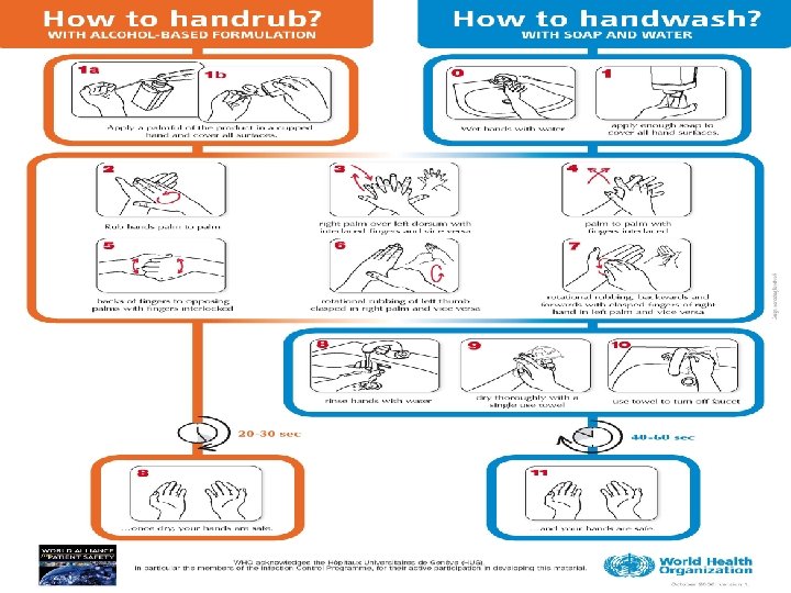
How to clean your hands • Handrubbing with alcohol-based handrub is the preferred routine method of hand hygiene if hands are not visibly soiled • Handwashing with soap and water – essential when hands are visibly dirty or visibly soiled (following exposure to body fluids)

Hand hygiene and glove use • The use of gloves does not replace the need to clean the hands • Remove gloves to perform Hand hygiene, when an indication occurs while wearing gloves • Wear gloves only when indicated, otherwise they become a major risk for germ transmission

Types of Isolation Precautions • Standard precautions • Transmission-based precautions – Contact precautions – Airborne precautions – Droplet precautions 49

TRANSMISSION-BASED PRECAUTIONS Contact Precautions • Infections spread by direct or indirect contact with patients or patient-care environment –C. difficle, MRSA, VRE, ESBL, CRE and MDR GNR • Limit patient movement • Private/SINGLE room or cohort with patients with same infection • Wear disposable gown and gloves when entering the patient room • Remove and discard used gown and gloves inside the patient room • Wash hands immediately after leaving the patient room • Use dedicated equipment if possible (e. g. , stethoscope) 50

Contact precautions signs 51

Droplet Precautions • Reduce the risk of transmission by large particle droplets (larger than 5 m in size). • Requires close contact between the source person and the recipient • Droplets usually travel 3 feet or less • E. g. , influenza, MERS-Co. V, other respiratory viruses, rubella, parvovirus B 19, mumps, H. influenzae, and N. meningitidis 52

Droplet Precautions cont. • A private/single room or • Cohort with patient with active infection with same microorganism • Use a mask when entering the room especially within 3 feet of patient • Limit movement and transport of the patient. Use a mask on the patient if they need to be moved and follow respiratory hygiene/cough etiquette 53

Droplet precautions signs 54
 • Place the patient in")
Airborne Precautions • Tuberculosis, measles, varicella, MERS-Co. V (severe) • Place the patient in an airborne infection isolation room (AIIR) • Negative Pressure should be monitored with visible indicator • Use of respiratory protection (e. g. , fit tested N 95 respirator) or powered airpurifying respirator (PAPR) when entering the room • Limit movement and transport of the patient. Use a mask on the patient if they need to be moved • Keep patient room door closed, do not open anteroom door till other door closed 55

Airborne precautions signs 56

Quiz 36 Y/O man with indwelling urinary catheter for past 3 months after a motor vehicle accident, his nurse noticed the urine output to be a little turbid. He has no fever, no dysuria, no lower abdominal pain. His CBC and renal functions are normal, Urine culture grew more than 100, 000 colonies of E. Coli, susceptible to all beta-lactams, fluoroquinolones and TMP-SMX. • You recommend: A) B) C) D) Start ceftriaxone 1 gm IV od Start ciprofloxacin 500 mg pp Q 12 h Remove catheter and start TMP-SMX 960 mg po q 12 h

Quiz • 30 Y/O man underwent knee ligament tear repair 2 weeks ago; he now presents to clinic with 5 days of opening gap of surgical scar with pussy discharge. He has no fever, WBC 15, platelets 450, ESR 80, creatinine 70, culture from pus grew MRSA resistant to tetracycline TMP-SMX and clindamycin • You recommend • A) Clindamycin 300 mg po q 8 h • B)TMP-SMX 960 mg po q 12 h • C) Ciprofloxacin 500 mg po q 12 h • D) Linezolid 600 mg po q 12 h

Quiz 22 years old lady in ICU for past 6 months has tracheostomy for ventilation, she is on 2 L O 2, with minimal sputum, has no fever, CXR normal, the RT send a sputum culture which grows Pseudomonas aeruginosa susceptible to ceftazidime, meropenem, ciprofloxacin • You recommend: A) Ceftazidime B) Meropenem C) Moxifluxacin D) Change tracheostomy tube

- Slides: 60