
Hormones Affecting Female Breast By the end of
Hormones Affecting Female Breast By the end of this lecture, students should be able to describe: 1. Know the physiologic anatomy of the breast. 2. Describe the physiological changes that occur in the breast during mammogenesis, lactogenesis, and galactopoeisis and the hormones involved. 3. Recognize the phases of lactogenesis. 4. Describe the endocrine and autocrine control of lactation. 5. Explain the physiological basis of suckling reflex and its role in lactation. 2
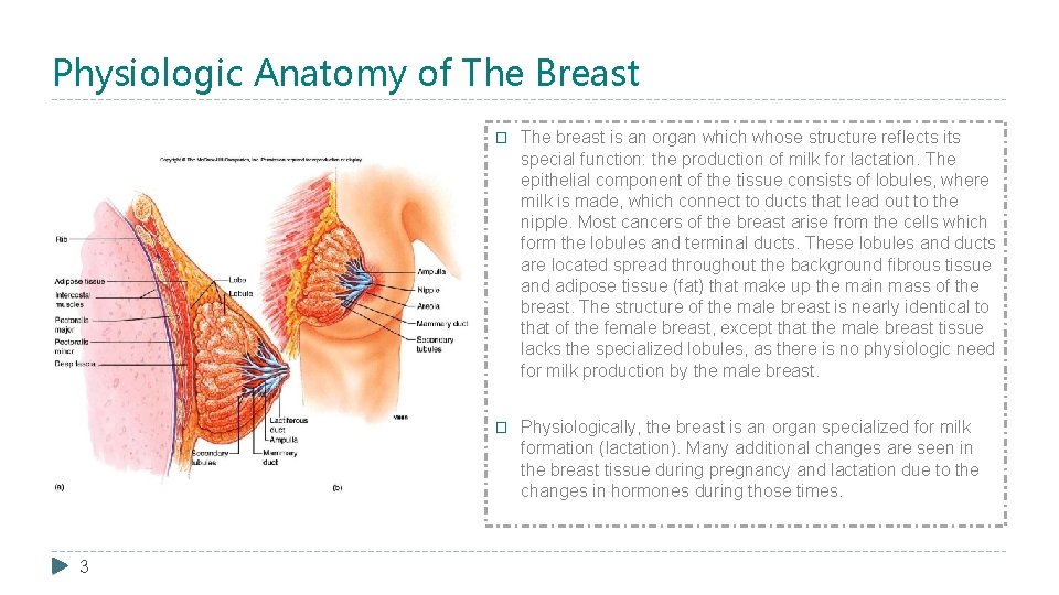
Physiologic Anatomy of The Breast 3 � The breast is an organ which whose structure reflects its special function: the production of milk for lactation. The epithelial component of the tissue consists of lobules, where milk is made, which connect to ducts that lead out to the nipple. Most cancers of the breast arise from the cells which form the lobules and terminal ducts. These lobules and ducts are located spread throughout the background fibrous tissue and adipose tissue (fat) that make up the main mass of the breast. The structure of the male breast is nearly identical to that of the female breast, except that the male breast tissue lacks the specialized lobules, as there is no physiologic need for milk production by the male breast. � Physiologically, the breast is an organ specialized for milk formation (lactation). Many additional changes are seen in the breast tissue during pregnancy and lactation due to the changes in hormones during those times.
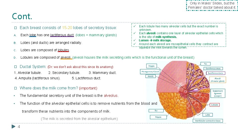
Only in Males’ Slides, but the Females’ doctor talked about it. Cont. ü Each lobule has many alveolar cells but the exact number is unknown. Each alveoli contains one layer of alveolar epithelial cells which is the site of milk synthesis. Lumen milk storage. Around each alveoli are myoepithelial cells they contract are squeeze the milk towards the lumen. � Each breast consists of 15 -20 lobes of secretory tissue: A. Each lobe has one lactiferous duct. (lobes = mammary glands) B. Lobes (and ducts) are arranged radially. C. Lobes are composed of lobules. D. Lobules are composed of alveoli. (alveoli houses the milk secreting cells which is the functional unit of the breast) � Ductal System: (Dr: we don’t ask about this since its anatomy) 1. Alveolar tubule. 2. Secondary tubule. 4. Ampulla (lactiferous sinus). ü ü ü 3. Mammary duct. 5. Lactiferous duct. � Where does the milk come from? (important) • The fundamental secretory unit of the breast is the alveolus. • The function of the alveolar epithelial cells is to remove nutrients from the blood and transform these nutrients into the components of milk. (The milk is secreted from the alveolar epithelium) 4
Only in Females’ Slides Theories � Uterine milk theory: Vessel connecting the uterus to the breast (diversion of menstrual blood to the breast). � Chyle theory: Milk is derived directly from chyle (milky fluid of emulsified fat absorbed from the intestinal tract into lymphatic system). � Synthesis theory: Milk is formed from substrates carried to the gland in the blood. 5
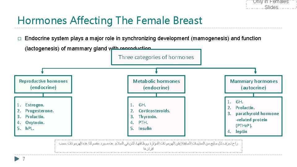
Only in Females’ Slides Hormones Affecting The Female Breast � Endocrine system plays a major role in synchronizing development (mamogenesis) and function (lactogenesis) of mammary gland with reproduction. Three categories of hormones Reproductive hormones (endocrine) 1. 2. 3. 4. 5. Estrogen. Progesterone. Prolactin. Oxytocin. h. PL. Metabolic hormones (endocrine) 1. 2. 3. 4. 5. GH. Corticosteroids. Thyroxin. PTH. Insulin Mammary hormones (autocrine) 1. GH. 2. Prolactin. 3. parathyroid hormone -related protein (PTHr. P). 4. leptin ﻟﻜﻦ ﻓﻲ ﺍﻟﺴﻼﻳﺪ ﻫﺬﻩ ﻣﺠﺮﺩ ﻣﻘﺴﻢ ﻟﻨﺎ ﻫﺬﻩ ﺍﻟﻬﺮﻣﻮﻧﺎﺕ ﺣﺴﺐ ، ﺭﺍﺡ ﻧﻌﺮﻑ ﻛﻞ ﺳﺘﻴﺞ ﻣﻦ ﺍﻟﺴﺘﻴﺠﺎﺕ ﺍﻟﺴﺎﺑﻘﺔ ﺇﻳﺶ ﺍﻟﻬﺮﻣﻮﻧﺎﺕ ﺍﻟﻤﺆﺜﺮﺓ ﻭﻭﻇﺎﺋﻔﻬﺎ ﺍﻓﺮﺍﺯﻫﺎ 7
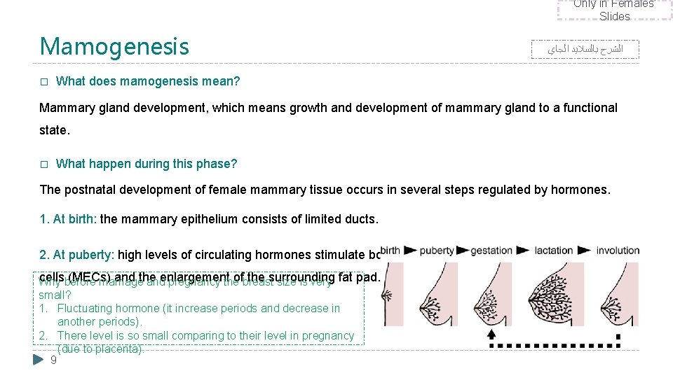
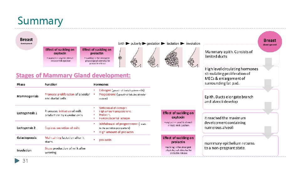
Only in Females’ Slides Mamogenesis � ﺍﻟﺸﺮﺡ ﺑﺎﻟﺴﻼﻳﺪ ﺍﻟﺠﺎﻱ What does mamogenesis mean? Mammary gland development, which means growth and development of mammary gland to a functional state. � What happen during this phase? The postnatal development of female mammary tissue occurs in several steps regulated by hormones. 1. At birth: the mammary epithelium consists of limited ducts. 2. At puberty: high levels of circulating hormones stimulate both the proliferation of the mammary epithelial cellsbefore (MECs) and the ofbreast the surrounding Why marriage andenlargement pregnancy the size is very fat pad. small? 1. Fluctuating hormone (it increase periods and decrease in another periods). 2. There level is so small comparing to their level in pregnancy (due to placenta). 9
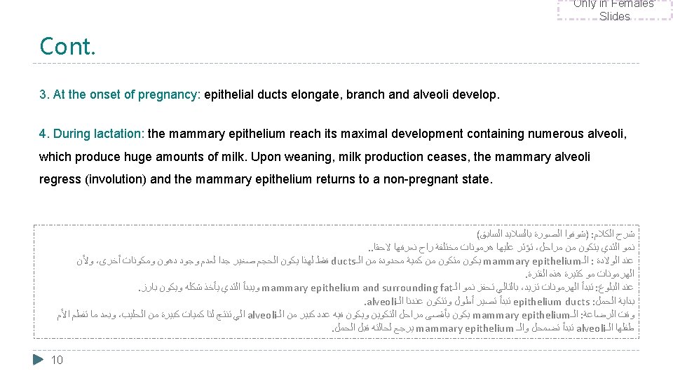
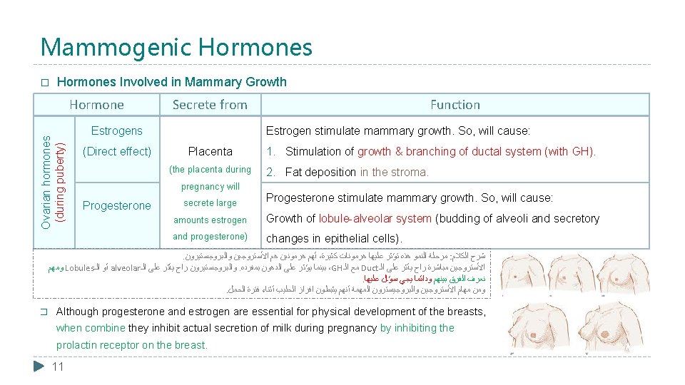
Mammogenic Hormones � Hormones Involved in Mammary Growth Ovarian hormones (during puberty) Hormone Secrete from Estrogens (Direct effect) Estrogen stimulate mammary growth. So, will cause: Placenta (the placenta during pregnancy will Progesterone Function secrete large 1. Stimulation of growth & branching of ductal system (with GH). 2. Fat deposition in the stroma. Progesterone stimulate mammary growth. So, will cause: amounts estrogen Growth of lobule-alveolar system (budding of alveoli and secretory and progesterone) changes in epithelial cells). . ﺃﻬﻢ ﻫﺮﻣﻮﻧﻴﻦ ﻫﻢ ﺍﻷﺴﺘﺮﻭﺟﻴﻦ ﻭﺍﻟﺒﺮﻭﺟﺴﺘﻴﺮﻭﻥ ، ﻣﺮﺣﻠﺔ ﺍﻟﻨﻤﻮ ﻫﺬﻩ ﺗﺆﺜﺮ ﻋﻠﻴﻬﺎ ﻫﺮﻣﻮﻧﺎﺕ ﻛﺜﻴﺮﺓ : ﺷﺮﺡ ﺍﻟﻜﻼﻡ ﻭﻣﻬﻢ Lobules ﺃﻮ ﺍﻟـ alveolar ﻭﺍﻟﺒﺮﻭﺟﺴﺘﻴﺮﻭﻥ ﺭﺍﺡ ﻳﺄﺜﺮ ﻋﻠﻰ ﺍﻟـ. ﺑﻴﻨﻤﺎ ﻳﺆﺜﺮ ﻋﻠﻰ ﺍﻟﺪﻫﻮﻥ ﺑﻤﻔﺮﺩﻩ ،GH ﻣﻊ ﺍﻟـ Duct ﺍﻷﺴﺘﺮﻭﺟﻴﻦ ﻣﺒﺎﺷﺮﺓ ﺭﺍﺡ ﻳﺄﺜﺮ ﻋﻠﻰ ﺍﻟـ . ﻧﻌﺮﻑ ﺍﻟﻔﺮﻕ ﺑﻴﻨﻬﻢ ﻭﺩﺍﺋﻤﺎ ﻳﺠﻲ ﺳﺆﺎﻝ ﻋﻠﻴﻬﺎ . ﻭﻣﻦ ﻣﻬﺎﻡ ﺍﻷﺴﺘﺮﻭﺟﻴﻦ ﻭﺍﻟﺒﺮﻭﺟﻴﺴﺘﺮﻭﻥ ﺍﻟﻤﻬﻤﺔ ﺃﻨﻬﻢ ﻳﺜﺒﻄﻮﻥ ﺍﻓﺮﺍﺯ ﺍﻟﺤﻠﻴﺐ ﺃﺜﻨﺎﺀ ﻓﺘﺮﺓ ﺍﻟﺤﻤﻞ � Although progesterone and estrogen are essential for physical development of the breasts, when combine they inhibit actual secretion of milk during pregnancy by inhibiting the prolactin receptor on the breast. 11
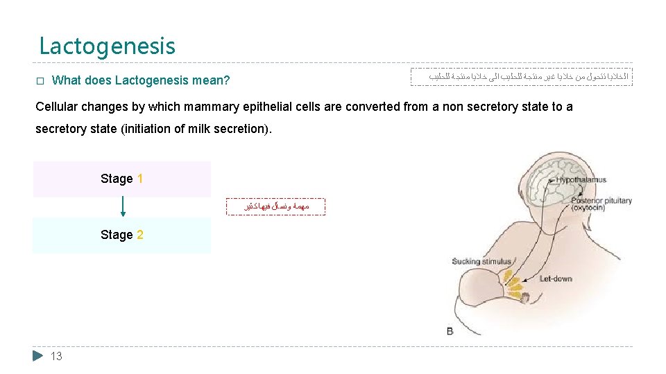
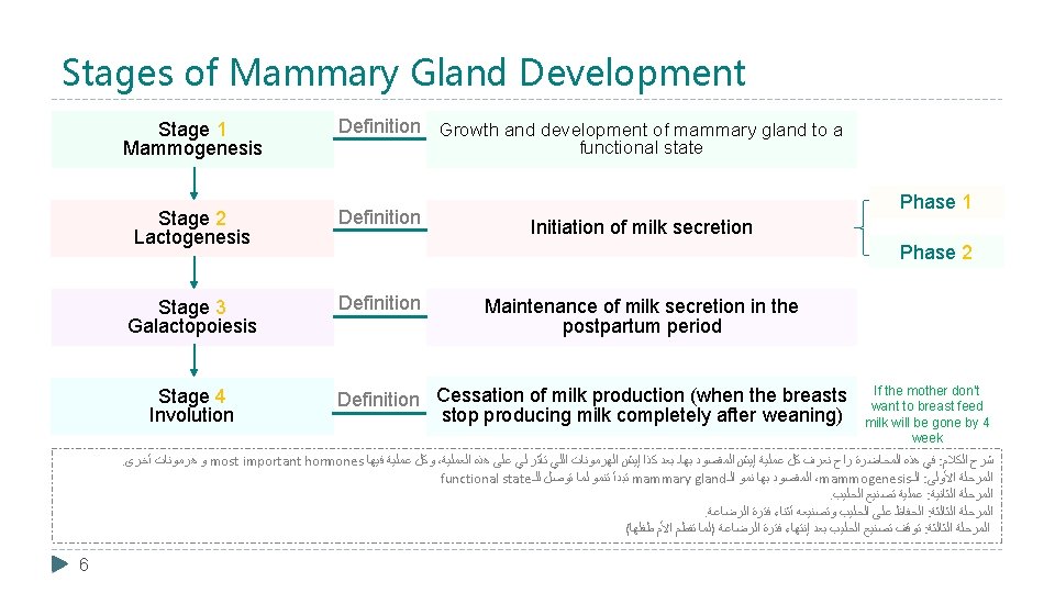
Lactogenesis � ﺍﻟﺨﻼﻳﺎ ﺗﺘﺤﻮﻝ ﻣﻦ ﺧﻼﻳﺎ ﻏﻴﺮ ﻣﻨﺘﺠﺔ ﻟﻠﺤﻠﻴﺐ ﺍﻟﻰ ﺧﻼﻳﺎ ﻣﻨﺘﺠﺔ ﻟﻠﺤﻠﻴﺐ What does Lactogenesis mean? Cellular changes by which mammary epithelial cells are converted from a non secretory state to a secretory state (initiation of milk secretion). Stage 1 ﻣﻬﻤﺔ ﻭﻧﺴﺄﻞ ﻓﻴﻬﺎ ﻛﺜﻴﺮ Stage 2 13
ﻣﻬﻤﺔ ﻭﻧﺴﺄﻞ ﻓﻴﻬﺎ ﻛﺜﻴﺮ Cont. Stage 1: : ﺷﺮﺡ ﺍﻟﻜﻼﻡ ﺍﻷﻮﻟﻰ ﻣﻦ ﺗﺼﻨﻴﻊ ﺍﻟﺤﻠﻴﺐ ﻳﺒﺪﺃ ﻓﻲ phase ﺍﻟـ � When does this phase start? . ﻣﻨﺘﺼﻒ ﺍﻟﺤﻤﻞ ﻭﻳﻜﻮﻥ ﻓﻴﻬﺎ ﺗﺼﻨﻴﻊ ﺍﻟﺠﻴﻨﺎﺕ ﺍﻟﻤﺨﺼﺼﺔ ﻟﺘﻜﻮﻳﻦ Starts in mid pregnancy until labor. (If abortion happen after mid stage there will be still milk) ﺍﻟﺤﻠﻴﺐ ﻭﻋﺸﺎﻥ ﻳﺼﻴﺮ ﻫﺬﺍ ﺍﻟﺘﺼﻨﻴﻊ ﺗﺤﺘﺎﺝ ، ﺍﻟﺨﻠﻴﺔ ﺍﻧﻬﺎ ﺗﻤﺘﺺ ﻛﻞ ﺍﻟﻤﻐﺬﻳﺎﺕ ﻣﻦ ﻛﺎﻟﺴﻴﻮﻡ � What happen during this phase? . amino acids ﺍﻟﺠﻠﻮﻛﻮﺯ ﻭﺍﻟـ 1. Histological and enzymatic differentiation of alveolar epithelial cells. 2. Expression of many genes involved in synthesis of milk components → increase in the uptake transport systems for amino acids, glucose, and calcium required for milk synthesis. � What are the hormone involved in this phase? 14 1. Progesterone (suppresses milk secretion): inhibit further differentiation by high levels of progesterone from the placenta. (and estrogen! as we said progesterone alone cannot cause inhibition) 2. Prolactin and/or placental lactogen: stimulates mammary secretory cells to produce milk. 3. Growth hormone 4. Glucocorticoids (Cortisol)
ﻣﻬﻤﺔ ﻭﻧﺴﺄﻞ ﻓﻴﻬﺎ ﻛﺜﻴﺮ Cont. . ﺑﺲ ﺍﻟﻔﺮﻕ ﺃﻦ ﺍﻷﻮﻟﻰ ﻣﺎﻓﻴﻬﺎ ﺇﻓﺮﺍﺯ ﺣﻠﻴﺐ ، ﺍﻟﻤﺮﺣﻠﺔ ﺍﻷﻮﻟﻰ ﻭﺍﻟﺜﺎﻧﻴﺔ ﻣﺘﺪﺍﺧﻠﻴﻦ Stage 2: � ﺍﻟﺸﺮﺡ ﺑﺎﻟﺴﻼﻳﺪ ﺍﻟﺠﺎﻱ When does this phase start? Around parturition (starts 2 -3 days postpartum). (postpartum = after labor) 1 st day after labor: milk secretion never occur due to still high estrogen and progesterone, these two hormones needs time for clearance (2 -3 days). � What happen during this phase? 1. Copious secretion of all milk components. 2. Withdrawal of progesterone + Estrogens (due to loss of placenta which was the main source) & increase of prolactin level which will lead to: • Further increase in expression of milk protein genes. • Glands absorb increased quantities of metabolic substrates from the blood. Due to the presence of • Movement of cytoplasmic lipid droplets and casein into alveolar Lumina. • Transfer of immunoglobulins. • 15 The colostrum is very good, why? immunoglobulin. ﺗﺨﻴﻠﻮﺍ ﺍﻟﺤﻠﻴﺐ ﻗﺎﻋﺪ ﻳﺘﺼﻨﻊ ﺑﺎﻟﺜﺪﻱ ﻓﺘﺮﺓ ﺍﻟﺤﻤﻞ ﻣﻦ ﻏﻴﺮ ﻣﺎ ﻭﻳﺒﺪﺃ ﺍﻟﺒﺮﻭﺗﻴﻦ ﻳﺘﻜﻮﻥ ﻭﻳﺘﺮﺍﻛﻢ ﻭﻳﺘﺼﻨﻊ ﺑﺮﻭﺗﻴﻦ ﺟﺪﻳﺪ ، ﻳﻄﻠﻊ ﺷﻬﻮﺭ ﻭﻫﻮ ﻣﺘﻜﺪﺱ ﻭﻳﺘﺮﺍﻛﻢ ﻓﺎﻟﻨﺘﻴﺠﺔ ﻟﻤﺎ ﻳﻄﻠﻊ ٥ ﺗﻘﺮﻳﺒﺎ Finally after all that steps there is secretion of colostrum followed by pure milk. . ﻳﻜﻮﻥ ﻟﺰﺝ
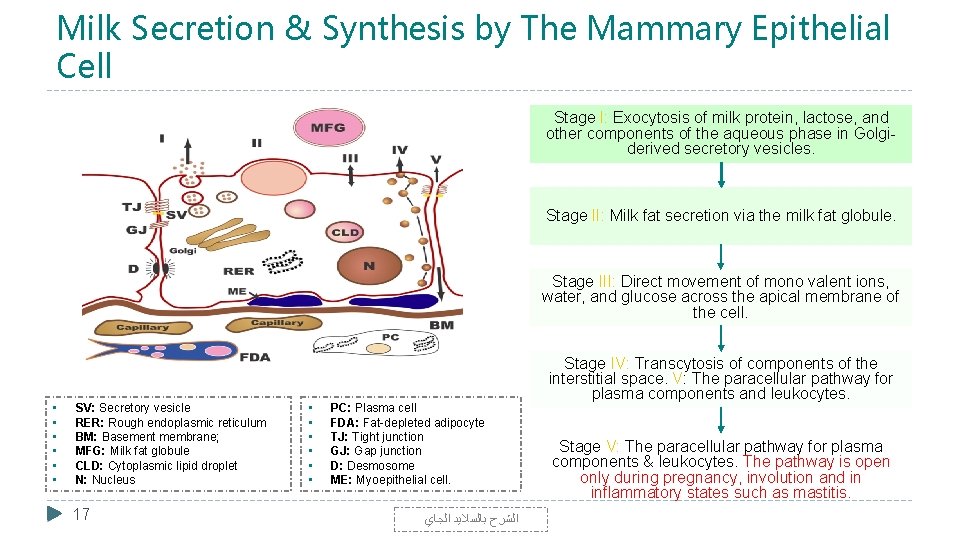
Milk Secretion & Synthesis by The Mammary Epithelial Cell Stage I: Exocytosis of milk protein, lactose, and other components of the aqueous phase in Golgiderived secretory vesicles. Stage II: Milk fat secretion via the milk fat globule. Stage III: Direct movement of mono valent ions, water, and glucose across the apical membrane of the cell. • • • SV: Secretory vesicle RER: Rough endoplasmic reticulum BM: Basement membrane; MFG: Milk fat globule CLD: Cytoplasmic lipid droplet N: Nucleus 17 • • • PC: Plasma cell FDA: Fat-depleted adipocyte TJ: Tight junction GJ: Gap junction D: Desmosome ME: Myoepithelial cell. ﺍﻟﺸﺮﺡ ﺑﺎﻟﺴﻼﻳﺪ ﺍﻟﺠﺎﻱ Stage IV: Transcytosis of components of the interstitial space. V: The paracellular pathway for plasma components and leukocytes. Stage V: The paracellular pathway for plasma components & leukocytes. The pathway is open only during pregnancy, involution and in inflammatory states such as mastitis.
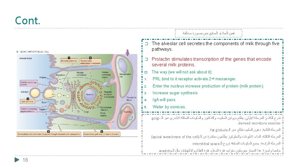
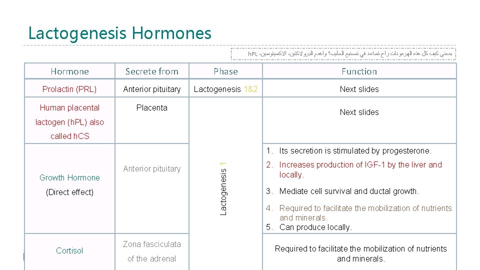
Lactogenesis Hormones h. PL ، ﺍﻻﻛﺴﻴﺘﻮﺳﻴﻦ ، ﺑﻤﻌﻨﻰ ﻛﻴﻒ ﻛﻞ ﻫﺬﻩ ﺍﻟﻬﺮﻣﻮﻧﺎﺕ ﺭﺍﺡ ﺗﺴﺎﻋﺪ ﻓﻲ ﺗﺼﻨﻴﻊ ﺍﻟﺤﻠﻴﺐ؟ ﻭﺍﻫﻤﻢ ﺍﻟﺒﺮﻭﻻﻛﺘﻴﻦ Hormone Secrete from Phase Function Prolactin (PRL) Anterior pituitary Lactogenesis 1&2 Next slides Human placental Placenta Next slides lactogen (h. PL) also called h. CS Anterior pituitary Growth Hormone (Direct effect) 19 Cortisol Zona fasciculata of the adrenal Lactogenesis 1 1. Its secretion is stimulated by progesterone. 2. Increases production of IGF-1 by the liver and locally. 3. Mediate cell survival and ductal growth. 4. Required to facilitate the mobilization of nutrients and minerals. 5. Can produce locally. Required to facilitate the mobilization of nutrients and minerals.
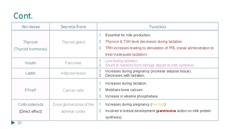
Cont. Hormone Secrete from Function 1. Essential for milk production. Thyroxin Thyroid gland 2. Thyroxin & TSH level decreases during lactation. 3. TRH increases leading to stimulation of PRL (nasal administration to (Thyroid hormones) treat inadequate lactation). Insulin Pancreas Leptin Adipose tissue 1. Low during lactation. 2. Shunt of nutrients from storage depots to milk synthesis. 1. Increases during pregnancy (increase adipose tissue). 2. Decreases with lactation. 1. Increases during lactation. PTHr. P Cancer cells 2. Mobilizes bone calcium. 3. Increase in alkaline phosphatase. Corticosteroids Zona glumerulosa of the (Direct effect) adrenal cortex 1. Increases during pregnancy (five fold). 2. Involved in breast development (permissive action on milk protein synthesis). 20
Cont. Prolactin (PRL) Hormone � Where the hormone secret from? Secreted from the anterior pituitary gland (Lactotrophs). � Level: • Its level rises steadily (gradually) from the 5 th week of pregnancy until birth (10 -20 times the nonpregnant level). Enhanced by Estrogen. • � Effect: ﺃﻲ ﻟﻪ ﺃﻜﺜﺮ ﻣﻦ ﻣﻬﻤﺔ ، ﺍﻟﺒﺮﻭﻻﻛﺘﻴﻦ ﻳﺸﺘﻐﻞ ﻋﻠﻰ ﺍﻟﻨﻤﻮ ﻭﺍﻟﺘﺼﻨﻴﻊ 1. It has mammogenic, lactogenic and galactopoietic effects (Its main function is milk production). 2. It stimulates expression of genes that encode several milk components: casein, lactalbumin, lactose and lipids. 3. Sudden drop in estrogen & progesterone after delivery allows milk production. 4. Required to facilitate the mobilization of nutrients and minerals. � Inhibited/ Stimulated by: 1. It is inhibited mainly by hypothalamic hormone (Dopamine). 2. It can be stimulated by Thyrotropin-releasing hormone (TRH). 21
Cont. Human Placental Lactogen (h. PL) � Where the hormone secret from? Secreted by the placenta at about the 5 th week of pregnancy. � Level: Its secretion increases progressively throughout the remainder of pregnancy in direct proportion to the weight of the placenta. � Effect: 1. Causes at least partial development of the animal’s breasts and in some instances causes lactation. 2. Facilitates growth of mammary glands, supports prolactin during pregnancy (lactogenic properties). 3. It has weak actions similar to those of growth hormone. 4. It decreases maternal insulin sensitivity, decreases utilization of glucose, and promotes the release of free fatty acids (metabolic effect). (takes glucose from mother to fetus which can cause gestational diabetes) 5. Required to facilitate the mobilization of nutrients and minerals. 22
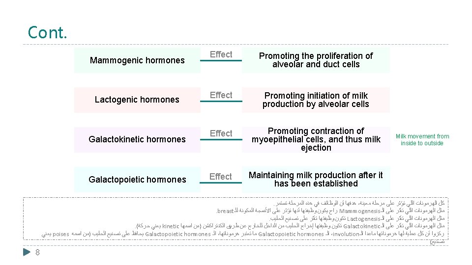
Galactokinetic & Galactopoeisis � What are the hormone involved hormone in Galactokinetic phase? 1. Oxytocin (OT) (mainly) Vasopressin (its has similar structure to oxytocin so it binds with oxytocin receptors and produces the same action) 2. � What does Galactopoeisis mean? Galactopoeisis is defined as the maintenance of lactation once lactation has been established. � When does the Galactopoeisis start? Starts 9 -15 days postpartum. � What are the hormone involved hormone in Galactopoeisis phase? Slide 25. 23
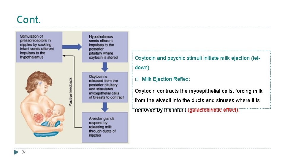
Cont. Oxytocin and psychic stimuli initiate milk ejection (letdown) � Milk Ejection Reflex: Oxytocin contracts the myoepithelial cells, forcing milk from the alveoli into the ducts and sinuses where it is removed by the infant (galactokinetic effect). 24
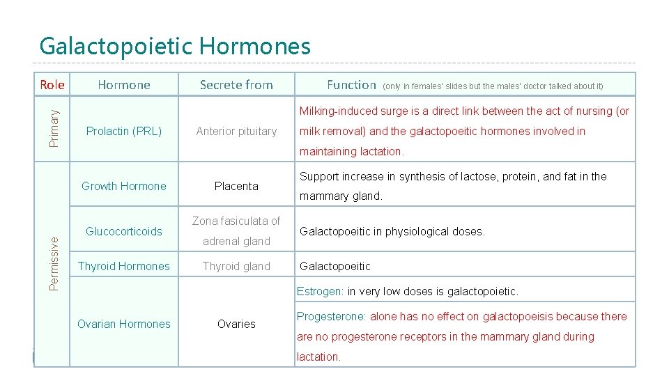
Galactopoietic Hormones Primary Role Hormone Function (only in females’ slides but the males’ doctor talked about it) Milking-induced surge is a direct link between the act of nursing (or Prolactin (PRL) Anterior pituitary milk removal) and the galactopoeitic hormones involved in maintaining lactation. Growth Hormone Permissive Secrete from Glucocorticoids Thyroid Hormones Placenta Zona fasiculata of adrenal gland Thyroid gland Support increase in synthesis of lactose, protein, and fat in the mammary gland. Galactopoeitic in physiological doses. Galactopoeitic Estrogen: in very low doses is galactopoietic. Ovarian Hormones Ovaries Progesterone: alone has no effect on galactopoeisis because there are no progesterone receptors in the mammary gland during 25 lactation.
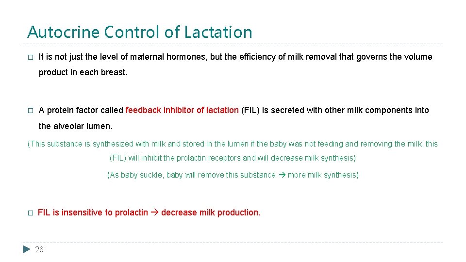
Autocrine Control of Lactation � It is not just the level of maternal hormones, but the efficiency of milk removal that governs the volume product in each breast. � A protein factor called feedback inhibitor of lactation (FIL) is secreted with other milk components into the alveolar lumen. (This substance is synthesized with milk and stored in the lumen if the baby was not feeding and removing the milk, this (FIL) will inhibit the prolactin receptors and will decrease milk synthesis) (As baby suckle, baby will remove this substance more milk synthesis) � FIL is insensitive to prolactin decrease milk production. 26
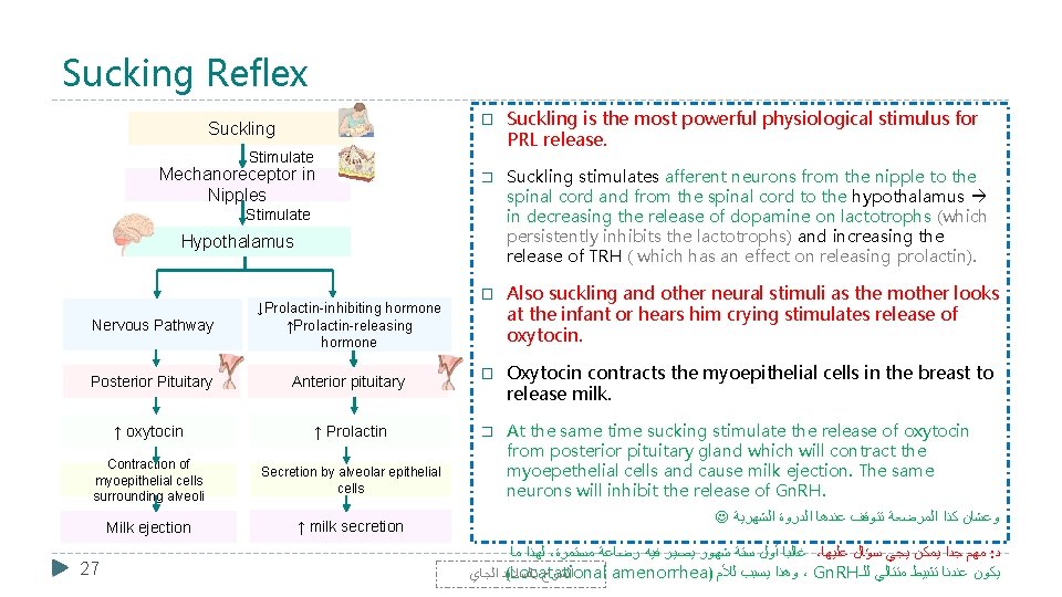
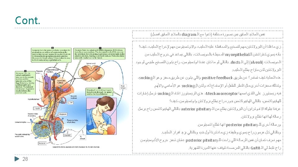
Sucking Reflex � Suckling Stimulate Mechanoreceptor in Nipples � Stimulate Hypothalamus Nervous Pathway ↓Prolactin-inhibiting hormone ↑Prolactin-releasing hormone Posterior Pituitary Anterior pituitary ↑ oxytocin ↑ Prolactin Contraction of myoepithelial cells surrounding alveoli Secretion by alveolar epithelial cells Milk ejection ↑ milk secretion 27 Suckling is the most powerful physiological stimulus for PRL release. Suckling stimulates afferent neurons from the nipple to the spinal cord and from the spinal cord to the hypothalamus in decreasing the release of dopamine on lactotrophs (which persistently inhibits the lactotrophs) and increasing the release of TRH ( which has an effect on releasing prolactin). � Also suckling and other neural stimuli as the mother looks at the infant or hears him crying stimulates release of oxytocin. � Oxytocin contracts the myoepithelial cells in the breast to release milk. � At the same time sucking stimulate the release of oxytocin from posterior pituitary gland which will contract the myoepethelial cells and cause milk ejection. The same neurons will inhibit the release of Gn. RH. ﻭﻋﺸﺎﻥ ﻛﺬﺍ ﺍﻟﻤﺮﺿﻌﺔ ﺗﺘﻮﻗﻒ ﻋﻨﺪﻫﺎ ﺍﻟﺪﺭﻭﺓ ﺍﻟﺸﻬﺮﻳﺔ ﻟﻬﺬﺍ ﻣﺎ ، ﻏﺎﻟﺒﺎ ﺃﻮﻝ ﺳﺘﺔ ﺷﻬﻮﺭ ﻳﺼﻴﺮ ﻓﻴﻪ ﺭﺿﺎﻋﺔ ﻣﺴﺘﻤﺮﺓ ، ﻣﻬﻢ ﺟﺪﺍ ﻳﻤﻜﻦ ﻳﺠﻲ ﺳﺆﺎﻝ ﻋﻠﻴﻬﺎ : ﺩ (Locatational amenorrhea) ﻭﻫﺬﺍ ﻳﺴﺒﺐ ﻟﻸﻢ ، Gn. RH ﻳﻜﻮﻥ ﻋﻨﺪﻧﺎ ﺗﺜﺒﻴﻂ ﻣﺘﺘﺎﻟﻲ ﻟﻠـ ﺑﺎﻟﺴﻼﻳﺪ ﺍﻟﺠﺎﻱ ﺍﻟﺸﺮﺡ
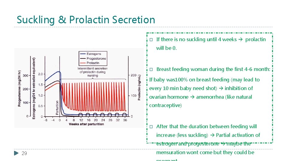
Suckling & Prolactin Secretion � If there is no suckling until 4 weeks prolactin will be 0. � Breast feeding woman during the first 4 -6 month: If baby was 100% on breast feeding (may lead to every 10 min baby need shot) inhibition of ovarian hormone amenorrhea (like natural contraceptive) � After that the duration between feeding will increase (less suckling) Partial activation of estrogen and progesterone maybe the 29 mensuration wont come but they could be
Milk Production � Milk production is a "use it or lose it" process. The more often and effectively the baby nurses, more milk will be produced. � Milk production <100 ml/day in day 1 postpartum. (because of levels of estrogen and progesterone) � Milk production by day 3 reaches 500 ml/day. (the levels of estrogen and progesterone have been strongly reduced) � Milk composition changes dramatically (↓Na+2 & Cl- ) due to closure of tight junctions that. use block pathway. it orparacellular lose it ﻳﺨﻀﻊ ﻟﻘﺎﻧﻮﻥ ﺇﻓﺮﺍﺯ ﺍﻟﺤﻠﻴﺐ AAP Recommendations about breast feeding: ﻳﻌﻨﻲ ﻓﻲ ﺣﺎﻝ ﺍﻷﻢ ﺭﺿﻌﺖ ﺍﻟﻄﻔﻞ ﻭﺣﺎﻓﻈﺖ ﻋﻠﻰ ﻭﺟﻮﺩ ﺍﻟﺤﻠﻴﺐ ﺭﺍﺡ ﻳﺴﺘﻤﺮ ﻭﺫﻟﻚ ﺑﺴﺒﺐ ﻟﻜﻦ ﻟﻮ ﺍﻷﻢ ﻣﺎ ﺭﺿﻌﺖ ﻟﻔﺘﺮﺓ ﺭﺍﺡ ، ﺍﻟﻠﻲ ﺗﻜﻠﻤﻨﺎ ﻋﻨﻪ positive feedback ﺍﺳﺘﻤﺮﺍﺭ ﺍﻟـ . ﻳﺘﻮﻗﻒ ﻋﻨﺪﻫﺎ ﺍﻟﺤﻠﻴﺐ ﻣﺎﺭﺍﺡ ﻳﺘﻮﻗﻒ ﻋﻨﺪﻫﺎ ﺍﻟﺤﻠﻴﺐ ﻟﻜﻦ ﺇﻓﺮﺍﺯﻩ ﺑﻴﻜﻮﻥ ، ﻭﻟﻮ ﻛﺎﻧﺖ ﺗﺮﺿﻊ ﻋﺸﻮﺍﺋﻲ ﺑﺲ ﺑﺪﻭﻥ ﺗﻮﻗﻒ . ﺑﻜﻤﻴﺎﺕ ﺃﻘﻞ � Exclusive breastfeeding for the first six months of life. � Continued breastfeeding for at least one year, ‘As long ﻥ ﺍﻟ ﺍ ﺍ ﺍ as ﺍﺍﺍ ﺍ is desired by mother and child’. . ﺻﺪﻕ ﺍﻟﻠﻪ ﺍﻟﻌﻈﻴﻢ ﻭﺗﻢ ﺑﻔﻀﻞ ﺍﻟﻠﻪ ﻭﺗﻮﻓﻴﻘﻪ ﻭﻓﻀﻠﻪ 30
Summary 31
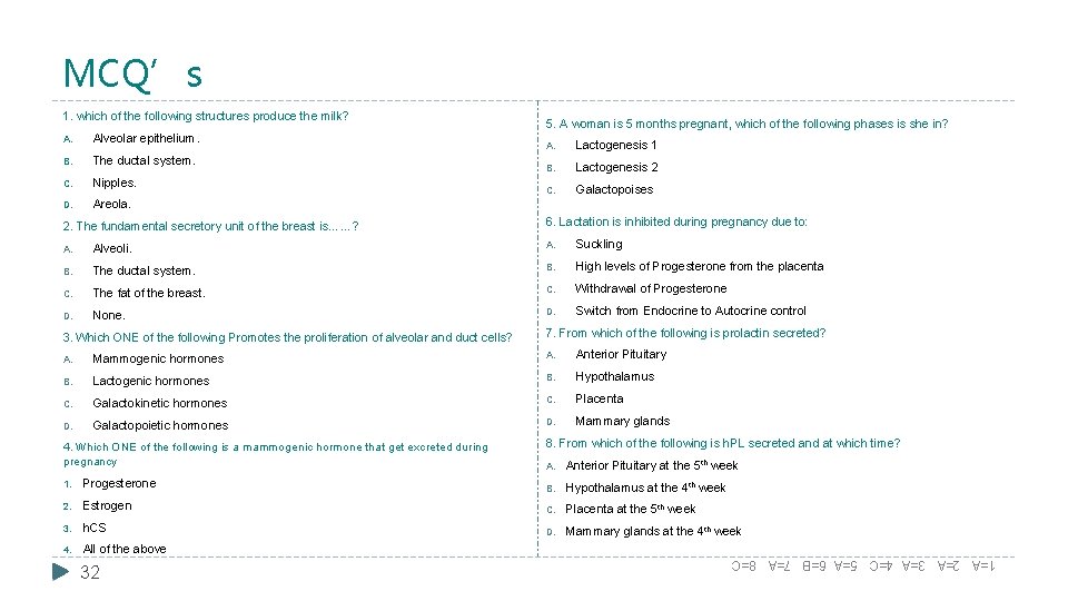
MCQ’s 1. which of the following structures produce the milk? A. Alveolar epithelium. B. The ductal system. C. Nipples. D. Areola. 2. The fundamental secretory unit of the breast is……? 5. A woman is 5 months pregnant, which of the following phases is she in? A. Lactogenesis 1 B. Lactogenesis 2 C. Galactopoises 6. Lactation is inhibited during pregnancy due to: A. Alveoli. A. Suckling B. The ductal system. B. High levels of Progesterone from the placenta C. The fat of the breast. C. Withdrawal of Progesterone D. None. D. Switch from Endocrine to Autocrine control 3. Which ONE of the following Promotes the proliferation of alveolar and duct cells? 7. From which of the following is prolactin secreted? A. Mammogenic hormones A. Anterior Pituitary B. Lactogenic hormones B. Hypothalamus C. Galactokinetic hormones C. Placenta D. Galactopoietic hormones D. Mammary glands 4. Which ONE of the following is a mammogenic hormone that get excreted during pregnancy 8. From which of the following is h. PL secreted and at which time? A. Anterior Pituitary at the 5 th week 1. Progesterone B. Hypothalamus at the 4 th week 2. Estrogen C. Placenta at the 5 th week 3. h. CS D. Mammary glands at the 4 th week 4. All of the above 1=A 2=A 3=A 4=C 5=A 6=B 7=A 8=C 32
- Slides: 34