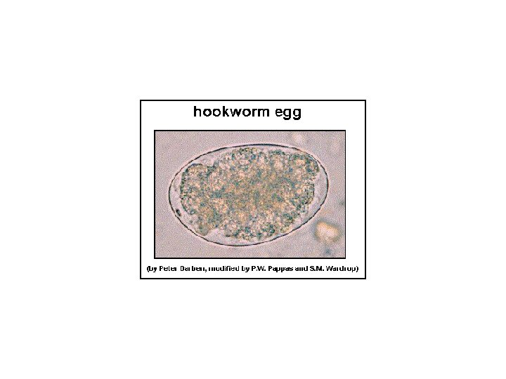
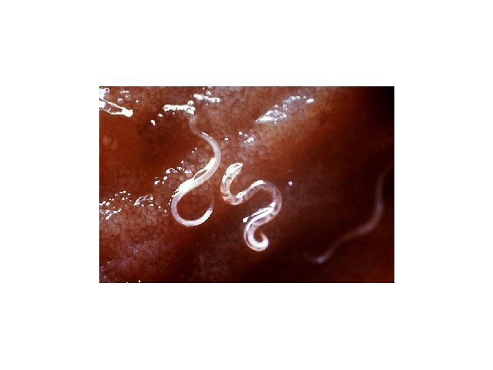
hookworm Hookworm hookworm hookworm Cutaneous larval migran hookworm


 C. Felis is the msot common flea found on")


 vertically or 13 inches")


")


")

")


 Rabbit Ear Mite")

 with")


, residing in the keratin layer of the skin and")

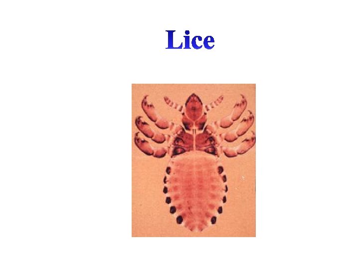
 and Anoplura (sucking lice) •")


")


- Slides: 88
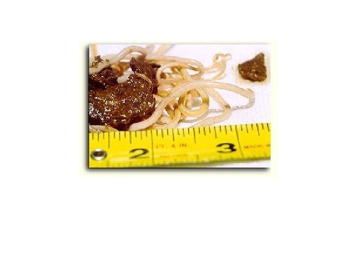
hookworm
Hookworm
hookworm
hookworm
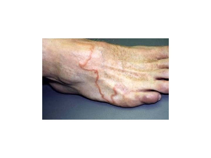
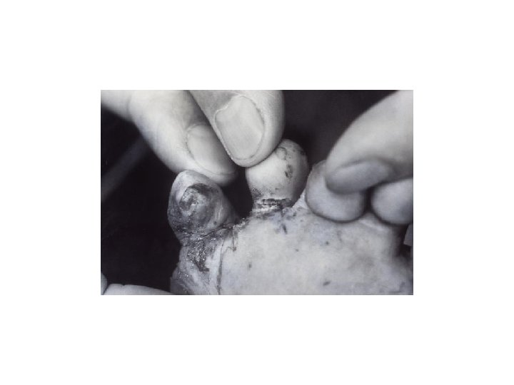
Cutaneous larval migran
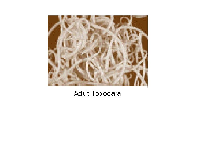
hookworm roundworm
roundworm hookworm
roundworm
roundworm
Roundworms
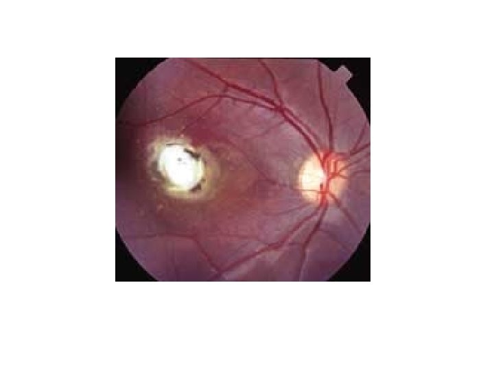
Ocular larval migran
Pulmonary larval migran
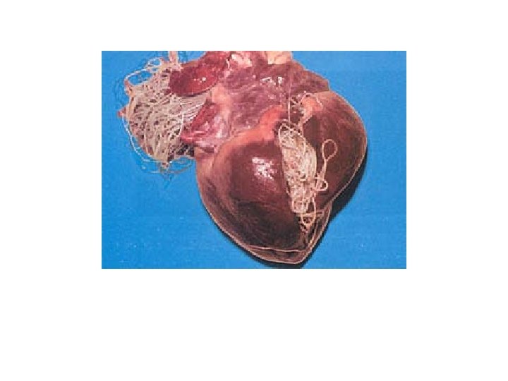
Dirofilaria immitis Microfilaria
whipworm
tapeworm
giardia
coccidia
coccidia
flea
Flea
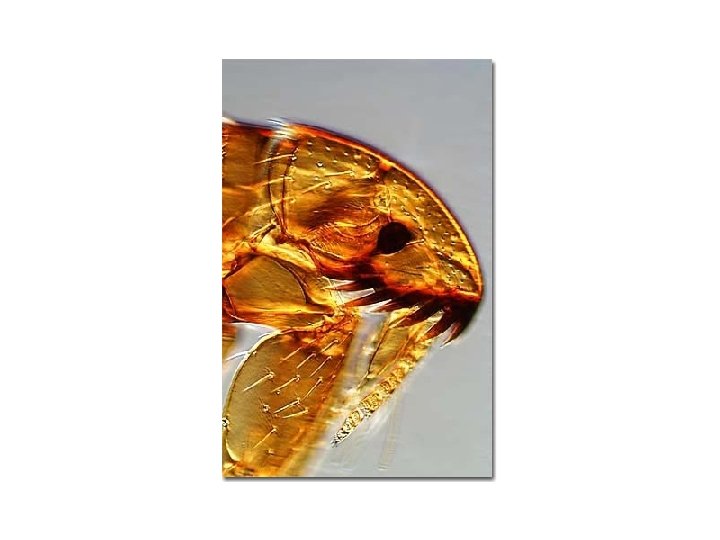
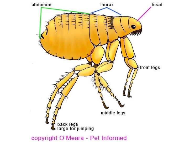
Ctenocephalides felis (the cat flea) C. Felis is the msot common flea found on dogs and cats. C. Canis is actually very uncommon and occurs less frequently on dogs than cats!!
• Fleas are tiny, but anyone who has seen one can usually recognize them with ease. • They're tiny, flat, wingless insects that have a knack for jumping away before you can catch them. • Their bodies are covered with hard plates called sclerites, so if you do catch one, squashing it can be a challenge. • Their hard outer shell protects fleas from everything from an animal's teeth to hitting the floor after a long jump. • Their flattened bodies and these backward-pointing hairs make it easy for fleas to crawl through their hosts' fur. If something tries to dislodge them, the hairs act like tiny Velcro anchors.
The mouth parts of the flea • Two sawlike laciniae cut the skin. They also fit together to form a saliva channel. • The epipharynx is like a needle. • The laciniae surround the epipharynx, and together they form the stylet, or puncturing organ. .
• As with all insects, a flea has three pairs of legs that attach to its thorax. • The back legs are very long, and the flea can bend them at several joints. • The flea bends its leg, and a pad of elastic protein called resilin stores energy the way a bowstring does. • A tendon holds the bent leg in place. When the flea releases this tendon, the leg straightens almost instantly, and the flea accelerates like a bolt from a crossbow. • As it lands, the flea uses tiny claws on the ends of its legs to grasp the surface under it.
A flea can jump about 7 inches (17. 8 centimeters) vertically or 13 inches (33 centimeters) horizontally. In human proportions, that's a 250 -foot (76 -meter) vertical jump or a 450 -foot (137 -meter) horizontal jump!!!!
The Life Cycle of the Flea
The Egg At any given time about one third of the flea population in someone’s home is present in the egg stage. The adult female flea lays up to 40 eggs daily. The eggs are laid on the host where they fall off to hatch in the environment. Eggs incubate best in high humidity and temperatures of 65 -80 degrees.
Larva At any given time about 57% of the fleas in someone’s home are in the larval stage. Larvae are like little caterpillars crawling around grazing on the flea dirt that is generally in their vicinity. (Flea eggs and flea dirt both fall off the host. When the eggs hatch, there is a bounty of food prepared lovingly by all the host’s fleas waiting for them). This is the stage that picks up tapeworm eggs (also likely to be in the vicinity) as they graze. The time between hatching and pupating (ie the time spent in the larval stage) depends on environmental conditions. It can be as short as 9 days.
Pupae By this life stage most young fleas have been killed off by an assortment of environmental factors. Only 8% make it to the pupal stage but once they have spun cocoons they are nearly invincible. The cocoon is sticky and readily picks up dust and dirt. Inside the developing cocoon, the pupa is turning into the flea that we are familiar with. They are especially protected under carpet, which is why carpet has developed such a reputation as a shelter for fleas. *The pupa can remain dormant in its cocoon for many months, maybe even up to a year as it waits for the right time to emerge.
Young Adult Flea After the pupa develops, it does not automatically emerge from its cocoon. Instead, it is able to remain in the cocoon until it detects a nearby host. The mature pupa is able to detect the vibrations of an approaching host, carbon dioxide gradients, and sound and light patterns. When the mature pupa feels the time is right, he emerges from the cocoon, hungry and eager to find a host.
A common scenario occurs when a dog is boarded while the owner is on vacation. The owner picks up the dog from the boarding kennel and returns home. The mature pupae have been waiting for a host and when the dog enters the home, a huge number of adult fleas emerge at once and attack the dog creating a sudden heavy infestation. Often the boarding kennel is blamed for giving the dog fleas. What really happened was that the pupae waited to emerge while there was no host present and then all emerged suddenly when the host arrived.
**The unfed flea is able to live for months without a blood meal but during that time it is aggressively using all its powers to locate a host. Once it finds a host, it will never purposely leave the host. Homeless & Hungry! Need a Hairy Dog
Flea allergy dermatitis
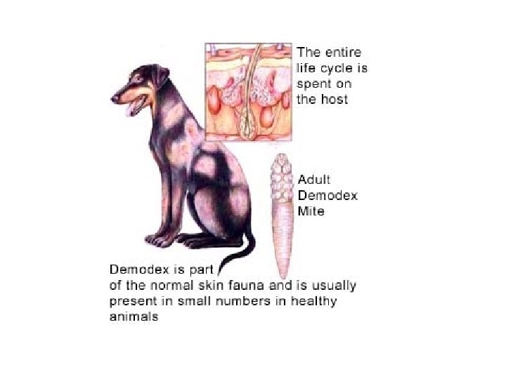
Demodectic Mange (Demodicosis or Red Mange)
• Demodicosis is caused by a microscopic, cigar-shaped skin parasite called Demodex. • This mite is normally found in low numbers in the hair follicles of all animals, including humans. • In dogs, the mites (Demodex canis) are passed from the dam to the puppy during the first few days of life. • Demodex becomes a problem when the mites multiply and proliferate inside the hair follicles. • Their massive numbers cause an inflammatory reaction and a secondary bacterial infection. • As a result, the hair follicles are destroyed.
• Demodex occurs in animals with depressed immune systems. • It is also seen in animals with serious underlying disorders. • Dogs with diabetes, Cushing's disease, and cancer often develop demodicosis. • Demodicosis is frequently seen in puppies and young dogs. • Ninety percent of the puppies diagnosed with Demodex improve spontaneously by eight months to three years of age. • This is the age when the dog's immune system reaches maturity. • Three forms of domodicosis are recognized in dogs: localized demodicosis, generalized demodicosis, and pododemodicosis.
Demodectic Mange Mite
Sarcoptes scabei (Scabies Mites)
Sarcoptse sacbei Mite • Almost every domestic species has its own distinct variety of this mite. • S. scabei var. canis (dogs) • Notedres cati (cats) • Very pruritic because burrow into skin. • Scaly, crusty, skin lesions develop on the ears, lateral elbows and ventral abdomen. Zoonosis Alert!!
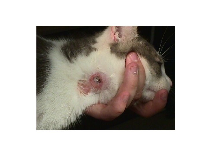
Ear mites (Otodectes cyonits)
Ear mite debris
• Common cause of otitis externa in dogs, cats and ferrets. • Occur primarily in the external canal, ear mites may be found anywhere on the body. • A common infestation site is the tail and head. • Mites are spread by direct contact. • Transmissible among and between canines and felines.
Ear Mite Egg
Ear Mite (Psorptes cuniculi) Rabbit Ear Mite
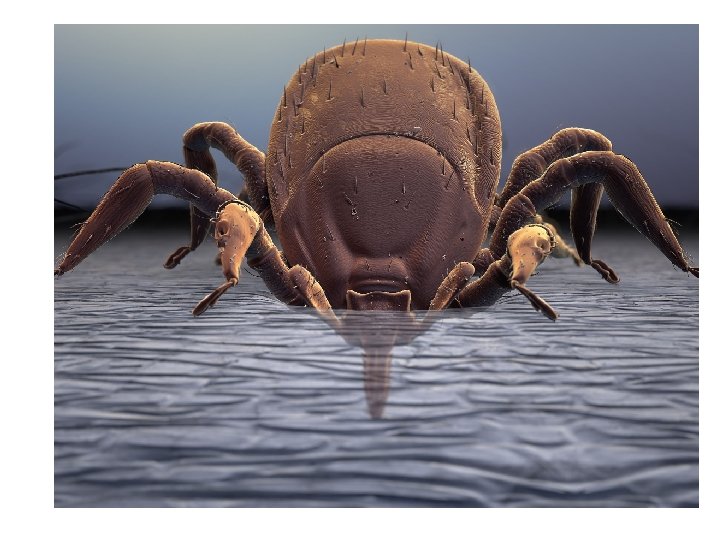
• Ticks are small to medium sized acarines (in the class Acarina) with dorsoventrally compressed, leathery bodies. • The head, or capitulum, serves as an organ of cutting and attachment. It is made of a penetrating anchorlike sucking organ, the hypostome; and four accessory appendages, two cutting chelicerae and two pedipalps, which act as sensors and supports when the tick fastens to the host’s body. • The body may be covered by a hard, chitinous plate, the scutum. • Most ticks are inornate; reddish or mahogany without markings. • Some species are ornate and have a distinctive white patterns on the dark scutum background • Adult ticks have 8 legs, with claws on the ends.
Ticks Engorged Tick
tick
• Ticks have a voracious blood-feeding activity. • They transmit many parasitic, bacterial, viral and other diseases, such as borreliosis (Lyme disease), among animals and from animals to humans. • The salivary secretions of some female ticks are toxic and can produce a syndrome known as “tick paralysis” in human beings and animals. • Tics are divided into two categories : Argasid – soft ticks Ixodid –hard ticks
Tick Life Cycle
• After engorging on a blood meal, the female ticks drop off the host and seek protected places, suck as within cracks and crevices or under leaves and branches to lay their eggs. • The six-legged larvar, or seed tick, hatch from the eggs and feed on a host. • The larva molts to the eight-legged nymphal stage, . • After one or two blood meals, the nymph matures and molts to the adult stage. • During the larval, numphal, and adult stages, ticks may infest many different host species. This plays an important role in the transmission of disease pathogens to many hosts.
Engorged Tick
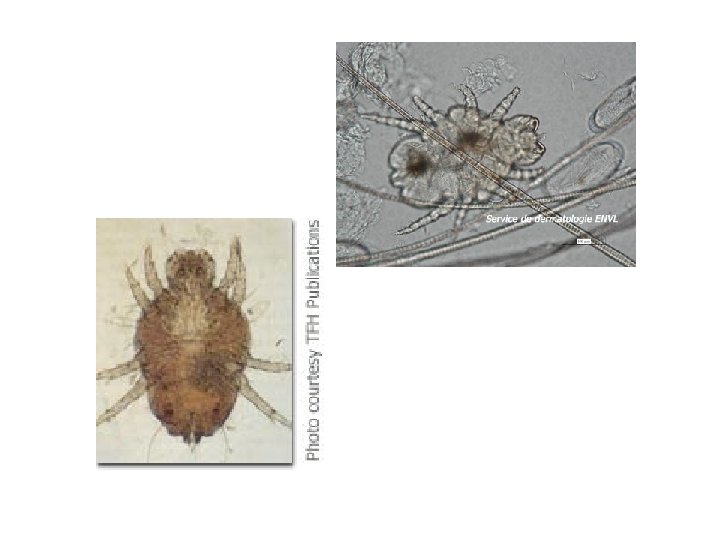
• Surface dwelling (nonburrowing), residing in the keratin layer of the skin and in the haircoat of the host. • Ingest keratin debris and fluids. • Hosts include the dog, cat and rabbit. • Distinct features : large, visible to the unaided eye. Microscopically, they have large hook-like mouthparts (palpi). • Body shape resembles a shield. • Appear as moving “dandruff” flakes alont the dorsal midline and head of the host.
These mites can temporarily infest humans causing skin irritation and some itching. In severe cases, some open lesions may occur. Zoonosis Alert!!
• Two orders exist : Mallophagia (biting lice) and Anoplura (sucking lice) • A “disease of neglect. ” • Some of the most prolific ectoparasites of domestic animals. • Infestation by lice is called pediculosis. • The eggs are called nits. • Dosoventraly flattened, wingless insects. • Transmitted by direct contact, but all life stages may be transmitted by fomites (inanimate objects such as blankets, brushes and other grooming equipment). • Species specific. Ex. Dog lice parasitize dogs only.
• Severe lice infestations can drop packed cell volumes as much as 10%to 20%, sometimes causing fatality. • Infested animals may be come more susceptible to other diseases and parasites and may succumb to stresses not ordinarily pathologic to uninfested animals.
Characteristically, the head of every chewing louse is wider than the widest portion of the thorax. The thorax contains 3 pairs of legs. They are smaller than sucking lice. Usually yellow and have a large, round head.
Hog Sucking Louse Sucking lice are red to gray (color depends on amount of blood ingested) Heads are narrower than widest portion of thorax. Have pincerlike claws.
Trichodectes canine biting louse
Hematopinus suis, the blood-sucking louse of swine
Note : Humans cannot get lice from pets. The pet cannot get lice from humans. Remember, SPECIES SPECIFIC!!
Flies
• The order Diptera includes many different species of flies. • Diptera are a large, complex order of insects. • As adults, most have only one pair of wings (di-(two), ptera(wings). • Diptera members vary insize, food source preference, and developmental stage that parasitizes the animal or produces lesions. • Dipterans may feed intermittently on vertebrate blood, saliva, tears of mucus. • As larvae, thay may develop in the subcutaneous tissues or within internal organs.
• Adult dipterans that make frequent visits to the vertebrate host to intermittently feed on blood are called periodic parasites. • Dipteran larvae that develop in the tissue or organs of the vetebrate host, produce a condition called myasis.
Cuterebra speceis (Wolf Warble)
• Larvae infest the skin of rabbits, squirrels, mice, rats, chipmunks, and occasionally, dogs and cats. • 2 nd stage larvae are grublike, 5 -10 mm ling and cream to grayish white in color. • 3 rd stage larvae are large, robust, black, and heavily spined. • Larval stages are usually found in swollen, cystlike subcutaneous sites, with a fistula or pore communicating to the outside environment. The larva breathes through this pore. • Larval sites are most commonly found on the neck and face, however they have been discovered in the eye.
**Important: Cuderebra larva must be carefully removed. Usually, the breathing hole is enlarged and the larva is removed with thumb forceps. If the larva is crushed during extraction, anaphylaxis may occur. http: //video. aol. com/video-detail/annabells-cuterebraremoval/2209805778