Homeostasis Homeostasis Ability to maintain stable favorable internal

Homeostasis

…. . Homeostasis - Ability to maintain stable, favorable internal conditions even though there are changes in the external environment. Required components for maintaining stable conditions (fig. 1. 4)

Types of homeostatic control systems Negative Feedback brings condition back to normal level

Blood Calcium Homeostasis Calcitonin released in response to HIGH blood Ca. PTH released in response to LOW blood Ca.

Types of homeostatic control systems Positive Feedback takes condition further from normal
 Your Own Personal Raincoat Guaranteed to last for life! Waterproof,")
The Integumentary System (covering) Your Own Personal Raincoat Guaranteed to last for life! Waterproof, stretchable, washable, if damaged can repair itself • Largest organ • Weighs 9 -11 pounds • Average 1 -2 mm thick (ranges 1. 5 – 4 mm)

Functions • Protection = barrier A. chemical barrier a. bacteriocides & acids inhibit microbial growth b. melanin prevents UV light from damaging DNA • • • c. lipids prevent dehydration & make us waterproof d. keratin prevent abrasions, resists chemicals • B. physical barrier • a. tight junctions inhibit microbial entry C. biological barrier a. contains immune cells Temperature regulation = sweat & dermal blood flow Sensory reception Vitamin D production = UV light converts cholesterol to a form of vitamin D Blood reservoir ~5% of blood Excretion - waste products with sweat
 Nervous structures")
Structure Hair shaft Epidermis Papillary layer Dermis Reticular layer Hypodermis (superficial fascia) Nervous structures • Sensory nerve fiber • Pacinian corpuscle • Hair follicle receptor (root hair plexus) Dermal papillae Subpapillary vascular plexus Pore Appendages of skin • Eccrine sweat gland • Arrector pili muscle • Sebaceous (oil) gland • Hair follicle • Hair root Cutaneous vascular plexus Adipose tissue Figure 5. 1
 • first degree = only epidermis is affected")
Homeostatic Imbalances Burns (fig. 5. 10) • first degree = only epidermis is affected should heal on own • second degree = epidermis + upper dermis is affected blisters, painful • third degree = epidermis + whole dermis affected may look white, red, or black not painful (receptors are gone) fluid loss & infection are a major concern
 Cancer (fig. 5. 8) • Basal cell carcinoma = basal layer")
Homeostasis (cont. ) Cancer (fig. 5. 8) • Basal cell carcinoma = basal layer keratinocytes divide and invade dermis, usually not malignant • Squamous cell carcinoma = spiny layer keratinocytes divide tends to metastasize to lymph nodes • Malignant melanoma = cancer of melanocytes often develops at the site of moles rapidly metastasize to lymph nodes and blood vessels, use the ABCD rule


The Urinary System 2 Kidneys 2 ureters bladder urethra

Functions of the Urinary System 1. 2. 3. 4. 5. 6. Filters the blood and excretes wastes Regulate chemical composition of blood, including p. H and osmolarity Regulates blood pressure by regulating blood volume and by producing renin Produces erythropoietin Produces calcitrol (a form of vitamin D) Gluconeogenesis during fasting
")
Circulation (Fig 25. 4)

Parts of a Nephron 1. Renal corpuscle – glomerular capsule, glomerulus 2. Proximal convoluted tubule 3. Loop of Henle – descending, loop, ascending 4. Distal convoluted tubule 5. Collecting tubule

Urine Formation • 3 parts of urine formation • Glomerular filtration • Tubular reabsorption (PCT & LOH) • Tubular secretion • Filtrate vs. Urine – Urine - What remains after filtrate is processed – Filtrate – everything that is reabsorbed = blood plasma – Kidneys process about 180 L (47 gal) of filtrate per day but only 1. 5 L is excreted as urine (3 bottles of water!!) • The rest is reabsorbed

1 - Glomerular Filtration • Occurs in Glomerular Capsule • Efficient – large surface area & very permeable to water, solutes • Fluids and small solutes move PASSIVELY from blood plasma into the capsular space • Filtration favored by pressure gradient

2. Tubular reabsorption Ø Ø Renal Tubule Glucose Amino acids Vitamins Na+ K+ HCO 3 Water Ca 2+ Mg 2+ Peritubular capillaries Ø Reclaims much of the water and useful solutes from the filtrate back into the circulatory system (about 99%) Occurs throughout tubules, most in the proximal tubules Amount of Ca 2+ depends on PTH Amount of Na+ K depends on aldosterone Amount of water depends on ADH For details see table 25. 1 Interstitial Fluid Ø

2. Tubular reabsorption

Peritubular capillary NH 4+ antibiotics H+ Urea K+ Interstitial Fluid ØSubstances are moved from the peritubular capillaries into the filtrate for disposal ØMain site is PCT (except K+) ØAllows removal of waste ØHelps control blood p. H ØAmount of K+ depends on aldosterone Inside Renal Tubule 3. Tubular Secretion
 suppresses Na+ and water")
Dilute Urine • Atrial Natriuretic Peptide (ANP – cardiac muscle) suppresses Na+ and water reabsorption which results in lower blood volume, bp and dilute urine
 ↓ Principal cells")
Concentrated Urine hypothalamus senses dehydration ↓ Pituitary releases ADH (antidiuretic hormone) ↓ Principal cells in collecting tubule become permeable to H 2 O ↓ Water diffuses from filtrate back to circulatory ↓ results in increased water in blood and concentrated urine

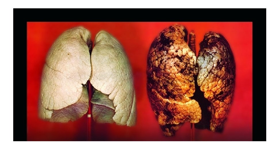
The Respiratory System Functions 1. 2. 3. 4. 5. Warm, moisten, and filter incoming air p. H Homeostasis Vocalization Olfactory reception Gas Exchange……

Gas Exchange 02 taken up and CO 2 is released Coupled to circulatory system Respiratory system responsibilities a. ventilation (air in & out of lungs) b. external respiration (gas exchange lungs – blood) Circulatory system responsibilities a. transport gases (between lungs and tissue) b. internal respiration (gas exchange blood – tissue)
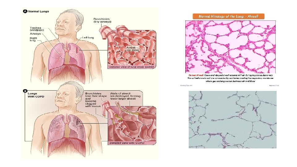
 conducting zone structures Ønasal cavity Øpharynx Ølarynx Øtrachea Øbronchial tree")
Anatomy (fig. 22. 1) conducting zone structures Ønasal cavity Øpharynx Ølarynx Øtrachea Øbronchial tree (note: open to external world , has MALT)
 As you move through the bronchi deeper into lungs: ØCartilage")
Bronchial Tree (cont. ) As you move through the bronchi deeper into lungs: ØCartilage changes from rings to plates to none ØCells of mucosal epithelium change from columnar to cubodial to simple squamous ØSmooth muscle increases
 ØRespiratory bronchioles have scattered alveoli ØAlveolar ducts walls")
Respiratory Zone Structures (fig. 22. 8) ØRespiratory bronchioles have scattered alveoli ØAlveolar ducts walls made of alveoli ØAlveolar sacs terminal ends, clusters of alveoli
 Ø Type I alveolar cells = simple squamous epithelium")
Alveolar Structure (fig. 22. 9) Ø Type I alveolar cells = simple squamous epithelium with thin basal membrane Ø Type II alveolar cells = secrete surfactant Ø Alveolar macrophages (Dust cells) = immunity Ø Pores = equalize pressure Ø Elastic fibers = allow expansion and recoil

The Reproductive System Functions Male § Make sperm § Deliver sperm into female’s reproductive tract Female § Make eggs § Nourish young until birth

Male Testes – paired, ovate, glands ~ 5 X 2. 5 cm § Sperm and hormone secretion § Develop near kidneys and descend into scrotum § Scrotum hang outside the body cavity so ~ 3 cooler than body temp (required for viable sperm) § can be drawn up, by cremaster muscle when cold § If cryptoorchidism (testes have not dropped) is left untreated, cells that normally produce sperm degenerate - male is infertile.

Testes sperm are made inside continuous with peritoneum invaginations form septa that divide inside of testes into lobules seminiferous tubules straight tubules rete testes efferent ductus epididymis ductus (vas) deferens ejaculatory duct urethra

Regulation - feedback Hypothalamus
 Accessory - fallopian tubes, uterus,")
Female Primary sex organ ovaries (make eggs and hormones) Accessory - fallopian tubes, uterus, vagina, and vulva (external genitals)

Ovary • Paired organs laying R/L of the uterus • ~ 2 X size of an almond • held in place by ovarian, suspensory, and broad ligaments
 • deep -")
Ovary capsule: • Superficial - germinal epithelium (simple squamous or cuboidal) • deep - tunica albuginea (dense irregular ct) inside: • cortex with follicles and eggs in different stages of development • medulla with loose ct and vessels

Ovaries Oocyte – eggs – sex cells - gametes • during fetal development the cells destined to be eggs migrate from the yolk sac to the developing ovary • these eggs start meiosis & arrest in prophase I • at birth a woman has ~ 1 -2 million • eggs die continually and by puberty she has ~ 250, 000 • Only 300 -400 will ever reach maturity

Recap: Error rate: as many as 20% of oocytes but only 3 to 4% of sperm have the wrong number of chromosomes.
 4 phases: menstrual, preovulation, postovulation")
Ovarian/uterine/hormonal cycle (fig. 27. 19, 27. 20) 4 phases: menstrual, preovulation, postovulation

Cycle Hierarchy of Control Hypothalamus Pituitary Ovaries Uterus Note that the ovaries also exert feedback control on the hypothalamus and pituitary

Hormones involved • Estrogens • Secreted by follicular cells • Functions: • Promote the development and maintenance of female reproductive structures • Increase protein anabolism, including building strong bones • Lower blood cholesterol level • Moderate levels of estrogens inhibit both the release of Gn. RH by the hypothalamus and secretion of LH
 • Progesterone • Secreted by cells of the corpus luteum")
Hormones involved (cont. ) • Progesterone • Secreted by cells of the corpus luteum • Function • Acts synergistically with estrogens to prepare and then maintain the endometrium for implantation of a fertilized ovum • Prepares mammary glands for milk secretion • High levels of progesterone inhibit secretion of Gn. RH and LH
 • Gonadotropin Releasing Hormone (Gn. RH) • Secreted by the")
Hormones involved (cont. ) • Gonadotropin Releasing Hormone (Gn. RH) • Secreted by the hypothalamus and controls the ovarian and uterine cycles • Stimulates the release of follicle stimulating hormone (FSH) and luteinizing hormone (LH) from the anterior pituitary
 • Follicle Stimulating Hormone (FSH) • Released by anterior pituitary")
Hormones Involved (cont. ) • Follicle Stimulating Hormone (FSH) • Released by anterior pituitary • Initiates follicular growth and the secretion of estrogens by the growing follicles • Luteinizing Hormone (LH) • Released by the anterior pituitary • Stimulates further development of ovarian follicles • Stimulates ovulation and then promotes the formation the corpus luteum

- Slides: 48