Holes Essentials of Human Anatomy Physiology Unit 6


 of a muscle fiber, there are two specialized")
 of a muscle fiber, there are two specialized")


 – 4")


 results in muscular")

- Slides: 36
Hole’s Essentials of Human Anatomy & Physiology Unit 6 Muscular System
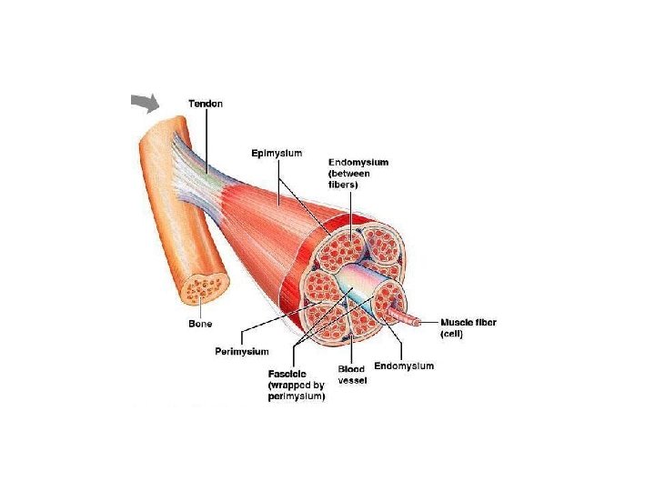
Microscopic Anatomy • Skeletal muscle is composed of a variety of layers of tissues • Muscle (gross muscles; Biceps brachii) • Fascicles (bundles of muscle fibers) • Muscle fibers (elongated muscle cells) • Myofibrils (bundles of myofilaments) • Myofilaments (protein filaments)
Microscopic Anatomy A muscle fiber is an elongated muscle cell. Each muscle fiber is composed of myofibrils which nearly fill its cytoplasm. Each myofibril is composed of two types of myofilaments, or protein filaments: 1. Thick filaments primarily composed of the protein myosin. 2. Thin filaments primarily composed of the protein actin. Striations are caused by the arrangement thick and thin filaments within the myofibrils: 1. A-Band = dark area = overlapping of thick and thin filaments; 2. I-Band = light area = thin filaments only.
Microscopic Anatomy The length of each myofibril is divided into sarcomeres: 1. Sarcomeres are repeating contractile units of a muscle fiber. 2. Regions within each sarcomere make distinctive repeating units called striations. 3. A chain of sarcomeres makes a myofibril. 4. Sarcomeres meet one another at an area called the Z-line (Zdisc). a. The Z-line is the attachment point of thin filaments. 5. Sarcomeres contain an H-zone (Bare zone), which is the central area of sarcomere consisting of thick filaments only 6. Sarcomeres also contain an M-line, which is a darkened area consisting of proteins holding thick filaments in place
Microscopic Anatomy The two types of myofilaments serve as the primary means for muscle contraction. Thick filaments = myosin filaments Rod-like myosin tail terminates in two globular “heads” or cross bridges Cross bridges interact with active sites on thin filaments Has ATPase enzymes to split ATP for contraction power Thin filaments = actin filaments coiled helical structure (resembles twisted strands of pearls) Anchored to Z-line Actin filaments interact with two additional proteins
Microscopic Anatomy a. Tropomyosin = rod-shaped protein spiraling around actin backbone to stabilize it; b. Troponin = complex of polypeptides 1. one binds to actin, 2. one binds to tropomyosin, 3. one binds to calcium ions; c. Tropomyosin and Troponin help control actin's interaction with myosin during contraction.
Label: epimysium; perimysium; endomysium; fascicle; muscle fiber
Add the following labels: sarcomere; A-band; I-band; H-zone; M-line; myosin; actin; zdisk; myosin cross bridge; Draw in tropomyosin and troponin
Microscopic Anatomy Within the sarcoplasm (cytoplasm) of a muscle fiber, there are two specialized organelles that reside at the membrane of the sarcolemma (plasma membrane): Sarcoplasmic reticulum Network of membranous channels that surround each myofibril Specialized smooth endoplasmic reticulum. SR has high concentration of calcium ions compared to the sarcoplasm (maintained by active transport calcium pump). When stimulated by muscle impulse, membranes become more permeable to calcium ions and calcium diffuses out of SR and into sarcoplasm.
Microscopic Anatomy Within the sarcoplasm (cytoplasm) of a muscle fiber, there are two specialized organelles that reside at the membrane of the sarcolemma (plasma membrane): Transverse tubule set of membranous channels that extend into the sarcoplasm as invaginations continuous with muscle cell membrane TTs are filled with extracellular fluid & extend deep into cell. Each TT runs between two enlarged portions of SR called cisternae. Structures form a triad near the region where actin & myosin overlap. SR and TT are involved in activating the muscle contraction mechanism.
Skeletal Muscle Contraction "Sliding Filament Theory“ is theory of muscle contraction 1. most popular theory concerning muscle contraction; 2. states that muscle contraction involves the sliding movement of the thin filaments (actin) past the thick filaments (myosin); 3. Sliding continues until the overlapping between the thin & thick filaments is complete. Changes in muscle cell during contraction: 1. The distance between the Z-lines of the sarcomeres decreases; 2. The I-Bands (light bands) shorten; 3. The A-Bands move closer together, but do not diminish in length.
https: //www. youtube. com/watch? v=f_t. Zne 9 ON 7 c – skeletal muscle structure https: //www. youtube. com/watch? v=MZJ 6 k. TKDFmw - Skeletal Muscle (Sarcomere, Myosin and Actin) https: //www. youtube. com/watch? v=Vs 0 t. ZV 35_pw - Skeletal Muscle Contraction https: //www. youtube. com/watch? v=BMT 4 Pt. XRCVA – muscle contraction process https: //www. youtube. com/watch? v=7 w. M 5_a. Un 2 qs - Muscle Contraction Part 1: Events at the Neuromuscular Junction https: //www. youtube. com/watch? v=HJj 3 j. UVDFFo - Muscle Contraction Part 2: Excitation-Contraction Coupling
Skeletal Muscle Contraction The role of calcium in contraction 1. In a resting muscle fiber (i. e. in the absence of calcium ions): Tropomyosin blocks or inhibits the myosin binding sites on actin. 2. When calcium ions (Ca++) are present (because of nervous system stimulation): Ca 2+ floods into the cell and binds to troponin causing a conformational change in the troponin-complex When the shape of troponin changes…. 1. Tropomyosin starts moving 2. which "opens" or exposes the myosin binding sites on actin; 3. This results in the interaction between the active sites on actin and the heads (or cross bridges) of myosin.
Skeletal Muscle Contraction Sequence of Events in Sliding of Actin filaments during Contraction 1. When calcium ions are present, the myosin binding sites on actin are exposed causing the “cocked” cross-bridge of myosin to bind to actin. a. ATP breakdown provides energy to “cocked” myosin head. b. “Cocked” myosin head attaches to exposed binding site on actin. 2. Power-stroke: Cross-bridge (myosin head) springs from cocked position and pulls on actin filament. a. ADP and P are released from myosin cross bridge 3. A new ATP binds to cross-bridge (but is not yet broken down) a. Cross bridges break bond with actin b. Myosin heads are released from actin. 4. ATP broken down into ADP + P “cocking” myosin cross-bridge * As long as calcium ions and ATP are present, this walking continues until the muscle fiber is fully contracted.
The Sliding Filament Theory of Muscle Contraction
Stimulation of Skeletal Muscle Fibers In order for a skeletal muscle to contract, its fibers must first be stimulated by a motor neuron. Neuromuscular Junction (NMJ) = the site where a motor nerve fiber and a skeletal Muscle fiber meet BUT DO NOT TOUCH; (also called a synapse) Motor Unit = one motor neuron and many skeletal muscle Each neuron may stimulate few muscle fibers or hundreds The neuron’s axon reaching the muscle fiber branches into multiple axon terminals Each axon terminal forms junctions with the motor end-plates of different muscle fibers The sarcolemma of the muscle fiber is specialized to form a motor end-plate
Stimulation of Skeletal Muscle Fibers Motor End-Plate = the specific part of a skeletal muscle fiber's sarcolemma directly beneath the NMJ Synaptic cleft = gap between the motor neuron and a muscle Neurotransmitter = chemical substance released from a motor end fiber, causing stimulation of the sarcolemma of muscle fiber; acetylcholine (ACh).
Stimulation of Skeletal Muscle Fibers a. Transmission of nerve impulse to muscle fiber. b. The process begins when a motor impulse is initiated by the brain, travels down the spinal cord, into a motor neuron, which branches into many motor nerve fibers/endings; Each motor nerve fiber extends to the motor end-plate of a skeletal muscle fiber forming a neuromuscular junction (NMJ) c. When the motor impulse reaches the end of the motor nerve fiber/ending, the membrane is depolarized and becomes permeable to calcium ions; calcium ions rush into motor nerve fiber, and d. Acetylcholine diffuses across the NMJ & stimulates/depolarizes the motor end-plate (sarcolemma) of a skeletal muscle fiber e. The sarcolemma of the skeletal muscle fiber becomes permeable to Na ions.
Stimulation of Skeletal Muscle Fibers f. The muscle impulse travels over the surface of the sarcolemma and deep into the muscle fiber by means of the transverse tubules g. The muscle impulse reaches the sarcoplasmic reticulum, which releases calcium ions into the sarcoplasm of the muscle fiber; This is termed “excitation contraction coupling” h. Calcium binds to troponin, moving tropomyosin and exposing myosin binding sites on actin filament (troponin conformational change); i. Cross bridges (linkages) form between actin and myosin; j. Actin filaments are pulled inward by myosin cross-bridges k. The muscle fiber shortens as contraction occurs.
Stimulation of Skeletal Muscle Fibers Relaxation Mechanism: Acetylcholinesterase is an enzyme present in the NMJ; It immediately destroys acetylcholine, so the motor end-plate is no longer stimulated (i. e. it cannot cause continuous muscle contraction). Calcium ions are transported from sarcoplasm back into sarcoplasmic reticulum. Linkages between actin and myosin are broken. Troponin and tropomyosin move and cover binding sites The muscle fiber relaxes.
The clostridial neurotoxins responsible for tetanus and botulism are proteins consisting of three domains endowed with different functions: neurospecific binding, membrane translocation and proteolysis for specific components of the neuroexocytosis apparatus. Tetanus neurotoxin (Te. NT) binds to the presynaptic membrane of the neuromuscular junction, is internalized and transported retroaxonally to the spinal cord. The spastic paralysis induced by the toxin is due to the blockade of neurotransmitter release from spinal inhibitory interneurons. In contrast, the seven serotypes of botulinum neurotoxins (Bo. NTs) act at the periphery by inducing a flaccid paralysis due to the inhibition of acetylcholine release at the neuromuscular junction. The remarkable specificity of Bo. NTs is exploited in the treatment of human diseases characterized by a hyperfunction of cholinergic terminals.
Energy for Muscle Contraction The energy used to power the interaction between actin and myosin comes from ATP stored in skeletal muscle lasts only about six seconds. a. ATP must be regenerated continuously if contraction is to continue. b. There are three pathways in which ATP is regenerated:
Energy for Muscle Contraction Direct phosphorylation Muscle cells contain creatine phosphate (CP) – 4 -6 x more abundant in muscle fibers than ATP CP is a high-energy molecule After ATP is depleted, ADP is left CP transfers energy to ADP, to regenerate ATP CP supplies are exhausted in about 10 -20 seconds
Energy for Muscle Contraction Aerobic respiration Series of metabolic pathways that occur in the mitochondria Glucose is broken down to carbon dioxide and water, releasing energy This is a slower reaction that requires continuous oxygen This can supply the muscles with ATP for hours
Energy for Muscle Contraction Anaerobic respiration Occurs during strenuous muscle use when respiratory and cardiovascular systems can’t supply sufficient oxygen Reaction that breaks down glucose without oxygen Glucose is broken down to pyruvic acid to produce some ATP Pyruvic acid is converted to lactic acid creating an oxygen debt
Muscle Fatigue and Oxygen Debt Muscle Fatigue a. Muscle fatigue is a state of physiological inability to contract; b. If no oxygen is available in muscle cells to complete aerobic respiration, pyruvic acid is converted to lactic acid, which causes muscle fatigue and soreness; c. Results from a relative deficit of ATP and/or accumulation of lactic acid (which decreases p. H). Oxygen Debt a. The oxygen debt is the amount of oxygen necessary to support the conversion of lactic acid to glycogen/glucose. b. needed to replenish spent glycogen stores in the liver by converting lactic acid into glucose. c. can take hours after exercise is complete to repay debt
Muscular Responses Threshold stimulus: the minimum strength of stimulation required to (generate an muscle impulse) cause a contraction A skeletal muscle fiber’s resting membrane potential must be depolarized from -100 m. V to -70 m. V before an impulse begins What is the threshold stimulus? +30 m. V Recording a contraction A myogram is a recording of a muscle contraction A twitch is a single contraction that lasts a fraction of a second, followed by relaxation. The delay between stimulation and contraction is called latent period. A muscle fiber must return to its resting state (-100 m. V) before it can be stimulated again. This is called refractory period.
Muscular Responses All or nothing response Muscle fiber brought to threshold twitch Sub-threshold stimulation no twitch In the whole muscle, not all muscle fibers may be stimulated during the same interval. Different combinations of muscle fiber contractions may give differing responses in the whole muscle. Graded responses are different degrees of skeletal muscle shortening. In the whole muscle the degree of tension depends on The frequency (how often) of individual muscle fiber stimulation The amount (how many) of individual muscle fibers taking part in whole muscle contraction
Types of Muscular Responses Twitch A single contraction that lasts a fraction of a second, followed by relaxation Summing of Contractions When several stimuli are delivered in succession to a muscle fiber, it cannot completely relax between contractions. The force of individual twitches combines in a process called summation. When the resulting sustained contractions lacks relaxation, it is called titanic contraction. Incomplete (unfused) Tetanus One contraction is immediately followed by another The muscle does not completely return to a resting state before the next stimulus arrives Complete (fused) tetanus a forceful sustained contraction that lacks even partial relaxation (titanic contraction)
Muscle Response to Strong Stimuli 1 nerve impulse = 1 contraction Motor units: a motor neuron and the many skeletal muscle fibers it stimulates Because the motor neuron branches into several motor nerve endings (axon terminals), it can stimulate may skeletal muscle fibers simultaneously. Stimulation causes all fibers of unit to contract together Each unit can contain 10 -100 s of fibers Recruitment: Muscle force depends upon the number of fibers stimulated (not all fibers may be stimulated at same time) Because a whole muscle is composed of many motor units, controlled by many different motor neurons, simultaneous contraction of all units does not necessarily occur. As the intensity of stimulation increases, recruitment of motor units increases, until all contract simultaneously (max contraction power) More fibers contracting results in greater muscle tension Muscles can continue to contract unless they run out of energy
Muscle Tone Even when a muscle is at rest, a certain amount of sustained contraction is occurring in its fibers. This is muscle tone. Muscle tone is very important in maintaining posture. The extensor and flexor muscles maintain a constant tone, even at rest, to maintain a normal posture. Requires unconscious, involuntary nerve impulses In other words, some fibers are contracted even in a relaxed muscle
Types of Muscle Contractions Isotonic contractions Myofilaments are able to slide past each other during contractions Tension in the muscle increases AND, the muscle shortens Isometric contractions Myofilaments are unable to slide past each other during contractions Tension in the muscles increases BUT, the muscle is unable to shorten
Fast and Slow Fibers Muscle fibers vary in contraction speed (i. e. slow or fast twitch) Slow twitch – red fibers Contain oxygen-carrying pigment, myoglobin Have a rich blood supply with many mitochondria Can contract for long periods of time without fatigue Can generate ATP quickly enough to keep up with demand Fast twitch – white fibers Contain less myoglobin Have less blood, and have fewer mitochondria Contain extensive SR to store and reabsorb Ca 2+ Contract rapidly but tire quickly (Lactic acid build-up)
Examples of Fast & Slow Muscle Fibers Forceful exercise (weight lifting) results in muscular hypertrophy These enlarged muscles produce more actin and myosin filaments, but not new fibers The enlarged muscle can produce stronger contractions. This exercise utilizes fast twitch fibers which are fatigable Sustained exercise (running, swimming) results in activating slow twitch fibers This increases the ability to resist fatigue during prolonged exercise but may not change strength or size Atrophy: the lack of use of a muscle results in decreasing its size and strength ; maintain muscle function through exercise As one ages: ATP, myoglobin and CP stores in muscle fibers decrease Muscle is replaced by connective and adipose tissue Reflexes reduced
Muscular System, Neuromuscular Junction http: //www. youtube. com/watch? v=Zsc. XOv. Dg. Cm. Q& feature=related Muscular System, Sliding Filament Theory (1) http: //www. youtube. com/watch? v=Ed. Hz. KYDxr. Kc&feature=related Sliding filament theory 2 (0. 49 sec) http: //www. youtube. com/watch? v=Vlchs 4 om. FDM Muscle Structure and Function http: //www. youtube. com/watch? v=ren_IQPOh. Jc&feature=related