History of the Microscope Before the Microscope About

History of the Microscope

Before the Microscope About 1590 • Zaccharias Janssen and his son Hans Ø two Dutch spectacle makers (what did they make? ) Ø discovered that nearby objects appeared enlarged. Ø Ø Made a crude instrument with two lenses (one on each end) That was the forerunner of the compound microscope and of the telescope.

The Earliest Known Compound Microscope In 1609, Galileo, father of modern physics and astronomy, heard Janssen’s early experiments, worked on the principles of lenses, and made IMPROVEMENTS
 l The father of microscopy l Started as an")
Anton van Leeuwenhoek (1632 -1723) l The father of microscopy l Started as an apprentice in a dry goods store in Holland l In the store magnifying glasses were used to count the threads in cloth l This led to the building of his microscopes for which he is famous. l He was the first to see – Bacteria yeast, – life in a drop of water – the circulation of blood cells in capillaries. –
 a screw for adjusting the height of")
Leeuwenhoek's microscope Consisted simply of: l A) a screw for adjusting the height of the object being examined l B) a metal plate serving as the body l C) a skewer to pierce the object and rotate it l D) the lens itself, which was spherical l
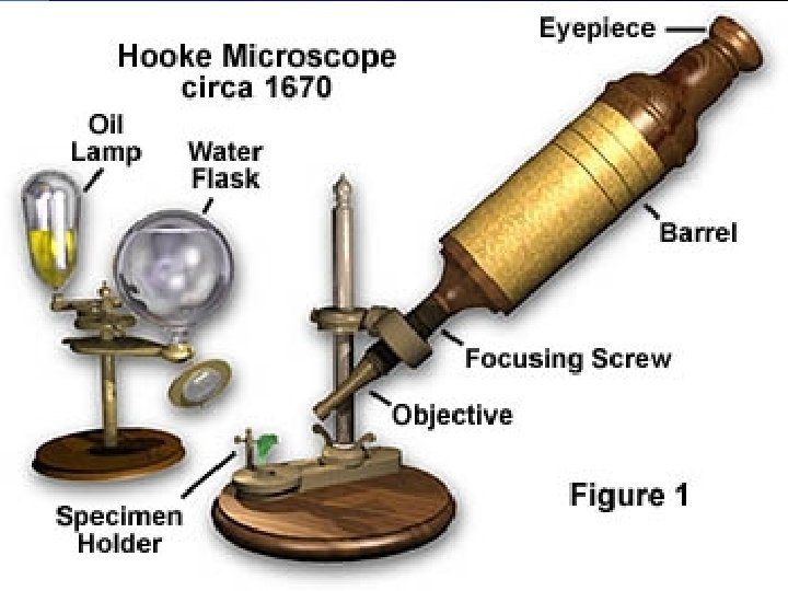
 l The English father of microscopy. l Hooke made a")
Robert Hooke (1635 -1703) l The English father of microscopy. l Hooke made a copy of Leeuwenhoek's light microscope and then improved upon his design. l First to observe lines and empty space in cork l First to use the term cells to describe the empty space l

Modern Microscope • Compound microscopes uses electric light or mirror • This microscope is the most commonly used. • The image seen is two dimensional. • You can see individual cells, even living ones. • It has high magnification. • However, it has a low resolution (sharpness). • Cost - $150 - $6000

Compound Light Microscope Monocular Microscope Stereomicroscope Can see 3 D Image Can not see 3 D Image

Phase Contrast Microscope can see live specimen without using stain. l Hydra dust mite

l. A dissection microscope is light illuminated. l l The image that appears is three dimensional. l It is used for dissection to get a better look at the larger specimen. l You cannot see individual cells because it has a low magnification. l Cost - $100 - $1500

Dissecting Microscope

Insect parts can be seen in 3 D with a dissecting microscope l Butterfly

Transmission Electron Microscope l Invented in 1931 l German scientist Ernst Ruska l Today TEM magnify over 2 million times

TEM l A focused beam of electrons are used to ‘see through’ the specimen l This gives a 2 -D view. l Thin slices of specimen are obtained. The electron beams pass through this. l It has high magnification and high resolution. l Cost - $50, 000 - $100, 000

TEM

Scanning Electron Microscope l l l Called SEM Scans the surface of an object w/ a beam of electron Produces a 3 -D image Can magnify up to 50, 000 times Produces a clear image

SEM l SEM uses electron lighting. The image is seen in 3 -D. l It has high magnification and high resolution. Cost - $50, 000 - $100, 000 l The first Scanning Electron Microscope (SEM) debuted in 1942.

SEM

SEM l Honeybee stinger RBC

Scanning Tunneling Microscope l 1981 invented by – Gerd Bining of Germany – Heinrich Rohrer of Switzerland l Can see 3 -D and living specimen l Magnify up to 100 million times l Used to see surface of atoms

Scanning Tunneling Microscope Surface of Platinum atoms
- Slides: 22