History of the Microscope and Cell Theory Mr


, an English scientist, improved on early compound microscopes around 1660.")

is widely credited to be “the father of microscopy”. .")

 building upon Leeuwenhoek’s research, began to develop what")
 also contributed to cell theory in")
 made the very important contribution to cell theory")


- Slides: 14
History of the Microscope and Cell Theory Mr. C. Frittenburg During the 1 st century AD (year 100), glass had been invented and the Romans were looking through the glass and testing it. Magnifiers and "burning glasses" or "magnifying glasses" are mentioned in the writings of Seneca and Pliny the Elder, Roman philosophers during the first century A. D.
In the 13 th century lenses are beginning to be used in spectacles. First recorded reference is made to eyeglasses from English friar and philosopher, Roger Bacon (1214? -1294).
The earliest simple microscope was merely a tube with a plate for the object at one end and, at the other, a lens which gave a magnification less than ten diameters -- ten times the actual size. Hans and Zacharias Janssen are given credit as the inventors of the first compound microscope. The first compound microscopes produced by the Janssen's was simply a tube with lenses at each end. The magnification of these early scopes ranged from 3 X to 9 X, depending on the size of the diaphragm openings.
Robert Hooke (1635 -1703), an English scientist, improved on early compound microscopes around 1660. In Micrographia (1665), he coined the word cell to describe the features of plant tissue (cork from the bark of an oak tree) he was able to discover under the microscope. The compartments he observed in the plant tissue appeared like little compartments, or “cells”
Hooke’s book, Micrographia, was a series of observations made with the aid of magnifying lenses; some of these on very small things, such as his famous drawing of the flea and tree bark lining
Antony van Leeuwenhoek (1632 -1723)is widely credited to be “the father of microscopy”. . . my work, which I've done for a long time, was not pursued in order to gain the praise I now enjoy, but chiefly from a craving after knowledge, which I notice resides in me more than in most other men. And therewithal, whenever I found out anything remarkable, I have thought it my duty to put down my discovery on paper, so that all ingenious people might be informed thereof. Antony van Leeuwenhoek. Letter of June 12, 1716
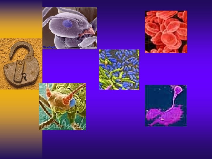
Leeuwenhoek came from a family of tradesmen, had no fortune, received no higher education or university degrees, and knew no languages other than his native Dutch. In 1648 he was apprenticed as a fabric merchant and learned to grind lenses in order to observe wools. Inspired by Hooke’s Micrographia he began to use his skill of grinding lenses to make compound microscopes that could magnify up to 300 x. Leeuwenhoek was the first to see and describe bacteria (1674), yeast plants, the life in a drop of water, and the circulation of blood corpuscles in capillaries. "No more pleasant sight has met my eye than this of so many thousands of living creatures in one small drop of water. . . " - Stated after his discovery of the microscopic world over three centuries ago.
German botanist, Matthias Schleiden (1804 -1881) building upon Leeuwenhoek’s research, began to develop what we now know as the “cell theory”. In 1838 he recorded that, “all plants are made of cells”.
Schleiden’s colleague, German physiologist Theodor Schwann (1810 -1882) also contributed to cell theory in 1839 by recording that, “all animals are made of cells”. In 1839 he also recorded, “all living things are composed of cells and cell products”.
German botanist Alexander Braun (1805 -1877) made the very important contribution to cell theory and observed that “the cell is the basic unit of life” in 1845.
German physiologist Rudolph Virchow made many great contributions to the medical world and is also the man responsible for discovering that "every cell originates from another cell“. Virchow founded the medical disciplines of cellular pathology , comparative pathology (comparison of diseases common to humans and animals) and anthropology.
Currently microscope capabilities have become almost limitless. Magnifying well over 1000 x.
Resources Science Power 9, Atlantic Edition http: //www. az-microscope. on. ca/history. htm http: //clendening. kumc. edu/dc/rv/ http: //www. crystalinks. com/bacon. html http: //www. denniskunkel. com/ http: //en. wikipedia. org/wiki/Anton_van_Leeuwenhoek http: //www. historic-glendale. net/microscope_history. htm http: //home. tiscalinet. ch/biografien/schleiden. htm http: //home. tiscalinet. ch/biografien/schwann. htm http: //inventors. about. com/library/inventors/blleeuwenhoek. htm http: //inventors. about. com/library/inventors/blmicroscope. htm http: //microscopy. fsu. edu/optics/timeline/people/janssen. html http: //www. olympusmicro. com/