History of DNA H Biology Winter 2018 What

History of DNA H Biology Winter 2018

What does DNA stand for? Deoxyribonucleic acid

What is DNA made of? DNA is a macromolecule Made up of nucleotides Covalently bonded together Double stranded Helix “Spiral” Which one?

What is a nucleotide? Molecule made of Deoxyribose sugar A phosphate group A nitrogenous base

How was DNA identified as genetic material? 1928 – Griffith notices that bacteria can transfer some type of material to other types of bacteria “Transforming principle”

How was DNA identified as genetic material? 1944 – Oswald Avery discovered that the transforming principle was DNA (genetic material) 1952 – Hershey and Chase studied a virus that affects bacteria (bacteriophage) and confirmed that DNA was genetic material

How was the structure of DNA discovered? 1950 – Erwin Chargaff Noticed that the same 4 bases are found in the DNA of all organisms Also noticed that: # of Adenine = # of Thymine # of Cytosine = # of Guanine “Chargaff’s Rule”

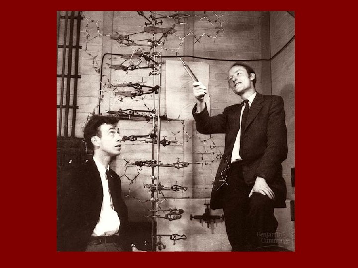
How was the structure of DNA discovered? 1950 s – James Watson and Francis Crick Worked to figure out DNA’s structure Thought that DNA might be a helix, but had no evidence Idea that DNA was a helix came from Linus Pauling

How was the structure of DNA discovered? 1951 -1952 – Rosalind Franklin Used x-ray diffractions to show DNA was truly a double helix Worked with Maurice Wilkins

How was the structure of DNA discovered? 1953 – Watson and Crick Wilkins (a colleague of Franklin) shows Watson and Crick the x-ray pictures This information gave Watson & Crick the evidence needed to conclude DNA has a helical shape Made a model of DNA which was made up of two chains of nucleotides

DNA Structure

DNA - Basics Deoxyribonucleic Acid Stores and transmits genetic info Tells the cells which proteins to make and when to make them
")
DNA - Basics Made up of nucleotides: Phosphate group Sugar Nitrogen bases (4 total) Adenine (A) Thymine (T) Cytosine (C) Guanine (G) Double helix structure (twisted ladder)

All about that base, ‘bout the base…

Nitrogen Bases 2 groups that bases are put in based on structure Purines → 2 carbon rings Adenine (A) Guanine (G) Pyrimidines → 1 carbon ring Cytosine (C) Thymine (T) Pure As Gold Cing Tut lived in a Pyramid
 always matches w/ Thymine (T) Cytosine (C) always matches")
Base Pairing Rules Adenine (A) always matches w/ Thymine (T) Cytosine (C) always matches w/ Guanine (G)

Base Pairing Rules Why these base pairings? Bases are specific! Sizes of the bases (rings) Number of H bonds formed with each other The sizes of the bases (and how they pair) also determine the structure of the larger DNA molecule

Size of Bases Purine + Purine = Too wide Pyrimidine + Pyrimidine = Too Narrow

NO NO YES! Purine + Pyrimidine = Perfect Fit

What makes up the “backbone” of DNA? The sides of the ladder are made up of: 1) Sugar 2) Phosphate Alternate along backbone

What Holds Everything Together!? Weak Hydrogen bonds connect the nitrogenous bases to each other Covalent bonds connect the sugars and phosphates to each other!
 Sides of ladder = sugar/ phosphate backbone")
DNA Structure Summary Double Helix (twisted ladder) Sides of ladder = sugar/ phosphate backbone Rungs of the ladder = nitrogen bases

DNA Replication

Protein Synthesis 3 major processes: Replication → DNA doubles to form 2 new DNA molecules Transcription → DNA info copied to RNA Translation → building a protein according to RNA instructions Uses m. RNA, r. RNA, and t. RNA

DNA Replication Occurs in nucleus DNA is copied Process: Enzyme helicase “unzips” strands of DNA by breaking H bonds at several places along segment of DNA called “origins of replication”

Point at which the strands separate is called the replication fork

primase

DNA Replication DNA Polymerase adds nucleotides to create two NEW identical daughter strands, by adding nucleotides (A to T and G to C) Also fills-in gaps between Okazaki fragments

DNA Replication Okazaki fragments are large chunks of nucleotides DNA Ligase joins Okazaki fragments and seals nicks in the backbone

Semi-Conservative DNA replication is said to be semiconservative Each new double-helix created contains one strand from the helix from which it was copied

Direction of Replication Enzyme DNA Polymerase helps build the new strands from the 5’ 3’ direction

Why Replication? • DNA replication is necessary to create identical copies of DNA, so it can be passed onto a new cell (cell division & reproduction) • Okazaki fragments are newly made DNA fragments that form on the lagging template
 because cells have enzymes (like DNA")
Accuracy of Replication Very low mistake rate (1/billion!) because cells have enzymes (like DNA Polymerase) that proofread, recognize, and fix mistakes! HOWEVER, mistakes can happen MUTATIONS (cancer)

Review: DNA Replication 1. A new sugar-phosphate backbone is made for each new strand 2. Base pairs are added (A-T and C-G) 3. Two strands are created in place of the original strand

Protein Synthesis Pt. 1 - Transcription

What is Transcription? DNA information is copied into an RNA message Occurs in the nucleus Base pair change: in RNA Thymine (T) is replaced with Uracil (U)
 base pair of DNA changes to Uracil (U)")
Transcription During transcription, the Thymine (T) base pair of DNA changes to Uracil (U) in RNA Uracil is a pyrimidine Uracil requires less energy to create vs. Thymine Uracil also prevents Nuclease from breaking down DNA during Protein Synthesis

Why RNA? RNA – Ribonucleic Acid How does DNA get out of the nucleus and go to the ribosomes where the proteins are made? It CAN’T! DNA cannot leave the nucleus! So, it copies itself into RNA and that leaves and goes to the ribosomes EXAMPLE – library book, photocopy Library book = DNA Photocopy = RNA

DNA vs. RNA Name Deoxyribonucleic Acid Ribonucleic Acid Sugar Deoxyribose Ribose Nitrogen Bases A, C, G & Thymine (T) A, C, G & Uracil (U) Location Inside nucleus only Stranded Double stranded In and out of nucleus Single Stranded

Steps of Transcription ² RNA polymerase “opens” a section of DNA (so the double helix strands are separate)

Steps of Transcription ² Using one strand of DNA as a template, RNA polymerase builds a complementary strand of RNA nucleotides ² This RNA strand is called m. RNA
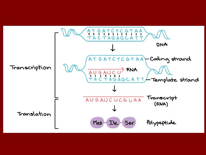

Steps of Transcription v Base pairing rules are the same as with DNA, except Uracil (not Thymine) pairs with Adenine v DNA: A C T G v RNA: U G A C v Once the entire gene (segment of DNA) has been transcribed, RNA polymerase releases new strand Strand exits the nucleus through the pores in the nuclear envelope goes to cytoplasm ribosomes (protein factories!

Practice Transcribing DNA Complementary – A T C DNA Template _____ TAG m. RNA _____ AUC _____ TCT AGA _____ UCU _____ TAG _____ ATC UAG **NOTE: Always create the m. RNA strand using the DNA Template**

Protein Synthesis Pt. 2 - Translation

Codons For every 3 bases copied from DNA to RNA, we have a codon Codons are important, because they “code” for specific amino acids These amino acids then build a larger protein molecule!

Practice Finding the Correct Amino Acid CCA AGA GUG UGA AUG CAG Remember, ALWAYS use m. RNA codons to find the correct amino acids!

Protein Synthesis Protein synthesis consists of 2 mains parts: Transcription – DNA is copied in the nucleus, the result is the formation of m. RNA Translation – m. RNA travels to the cytoplasm and attaches to a ribosome; with the help of t. RNA a protein is made Cells translate an RNA message (English) into amino acids (Spanish) Canada – French – Bonjour! – DNA – TACGCT USA – English –Hello! –m. RNA –AUGCGA Mexico – Spanish – Hola! –Protein – MET-ARG

Three types of RNA Transcription makes 3 types of RNA: 1. r. RNA – ribosomal RNA - Makes up part of the ribosomes (protein factories) 2. t. RNA – transfer RNA - Brings the amino acids from the cytoplasm to the ribosomes to make the growing protein 3. m. RNA – messenger RNA - A message (genetic code) that goes to the ribosomes and is translated to form a protein

Messenger RNA m. RNA is created by copying a segment of DNA code DNA cannot leave the nucleus, but m. RNA can! m. RNA is organized into codons A codon = 3 consecutive bases code for a specific amino acid Remember a chain of amino acids = a polypeptide = PROTEIN
 Ribosome attaches to m. RNA Contains the enzymes necessary for")
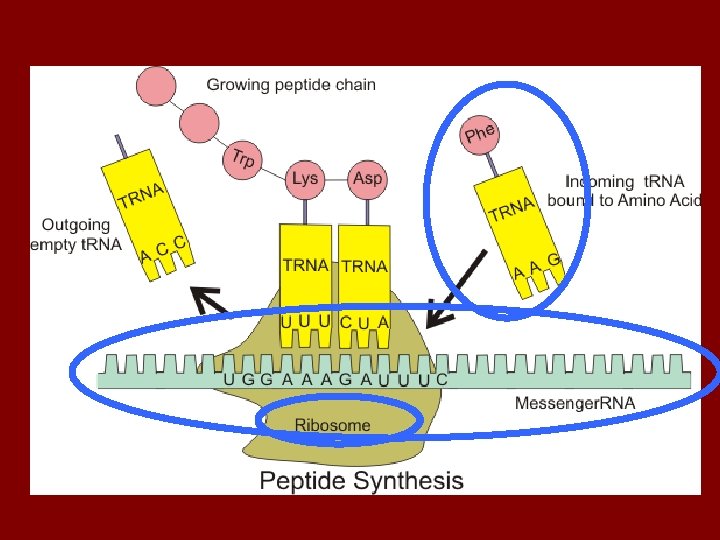
Ribosomal RNA (r. RNA) Ribosome attaches to m. RNA Contains the enzymes necessary for protein synthesis Ribosome: 3 t. RNA binding spots: E – exit P – current amino acid A – on deck amino acid Has a large & small subunit E P A
 Reads the m. RNA Carries the amino acid that will")
Transfer RNA (t. RNA) Reads the m. RNA Carries the amino acid that will be added to the growing protein chain The 3 bases at the bottom of t. RNA = anticodon The anticodon pairs with the m. RNA codon This ensures that each AA is delivered to the correct place on m. RNA At the top of t. RNA is an amino acid attachment site
 the m.")
What is Translation? Process of building a protein chain by translating (reading) the m. RNA code Occurs on ribosomes Uses codons

Translation: The Basics Translation can be divided into three stages: Initiation Elongation Termination

Initiation ² m. RNA binds to small subunit ² Anticodon on the t. RNA molecule bind to the complementary m. RNA codon ² Each amino acid chain starts with methionine (AUG) = anticodon (UAC) A to U and G to C ² Initiator t. RNA fits into the P site and holds the growing protein ² The A site is empty and ready for the second A. A.

Elongation After initiation, amino acids are added to the first amino acid… • Incoming t. RNA anticodon binds with m. RNA codon • t. RNA in the P site “hands over” the growing protein to the t. RNA molecule in the “A site”

Termination Protein continues to grow until a STOP codon reaches the “A site” Each protein is told to stop by 1 of 3 stop codons: UAA, UAG, UGA The protein is released from the ribosome

Coding Cheat Sheet #2 DNA Template. ……… #1 ATC CAG CAT DNA Complementary…. TAG GTC GTA m. RNA…………………. #3 t. RNA………… AUG GUC GUA UAC CAG CAU Amino Acid……………. MET- VAL #4
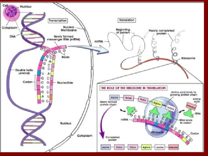

Summary of Protein Synthesis Replication → DNA to DNA, occurs in nucleus Transcription → DNA to RNA, occurs in nucleus Translation → RNA to Protein, occurs in ribosome DNA RNA PROTEIN! https: //www. youtube. com/watch? v=g. G 7 u. Csk. UOr. A
 May")
Mutations Change in the nucleotide sequence of a gene (and thus, overall DNA) May be due to copying errors (DNA replication), chemicals, UV radiation, viruses, etc. Many mutations are repaired by enzymes Almost all mutations are neutral and do not cause any physiological change We call these silent mutations

Gene Mutations Several types of chromosomal and gene mutations exist Can cause cancer, CF, Crohns’ disease, tay sachs Gene mutations can happen in: Somatic (body) cells affect only that individual Germ cells (gametes) affect future offspring Two main types of gene mutations: Point (substitutions) missense, silent, nonsense Frameshift deletion, insertion

Point Mutations Point mutations are also called substitutions One nucleotide is substituted for another Usually fixed by DNA Polymerase Three types: Missense mutation changes the amino acid Silent mutation does not change the amino acid Nonsense mutation changes the amino acid to a “STOP” codon prematurely stops the production of a protein

Point Mutation Examples
")
Sickle Cell Anemia Caused by a missense point mutation in hemoglobin (protein in RBC) Causes blood cells to be shaped like sickles Mutated RBC cannot transport O 2 as well as “normal” RBC

Frameshift Mutations A more serious type of gene mutation is called a frameshift mutation A nucleotide is inserted or deleted Alters the sequence of the gene changes the entire “frame of reading” Affects every codon beyond the point of mutation and thus may dramatically change the amino acid sequence protein Ilo veb iol ogy ilo ebi olo gy…

Frameshift Mutation Examples

Analogies of Gene Mutations
- Slides: 71