Histology Tissues Groups of cells similar in structure


- Slides: 74
Histology
Tissues • Groups of cells similar in structure and function • - the structure plays a role into the function • The structure and function complement each other
Types of Tissues • • Epithelial tissue Connective tissue Muscle tissue Nerve tissue • Atoms to Molecules to Cells
Tissue Characteristics Differences between the tissue classes Types and functions of cells Characteristics of matrix (extracellular matrix) Fibrous proteins Ground substance – thick or thin Clear gels (ECF, tissue fluid, interstitial fluid, tissue gel) • Rubbery or stony in cartilage or bone • • •
Tisssue Characteristics • Space occupied by cells versus matrix • Connective tissue cells are widely separated • Little matrix between epithelial and muscle cells
Cardiac Muscle - one nucleus in cardiac muscle
Bone tissue
Skeletal muscle - alternating dark and white patterns in skeletal muscles
Smooth Muscle - they do not have alternating dark and light muscles
Smooth Muscle
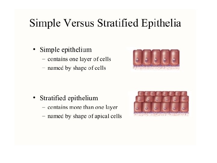
Epithelial Tissue • Two main types • - Covering and lining epithelia • - on external and internal surfaces • Glandular epithelia • Secretory tissue in glands
Glandular Epithelia ge – glandular epithelia lp – lamina propria
Epithelial Tissue • Layers of closely adhering cells • Flat sheet with upper surfaces exposed to the environment or an internal body cavity • No blood vessels • Underlying connective tissue supplies oxygen • Rests on basement membrane • Thin layer of collagen and adhesive proteins • Anchors epithelium to connective tissue
Basement Membrane
Basement Membrane • Not only an anchoring gel but also provides nutrients to the epithelium
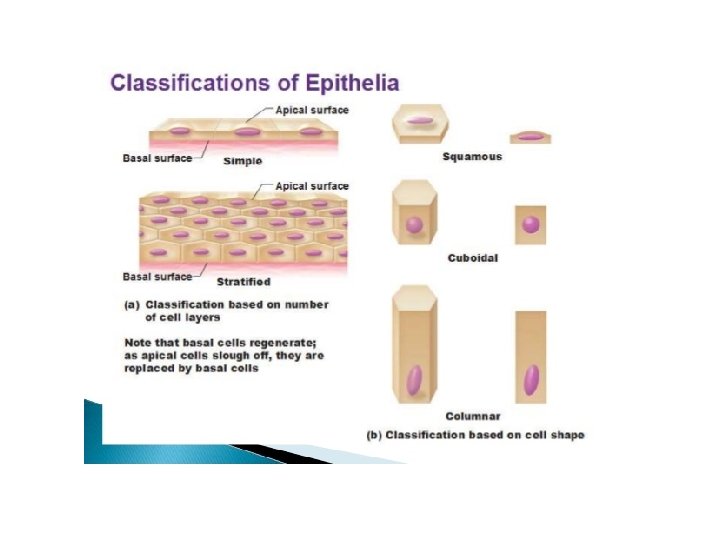
• Squamous – covering • Cuboidal and Columnar – absorption • The nucleus of columnar cells are found on the lower third of a cell
Squamous
Simple Squamous • 1 layer of flat cells
Serous Membranes – watery membrane
• Small intestine – has epithelial tissue lining the inside and outside • Outside of small intestine – serous membrane • Outer layer of the heart – serous membrane • What is the common feature of the small intestine and the heart?
• Heart is constantly pumping – the serous membrane also reduces wear and tear • Small intestine – produces watery secretions • - the water in the serous membrane reduces friction and therefore reduces wear and tear
Simple Squamous – Alveoli
Simple squamous – alveoli
• Surrounding the alveoli air sacs are capillaries • Alveoli – capillaries for oxygenation and removal of carbon dioxide • Allows for rapid exchange if you have simple squamous epithelium
Simple Cuboidal Picture
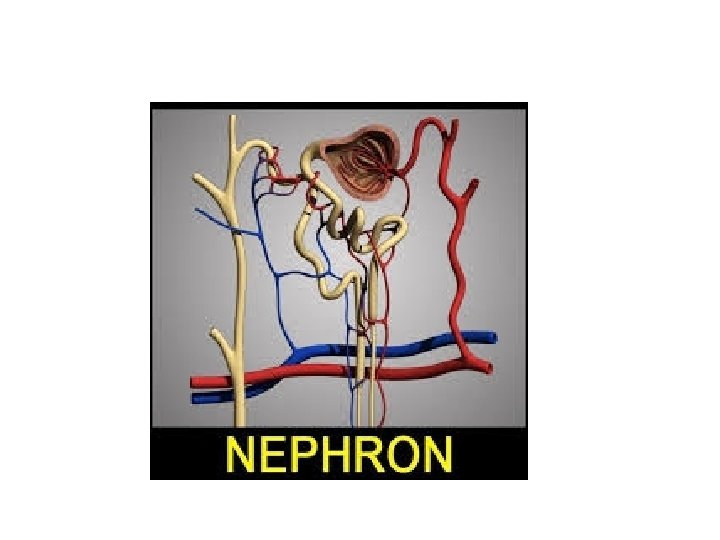
Simple Cuboidal Epithelium • Found in areas of secretion and excretion • Kidneys
Renal Tubules
Renal Tubules
Simple Cuboidal Epithelium • One layer of cube-shaped cells
Simple Columnar Epitheliumm
Simple Columnar Epithelium • Tallest of the epithelial cells • Found in small intestines – almost all of absorption of nutrients is found here
Small Intestine: Villi
Villi – finger-like extensions
Lumen – hollow opening within an organ
Crypts
Villi – in the apical surface are microvilli
• Microvilli – they form the brush border • Small extensions of the cell
• The taller the cells the higher the chance of absorption • The microvilli increases the absorptive area by increasing the height of cells • Whenever you see cells with microvilli you should think that these cells are used to absorb everything
Villi and Goblet Cells
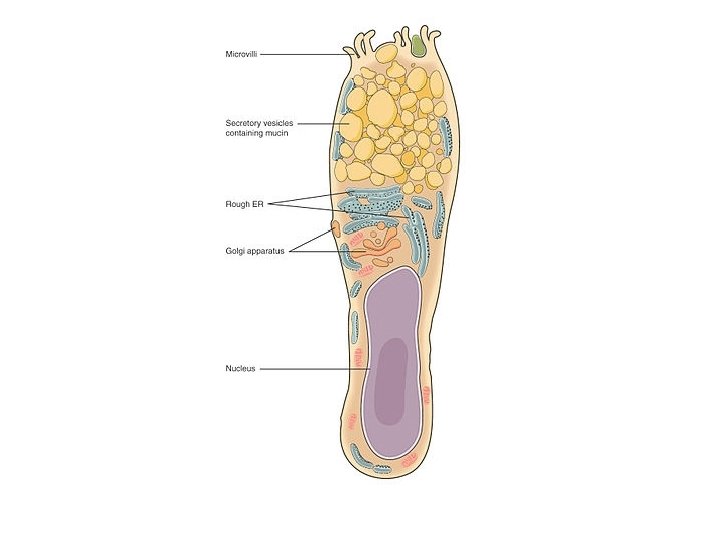
Goblet cells – produce mucus
Pseudostratified Columnar Epithelium • Stratified – multi-layered • - every single cell originates on one exact membrane but they are of different heights that is why they look stratified • Respiratory mucosa
Ciliated Epithelium – respiratory mucosa - lines the conducting airway of the lungs
Respiratory Mucosa Goblet Cells Secrete mucus and line the conducting airways Very sticky secretion Release a protein called Mucin combines with water and it becomes mucus • We want to clear the air before it reaches the our air sacs so that it would not damage our lungs • •
Fallopian tubes in females
• Which one moves? Cilia or microvilli?
Stratified Squamous Epithelium • 2 major types • a. keratinized • b. non-keratinized
Stratified Squamous Non Keratinized – wet areas of the body
Stratified Squamous Epithelium Non Keratinized – all cells are alive • Oral mucosa • Inner lining of the vagina • Lining of the esophagus • Wet, slippery areas
• Epithelial Tissue – high turnover meaning rapid change of cells like every 2 -3 days • - mitosis is fast
Stratified Squamous Keratinized
• Melanin – shields us from ultraviolet rays
Keratinocytes • The most abundant type of cells • Dead tough cells • Dead cells will exfoliate off
Stratified Cuboidal
Stratifed Cuboidal • • Quite rare in the body Found in some sweat and mammary glands Testicles Typically two cell layers thick
Stratified Cuboidal: Sweat Glands
Stratified Cuboidal: Seminiferous Tubules of the Testes
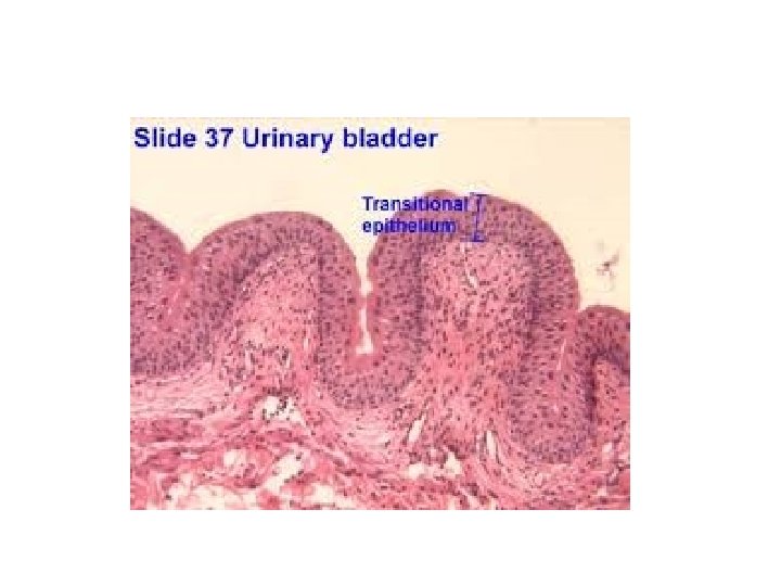
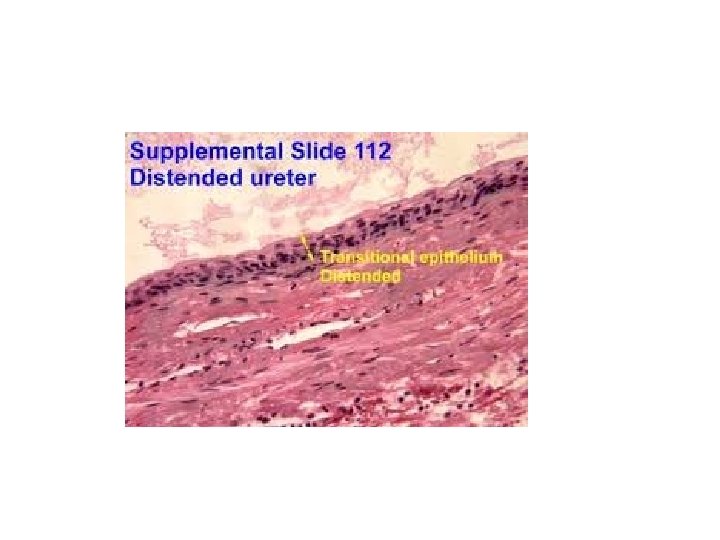
Transitional Epithelium • Coolest of them all • Either round or flat
Transitional Epithelium • Change depending on how much stress you apply • Found in the urinary system • - bladder, ureters, urethra
Glandular Epithelia • A gland is one or more cells that makes and secretes an aqeuous fluid • Classified by: • - Site of product released • - Structure of the gland (endocrine, exocrine) • - Distance travelled by secretion • - Relative number of cells forming the gland – unicellular (e. g. , goblet cells) or multicellular
• Endocrine glands – secrete into blood vessels • Exocrine glands – have ducts – ducts open into body surfaces
Pancreas • • Is a mixed gland It has endocrine and exocrine functions Most of it is exocrine gland 98% is exocrine in function, secretes digestive enzymes – pancreatic secretions go into the stomach
Pancreas
• Pancreatic secretion should not be going outside of the body • 2% of the pancreas is endocrine – secretion of insulin and glucagon
• Main job of a cell is to produce proteins • For every amino acid you make you have to burn ATP • Golgi Apparatus – packages proteins • for goblet cells it is the packaged mucin
• Goblet Cell – endocrine or exocrine? • - short distance of travel of mucin
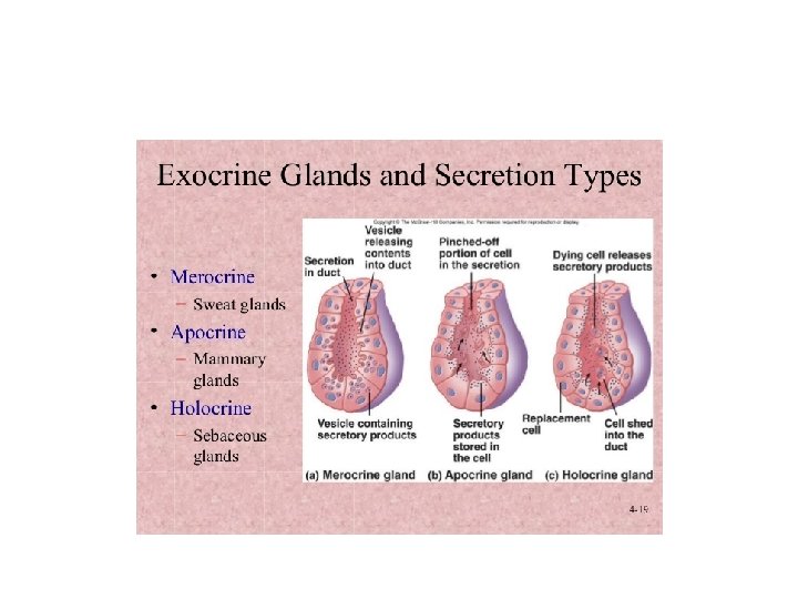
Exocrine Glands
• Sebaceous glands – oil releasing glands