Histology Study of Tissues Tissue Group of closely


 Secretion - glandular epithelium secretes hormones, mucus, digestive juices, and sweat D) Absorption")


 epithelium - forms epidermis of skin, surface of cornea, lining of")
 epithelium")


 - collagen is the most")


 - widely distributed - ground substance is")


 connective tissue - mostly fibers packed densely in the matrix")
 connective tissue - dermis")


 - produce a liquid")


- Slides: 45
Histology – Study of Tissues Tissue - Group of closely associated cells that are similar in structure and work together to perform a function. - Cells are nearly always embedded in or surrounded by nonliving matrix - Most organs contain some of each of the four main tissue types: - Epithelial, connective, muscle, nervous
Embryonic Germ Layers 1. Endoderm 2. 3. - inner layer which forms the lining of the digestive system Mesoderm - middle layer which forms muscles, bones, and blood vessels Ectoderm - outer layer that forms the skin and nervous system
Primary Germ Layers
Epithelial Tissue Serve to cover a body surface or line a cavity Two main types: 1. Covering and lining epithelium - all substances entering and leaving the body must pass through 2. glandular epithelium makes-up the glands of the body Functions A) Protection - from mechanical and chemical injury - from invasion by bacteria and viruses B) Sensory - specialized epithelium found in skin, nose, eye, and ear
C) Secretion - glandular epithelium secretes hormones, mucus, digestive juices, and sweat D) Absorption - lining epithelium of intestines and respiratory systems allow absorption of nutrients and gases E) Excretion - in kidneys and skin waste products are excreted F) Filtration - specialized epithelium in the kidneys filter waste products from the blood
Characteristics of Epithelial Tissue 1. Cellularity - composed almost entirely of cells - very little extracellular material 2. Specialized contacts - are held together in continuous sheets by many lateral contacts 3. Polarity - free surface = apical surface - exposed surface - cells often have microvilli or cilia attached to increase surface area - basal surface = attached
4. Basement Membrane - basal lamina is a noncellular adhesive sheet that attaches epithelial tissue to underlying structures - serves as a selective filter - important in healing of wounds - reticular lamina is connective fibers produced by underlying connective tissue - cancerous epithelial cells can pass through the basement membrane to underlying tissues 5. Innervated but avascular - contains nerve endings, but no blood vessels - nutrients must diffuse into the tissue from underlying tissues 6. Regeneration - as long as they receive nutrients they can regenerate by mitosis
Classification of Epithelia Each epithelium is given two names: First name indicates number of cell layers: A. Simple – single layer of cells - often found in areas of absorption and filtration B. Stratified – consists of two or more layers of cells - found in areas of abrasion Second name indicates the shape of the cells: A. squamous – flattened and scale-like B. cuboidal – cells are about as tall as they are wide C. columnar – cells are taller and column shaped
Classification of Epithelial Tissues
Simple Squamous - single layer of flattened cells - linings of alveoli, blood vessels, and many membranes - absorption, filtration, secretion
Simple Squamous lining of lung.
Simple cuboidal epithelium
Simple columnar epithelium
Pseudostratified ciliated epithelium - lines trachea, bronchi, epididymis, and vas deferens - protection and movement of mucus
Stratified squamous (keratinized) epithelium - forms epidermis of skin, surface of cornea, lining of mouth, esophagus, anus - protection
Stratified Squamous (nonkeratinized) epithelium
Transitional epithelium - often found in areas that are subjected to stress and tension changes, such as urinary bladder - usually composed of 10 or more layers of cuboidal cells
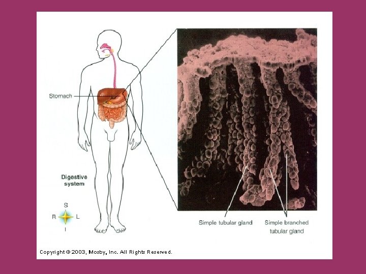
Glandular Epithelium - specialized for secretory activity - cells may function as unicellular or multicellular glands Glands are classified as: 1. Exocrine - discharge through ducts - ex. : salivary glands 2. Endocrine - secrete products directly into the blood or interstitial fluid - ex. : pituitary, thyroid, adrenals
Connective Tissues Functions: A. Binding and support - tendons and ligaments hold bones and muscles to each other - in soft organs, connective tissue supports and binds cells together B. Protection - bone and cartilage support and protect body organs C. Insulation - fat cushions, insulates, and protects body organs as well as providing reserve energy fuel D. Transportation - blood transports materials within the body
Characteristics of Connective Tissue 1. Intercellular Matrix - non-living material that makes-up most of the tissue - cells are embedded in the ground substance - gives different connective tissues their varying qualities - allows it to bear lots of weight, withstand tension, and endure physical abuse 2. Common origin - all originate from mesoderm 3. Various degrees of vascularity Fibers in connective tissue: A. Collagenous (white) - tough, often in bundles, high tensile strength, nonelastic
- found in tendons and ligaments (tough, flexible support) - collagen is the most abundant protein in our bodies B. Reticular fibers - delicate fibers found in networks with elastic and collagenous fibers - slightly elastic - found in lymphatic tissues (nodes) and supporting small structures C. Elastic fibers (yellow) - tough, elastic fibers that are not found in bundles - tough, expandable support - in aorta, arteries, trachea, and bronchi
Four main categories of connective tissues based on the matrix: 1. Fibrous - extracellular fibers are predominant features 2. Bone - has fibers and a hard mineral ground substance 3. Cartilage - fibers with a specialized ground substance that traps water to form a firm gel 4. Blood - lacks fibers - liquid matrix - plasma
Cells in connective tissues are designated by their suffix as to what they do: - blast – creates the matrix - cyte – maintains the matrix - clast – breaks down the matrix
Examples of Connective Tissues A. Loose (Areolar) - widely distributed - ground substance is a soft viscous fluid - collagenous and elastic fibers in a loose network - elastic glue – holds, but allows movement - found under skin – superficial fascia
B. Adipose Tissue - same matrix and fibers as areolar, but majority of tissue is composed of fat cells - fat cells are large, round, and delicate w/ nucleus - act as padding around organs, insulation for heat conservation, and as a reserve energy storage site
Fat storage areas – males versus females
C. Reticular connective tissue - spleen and lymph nodes - function in filtering and cleaning the blood
D. Dense fibrous (regular) connective tissue - mostly fibers packed densely in the matrix - found in areas needing tensile strength and flexibility - tendon, ligaments, dermis, scar tissue
E. Dense fibrous (irregular) connective tissue - dermis
F. Bone tissue - matrix of inorganic calcium salts and collagenous fibers - osteocytes – bone maintaining cells - organized in Haversion Systems - Osteons osteocytes in lacunae - lamellae – concentric circles of matrix - canaliculi – small canals connecting lacunae -
G. Cartilage - fiber matrix of collagenous and elastic fibers - chondrocytes – cartilage generating cells that secrete matrix and ground substance - firm, tough and flexible - lacks blood vessels Fibrocartilage Elastic Cartilage
H. Blood - matrix = plasma - 90% water w/ dissolved proteins, food, metabolic wastes and dissolved gases - formed elements made in bone marrow - erythrocytes – red blood cells – carry oxygen - leukocytes – white blood cells – defense against microbes - thrombocytes/platelets – help in clotting
Muscle Tissue - highly cellular and well vascularized - composed of contractile protein fibers called myofilaments Functions - provide movement - squeeze contents of internal organs - generate heat Striated vs. Nonstriated - striated muscle is arranged so that it has a striped appearance under the microscope Voluntary vs. Involuntary - voluntary muscles can be consciously controlled
Smooth muscle - found in internal organs - normally involuntary - spindle-shaped cells w/ one nucleus
Skeletal muscle - voluntary muscles responsible for movement - long, cylindrical fibers - striated multinucleate Cardiac muscle - muscle of the heart - branching fibers with single nucleus - intercalated discs connect cells - striated
Nervous tissue – functions in control mechanisms of the body and in coordinating responses to the environment
Neurons - nerve cells which transmit electrochemical signals - soma = cell body - axon – carries impulse away from soma - dendrite – carries impulse toward soma 1. Afferent = sensory - transmit impulses to CNS from sense organs 2. Efferent = motor, secretory, accelerator, inhibitory - transmit impulses from CNS to muscles or effector organs 3. Association - within CNS - run between afferent and efferent neurons Neuroglia - helper cells which nourish, protect, and insulate neurons
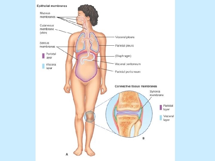
Membranes - thin sheet of tissue that covers a structure or lines a cavity - most are derived from epithelial and connective tissues 1. Serous membranes - line cavities that do not open to outside of body - normally composed of simple squamous epithelium - secretes fluid to decrease friction between structures - parietal layer – covers the body wall - visceral layer – covers the outer part of the organ 2. Mucous membranes - line cavities that do open to the outside - stratified squamous epithelium w/ connective tissue below - produces a fluid to trap particles or to keep underlying tissues moist
3. Synovial membranes - line synovial joints (mostly movable joints) - produce a liquid that lubricates and cushions the joint
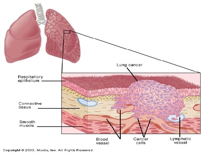
Tumors and Cancer Neoplasms, also called tumors, are any abnormal growth of cells Benign tumors do not spread to other tissues and normally grow slowly. - cells tend to stay together and are often in a capsule - normally not life threatening unless they interfere with organ function Malignant tumors, or cancers, are not encapsulated and tend to spread to other regions. - spread to other regions in blood or lymph - metastasis – spread of a cancer Cancer factors: - genetic factors, carcinogens, age
Detection and Treatment Cancer specialists are Oncologists - early detection is important 1. Self-examination - some cancers (breast and testicular for example) can be detected by self-examination 2. Medical imaging - radiography – x-ray photographs can find many - mammography - computed tomography (CT) – x-ray scanning - magnetic resonance imaging (MRI) - ultrasonography 3. Blood tests can detect abnormalities 4. Biopsy is removal and examination of living tissue
Without treatment cancer usually results in death. A. Surgical removal B. Chemotherapy - uses cytotoxic compounds and antineoplastic drugs to kill malignant cells C. Radiation therapy - uses x-ray or gamma radiation to destroy cancer - used alone or with chemotherapy D. Laser therapy - uses an intense beam of light to destroy a tumor E. Immunotherapy - attempts to bolster the body’s own immune system against cancer