HISTOLOGY OF TONGUE By Dr Sobia Ibrahim Assistant

HISTOLOGY OF TONGUE By Dr. Sobia Ibrahim Assistant Professor Anatomy, KEMU

Tongue is a muscular organ projecting into the oral cavity from its inferior side • Organ of speech • Organ of taste • It helps in swallowing and plays a role in digestion •

• • Three subdivisions Root/base Body Tip

Ø Tongue is basically a mass of striated muscle covered by mucous membrane Ø Lingual muscles are arranged in bundles that run in three planes; longitudinal, transverse, vertical Ø Muscles fibers firmly adherent to mucous membrane. Ø Between muscle fibers are gland Ø Mucous- at base Ø Serous- in body Ø Mixed- near tip
 § Ventral (inferior) Dorsal is roughened bcz")
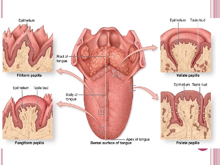
It has two surfaces § Dorsal (superior) § Ventral (inferior) Dorsal is roughened bcz of papillae Ventral is smooth Grossly dorsal surface is divided into anterior 2/3 and posterior 1/3 by sulcus terminalis

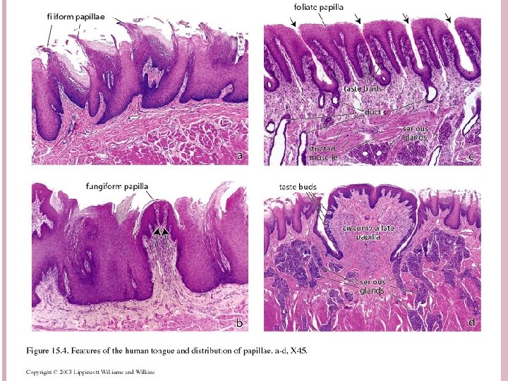
PAPILLAE Mucosal irregularities and elevations in different forms and functions Present anterior to sulcus terminalis on dorsum of tongue These papillae and associated taste buds constitute the specialized mucosa of oral cavity Filliform Ø Fungiform Ø Circumvallate Ø Foliate Ø

PAPILLAE OF TONGUE

FILLIFORM PAPILLAE Ø Smallest and most numerous in humans Ø Conical Ø Heavily keratinized Ø Lacks taste buds Ø Distributed over entire anterior dorsal surface Ø Provides rough surface

FUNGIFORM PAPILLAE Ø Less numerous Ø Mushroom shaped Ø Lightly keratinized Ø Covering epithelium thin Ø CT core with blood vessels Ø Taste buds on upper surface Ø Rich blood supply give its red color
 PAPILLAE Ø Least numerous Ø Largest in size Ø Abundance of taste")
VALLATE (CIRCUMVALLATE) PAPILLAE Ø Least numerous Ø Largest in size Ø Abundance of taste buds Ø Size = 1 -3 mm & number is 8 -12 Ø Lie anterior to sulcus terminalis Ø Surrounded by groove Ø Ducts of deeply located serous glands (von Ebner’s) open into groove

FOLIATE PAPILLAE Ø Poorly developed in humans Ø Forms furrows posterolateral aspect tongue Ø Contain taste buds on of

TASTE BUDS Ø Ø Ø Receptors for taste sensation Oval, pale staining bodies Extends through thickness of epith A small opening on epithelial surface at the apex of taste bud is Taste pore Sensations Sweet & salt- tip Ø Sour- sides Ø Bitter - back Ø

Ø Ø Ø Ø Consists of 50 -75 cells Three principal cells Neuroepithelial/ Gustatory cells having microvilli Supportive cells Stem cells Rest on basal lamina Gustatory cells supplied by sensory axon
 Crypts")
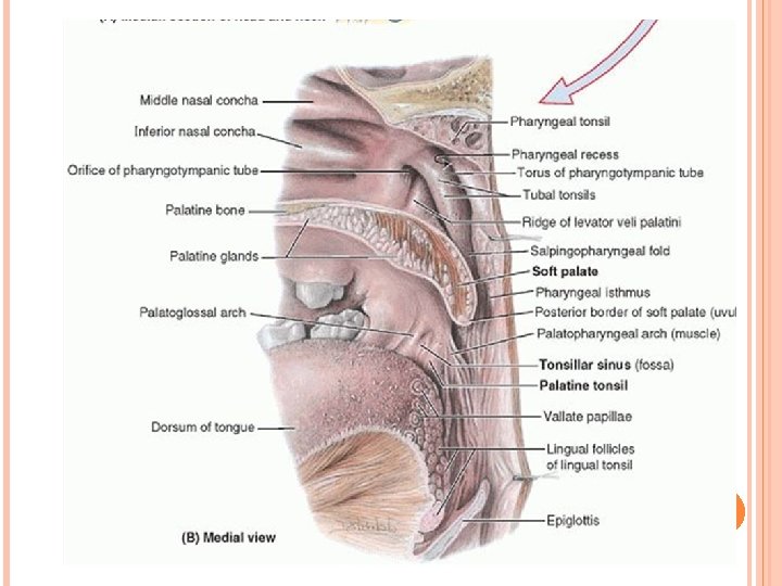
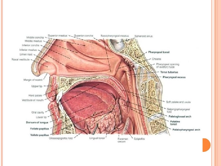
POSTERIOR 1/3 Irregular, nodular surface Lingual tonsil (Waldeyer’s ring) Crypts
- Slides: 18