Histology of the urinary tract Dr Stuart Brown

Histology of the urinary tract Dr Stuart Brown Histopathologist stuart. brown@sth. nhs. uk

Outline Kidneys Ureters Bladder Urethra

Kidney - surface anatomy Retroperitoneal Between T 12 and L 3 Right lower than left

Kidney - gross anatomy Three distinct structures Cortex Medulla Pelvis

A quick look at the different areas in the kidney

Cortex

Medullary Ray

Medullary Rays

Medulla

Pelvis

The kidney’s blood supply Abdominal aorta Renal artery at L 1 Anterior & posterior division Interlobar artery Arcuate artery (corticomedullary) Interlobular artery Afferent arteriole

Renal microvasculature

A lobulated kidney

A brief history of the kidney Pre 550 M 2 germ layers Cell surface transport No vascular system

Mesoderm and the Cambrian explosion 550 M 3 germ layers Mesoderm

Protonephridia in basal Chordates Salty marine environment Blood hypo-osmotic to environment Body gains salt and loses water by osmosis Aglomerular pronephridium excretes nitrogen without the loss of excess water

Freshwater, ammonia and the glomerulus Freshwater environment Blood hyperosmotic to environment Body gains water and loses salt by osmosis Glomeruli cause loss of greater volumes of fluid and help flush out ammonia

Land the loop of Henle 150 M - Land based life Limited access to water Need to conserve water Loop of Henle Concentrates urine

The kidney show us a map of our evolutionary heritage

Renal structure – the nephron There are millions of nephrons in the cortex and medulla Cortex Prox / Dist. convoluted tubules Renal corpuscles Medulla Loop of Henle Collecting ducts Pelvis Receives the collecting ducts

The nephron in gross anatomy

The nephron - the basic unit of the kidney Renal Corpuscle Proximal convoluted tubule Loop of Henle Distal convoluted tubule Collecting duct

Renal corpuscle A filter Tuft of convoluted capillaries with fenestrated walls Glomerular basement membrane Lined by podocytes Supported by mesangial cells Encased in Bowman’s capsule

Glomerulus

Glomerulus

Glomerular filter

Glomerular basement membrane

Podocytes

Review of ultrafiltration Form of barrier Mechanical barrier Repulsive negative charge Capillary fenestration + GBM + + Podocyte slit membrane + +

Juxtaglomerular apparatus 2 Components: 1. Afferent arteriole 2. Distal convoluted tubule 1. Afferent arteriole Contributes Granular cells Secrete renin in response to: Systemic blood pressure

Juxtaglomerular apparatus 2. Distal convoluted tubule Contributes the Macula Densa Patch of closely packed endothelial cells along tubule Senses Na. Cl concentration and regulates tubuloglomerular feedback

JGA

Proximal convoluted tubule Cuboidal epithelium Round central/basal nuclei Brush border of microvilli at apical end Many mitochondria so appears eosinophilic Reabsorption of Na. Cl, proteins, polypeptides, amino acids, glucose

Proximal convoluted tubule EM Lysosomes in the PCT are involved in degradation of small protein molecules reabsorbed from the urinary space

Loop of Henle Descending and ascending limbs Both with thick and thin segments Thin – simple squamous Thick – low cuboidal Supplied by a rich vasa recta

Tamm-Horsfall Protein Produced in the thick ascending tubule Low levels in urine associated with calculi Mutations associated with renal failure Absence associated with severe infection
 Peritubular capillaries Descends")
Loop of Henle and vasa recta Afferent arteriole (from renal corpuscle) Peritubular capillaries Descends into medulla Vasa recta Renal veins Inferior Vena Cava

Distal convoluted tubule Low cuboidal epithelium Scanty microvilli Numerous mitochondria Regulates acid base by Secreting H+ and absorbing HCO 3 - (via cellular carbonic anhydrase) Regulates Na level by exchanging Na for K

Collecting duct Cuboidal epithelium Principal cells Respond to aldosterone Reabsorbing Na Secreting K Also respond to ADH Inserting Aquaporin 2 Intercalated cells Exchange H+ for HCO 3 -

Collecting Duct

Aquaporin 2
 Transmits filtrate from nephron to ureter")
Renal pelvis Transitional epithelium (urothelium) Transmits filtrate from nephron to ureter

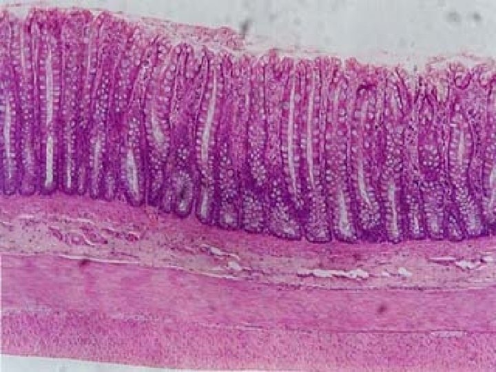
Urothelium AKA Transitional epithelium About 5 cells thick Changes shape and stretches

Urothelium Surface layer – large dome-shaped umbrella cells Intermediate layer - note nuclear orientation Basal layer – cuboidal cells

Urothelium The umbrella cells are large and cover several underlying intermediate cells. They have tight junctions at their surface

Tight Junctions - Zonula Occludens Prevent liquid movement around cells Composed of Claudins And Occludins Anchored to Actin in the cytoskeleton
")
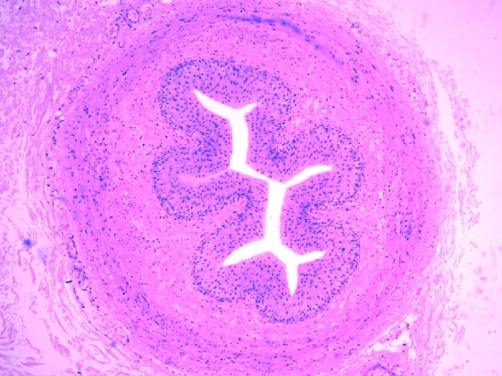
Ureters Transitional epithelium Spiral muscular tube Inner “longitudinal” Outer “circular” (different to GI arrangement) No serosa Loose adventitia

Bladder Transitional epithelium Lamina propria Muscularis Mucosa Submucosa Muscularis Propria Subserosa and Serosa Functional valve prevents reflux into ureter

Urethra Different anatomy in males and females Two sphincters Internal sphincter composed of smooth muscle from the bladder External sphincter composed of skeletal muscle from the pelvic floor

Urethra - female 4 -5 cm long Proximally transitional epithelium Distally squamous epithelium Paraurethral and periurethral glands open into the urethra

Urethra - male 20 cm long 3 parts 1. Prostatic urethra 2. Membranous urethra Transitional epithelium 3. Penile urethra Pseudostratified epithelium proximally Stratified squamous epithelium distally

Summary The kidney has a complicated histology reflecting its complicated function and our evolutionary history The renal pelvis, ureter, bladder and urethra all have the same transitional epithelium Transitional epithelium is unique to the urinary tract

Quiz time!

Q 1. A 5 year old child has nephrotic syndrome. A renal biopsy shows normal glomeruli. Electron microscopy shows this following appearance: What is the abnormality here?

Q 2. An adult has nephrotic syndrome. A renal biopsy shows the following appearance. What is the abnormality here?

Q 2. A silver stain of the tissue demonstrates the following: What is the diagnosis?

Q 3. Another adult has nephrotic syndrome. A renal biopsy shows the following appearance. What is the abnormality here?

Q 3. A silver stain of the tissue demonstrates the following: What is the diagnosis?

Q 4. Another adult is coughing up blood and has blood in their urine. They have antibodies to the glomerular basement membrane. This is their kidney biopsy: What is the diagnosis?

Do you have any questions?
- Slides: 62