Histology of the Urinary System Lecture Objectives Describe

Histology of the Urinary System

Lecture Objectives • Describe the normal microscopic appearance of the different parts of the kidney including cortex, medulla, juxtaglomerular apparatus and the distribution of the vasculature within the kidney. • List the different parts of the nephron with the details of each part. • Describe the microscopical appearance of the ureter, urinary bladder and urethra. • Compare the histological appearance of the distended and contracted bladder.

Internal Anatomy of the Kidneys • Parenchyma of kidney – Renal cortex = superficial layer of kidney – Renal medulla • inner portion consisting of 8 -18 cone-shaped renal pyramids separated by renal columns • renal papilla point toward center of kidney – Medullary rays, parallel arrays of tubules penetrate the cortex from the base of the medullary pyramids • Drainage system fills renal sinus cavity – minor & major calyces empty into the renal pelvis which empties into the ureter – minor calyces collect urine from the papillary ducts of the papilla
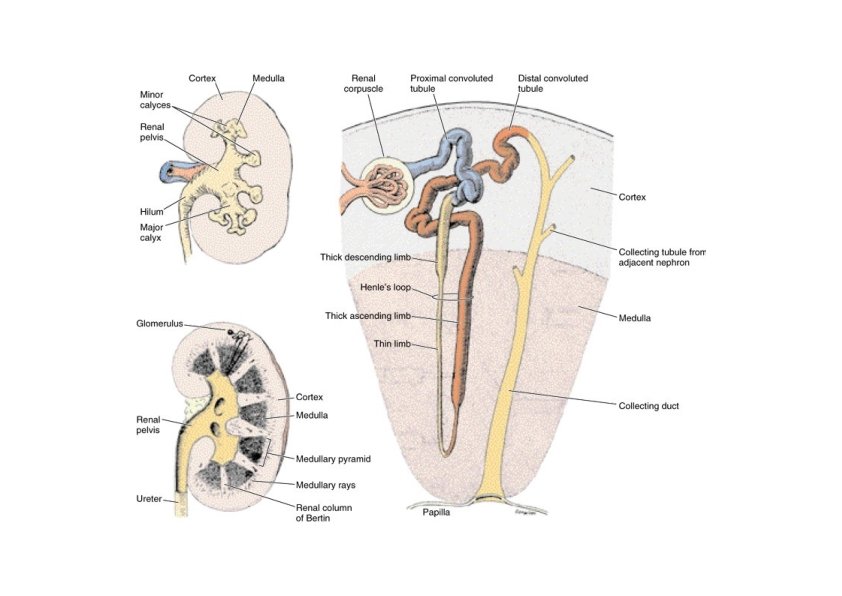

Nephrons • A nephron consists of a renal corpuscle where fluid is filtered, and a renal tubule into which the filtered fluid passes • Renal corpuscle = site of plasma filtration – glomerulus is capillaries where filtration occurs – glomerular (Bowman’s) capsule is double-walled epithelial cup that collects filtrate
 – loop")
Nephrons • A renal tubule consists of: – proximal convoluted tubule (PCT) – loop of Henle (nephron loop) – distal convoluted tubule (DCT) • Distal convoluted tubules of several nephrons drain into to a single collecting duct and many collecting ducts drain into a small number of papillary ducts.

Nephrons • The loop of Henle consists of: – – thick descending limb thin ascending limb thick ascending limb

Nephrons: Types There are two types of nephrons that have differing structure and function • A cortical nephron – glomerulus in the outer portion of the cortex – short loop of Henle • penetrates only into the outer region of the medulla • A juxtamedullary nephron – glomerulus deep in the cortex close to the medulla – long loop of Henle stretches through the medulla and almost reaches the renal papilla

Cortical Nephron • 80 -85% of nephrons are cortical nephrons • Renal corpuscles are in outer cortex and loops of Henle lie mainly in cortex

Juxtamedullary Nephron • 15 -20% of nephrons are juxtamedullary nephrons • Renal corpuscles close to medulla and long loops of Henle extend into deepest medulla enabling excretion of dilute or concentrated urine

Renal Corpuscle • Renal Corpuscle is about 200 μm in diameter • Consists of: – Bowman’s capsule surrounds capsular (urinary) space • podocytes cover capillaries to form visceral layer • simple squamous cells form parietal layer of capsule – Glomerular capillaries arise from afferent arteriole & form a ball before emptying into efferent arteriole • Fenestrated endothelial cells constitute the capillaries • Renal corpuscle have a vascular pole where the afferent arteriole enters and the efferent arteriole leaves, and a urinary pole where proximal convoluted tubule begins

Glomerular Capsule • • The glomerular capsule consists of visceral and parietal layers The parietal layer consists of simple squamous epithelium supported by basal lamina and thin layer of reticular fibers • • Near the urinary pole the epithelium becomes cuboidal or low columnar The visceral layer consists of modified simple squamous epithelial cells called podocytes.

Glomerular Capsule • Podocytes have cell body from which arise several primary processes – – – Each primary process gives rise to many secondary processes (pedicels) Only the secondary processes have direct contact with the basal lamina Filtration slits , is a 25 nm wide elongated spaces between the secondary processes • • Filtration slits are covered by 6 nm thick diaphragm A thick basement membrane (0. 1 μm) separates the capillaries endothelium and the podocytes – – Lamina densa covered by lamina rara on each side Basement membrane is selectively permeable for macromolecules

Glomerular Capillaries • Glomerular capillaries have mesangial cells adhere to their walls • mesangial cells are contractile cells and have receptors for angiotensin II, so it can reduce the glomerular flow • mesangial cells contains also receptors for natriuretic factor produced by the cardiac atria cells causing vasodilation and relaxes mesangial cells • mesangial cells also have several functions – – Support the glomerulus Produce extracellular matrix Immune response Outside the glomerulus in the vascular pole, form the extraglomerular mesangial cells, which is part of the extraglomerular apparatus

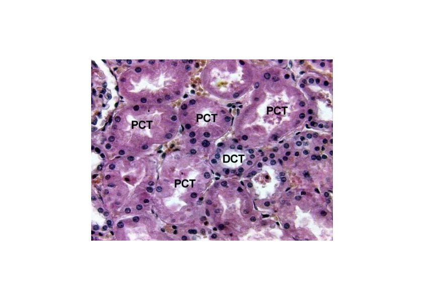
Proximal convoluted tubules • Longer than the distal convoluted tubule, thus seen more near the renal corpuscle in the cortex • Consists of cuboidal cells • Acidophilic cytoplasm , numerous elongated mitochondria • Brush border, abundant microvilli • Large cells, 3 -5 surround each tubule • Lateral interdigitation connecting lateral walls of cells, difficult to distinguish boundaries between adjacent cells • Basal infoldings with mitochondria (basal striations), characteristic of cells with active ionic transport

Proximal convoluted tubules • Functions – Resorption of 80% water, electrolytes through Na+, Ka+ATPase – Resorption of 100% glucose and amino acids – Protein resorption: apical canaliculi connecting microvilli increase the absorption of macromolecules by pinocytosis. • Vacuoles and lysosomes are more abundant in the apical portion of the cytoplasm – Secretion of creatinine and organic acids and bases, so p. H modification
 – Cytology • Similar to")
Loop of Henle • Thick descending limb (60 μm) – Cytology • Similar to proximal convoluted tubules • Slightly fewer microvilli • Less basolateral interdigitation • Mitochondria are smaller and more randomly oriented – Function: Na+ and H 2 O reabsorption • Thin tubule (12 μm) – Cytology: Simple squamous epithelium – Function • Descending limb: permeable to H 2 O and salt • Ascending limb: impermeable to H 2 O, permeable to salt and urea • Concentration of the urine
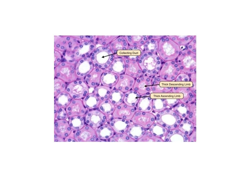

Loop of Henle • Thick ascending limb – Cytology • Cuboidal cells with apical nuclei • Numerous basal infoldings with larger mitochondria • Few and short microvilli • Macula densa, part of juxtaglomerular apparatus – Function • Cl- and Na+ transport into basolateral spaces • Establish a salt concentration gradient in medulla for urine concentration

Distal convoluted tubule • Cytology – Same as thick ascending limb of loop of Henle – Cells are flatter and smaller than those of proximal convoluted tubules, so more cells will appear in cross section • Function – Na+ reabsorption, Ka+ secretion (aldosterone) – Reabsorption of bicarbonate, hydrogen secretion, p. H increases – Conversion of ammonia to ammonium ions

Collecting tubules and ducts • Types – Connecting tubule: 40 µm in diameter, in cortex – Cortical duct: medullary rays – Medullary duct: medulla – Papillary duct (of Bellini): 200 µm in diameter, apex • Epithelium: cuboidal to columnar cells – Principal cells: light stained cells, cilium, short microvilli, basal infoldings, permeable to H 2 O (H 2 O channel, in the presence of antidiuretic hormone) – Intercalated cells: dark stained cells, microplicae (folds), H+ secretion, bicarbonate reabsorption. Absent in inner medulla

Juxtaglomerular Apparatus • Senses and regulates blood flow and composition • Structure where afferent arteriole makes contact with ascending limb of loop of Henle • Consists of three cell types – juxtaglomerular cells are modified muscle cells in arteriole, renin secretion – macula densa: sense Na. Cl concentration, regulates renin release. It is thickened part of ascending limb – the extraglomerular mesangial cells: unknown function

• Regulation of blood flow by renin Stimulation → JG cells → Renin → Angiotensinogen → Angiotensin I (lung) → Angiotensin II → Aldosterone (Adrenal cortex) → Na+, Cl- uptake (DCT)

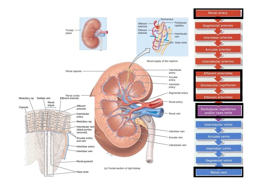
Blood & Nerve Supply of Kidney • Abundantly supplied with blood vessels – receive 25% of resting cardiac output via renal arteries • Functions of different capillary beds – glomerular capillaries where filtration of blood occurs • vasoconstriction & vasodilation of afferent & efferent arterioles produce large changes in renal filtration – peritubular capillaries that carry away reabsorbed substances from filtrate – vasa recta supplies nutrients to medulla without disrupting its osmolarity form • The nerve supply to the kidney is derived from the renal plexus (sympathetic division of ANS). Sympathetic vasomotor nerves regulate blood flow & renal resistance by altering arterioles

Urinary bladder & Urinary passages • Bladder and urinary passages store and conduct urine to the exterior • All have the same histological structure: • The mucosa – Transitional epithelium – Lamina propria • Dense woven sheath of smooth muscle – – Calyces, renal pelvis and ureters: helical arrangement Distal part of ureter: longitudinal Bladder: run in every direction Bladder neck: • Internal longitudinal. Distal to bladder neck become circular – Surround prostatic urethra – Extend to external urethral meatus in women • Middle circular: ends at the bladder neck • Outer longitudinal layer: continue to the prostate in men and to the external urethral meatus in women • Adventitia • Serosa: upper part of the bladder

A. Empty bladder B. Full bladder ureter

Urethra • Male urethra: consists of four parts – Prostatic urethra • Close to bladder, and ducts of prostate gland opens into it • Transitional epithelium • Dorsal and distal part have verumontanum (elevation protrudes into its interior) – Closed tube (prostatic utricle) opens into the tip with no known function • Ejaculatory ducts open on the sides of the verumontanum – Membranous urethra • 1 cm, surrounded by the external urethra sphincter (striated muscle) • Stratified or pseudostratified columnar epithelium – Bulbous and pendulous parts of urethra • • Located in the corpus spongiosum of the penis Lumen dilates distally forming the fossa navicularis Pseudostratified columnar with stratified squamous areas Litter’s glands: mucous glands found along the entire urethra and mostly in the pendulous part • Female urethra – 4 -5 cm long tube, lined by stratified squamous epithelium and areas of pseudostratified columnar epithelium – The mid part is surrounded by external urethral sphincter
- Slides: 29