
Histology of the respiratory system review D L
- Slides: 27
Histology of the respiratory system - review D. L. Kiss Anna Semmelweis University Department of Anatomy, Histology and Embryology 2019.
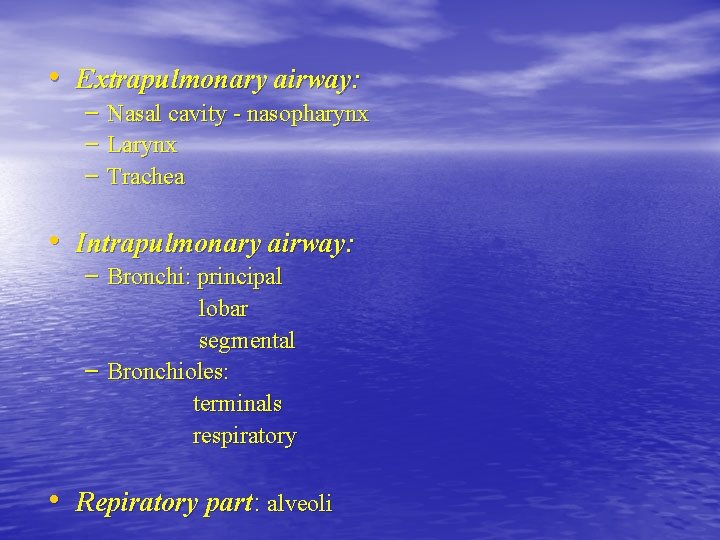
• Extrapulmonary airway: – Nasal cavity - nasopharynx – Larynx – Trachea • Intrapulmonary airway: – Bronchi: principal lobar segmental – Bronchioles: terminals respiratory • Repiratory part: alveoli
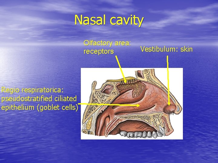
Nasal cavity Olfactory area: receptors Regio respiratorica: pseudostratified ciliated epithelium (goblet cells) Vestibulum: skin
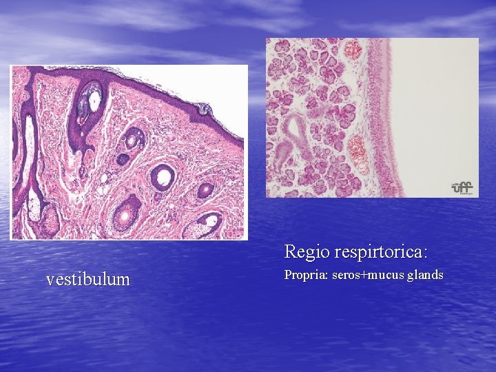
Regio respirtorica: vestibulum Propria: seros+mucus glands
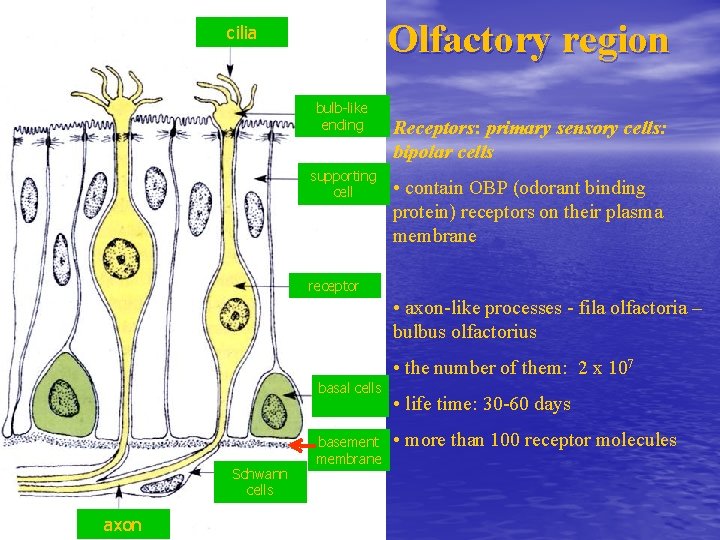
Olfactory region cilia bulb-like ending supporting cell Receptors: primary sensory cells: bipolar cells • contain OBP (odorant binding protein) receptors on their plasma membrane receptor • axon-like processes - fila olfactoria – bulbus olfactorius • the number of them: 2 x 10⁷ basal cells Schwann cells axon basement membrane • life time: 30 -60 days • more than 100 receptor molecules
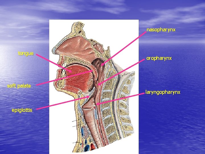
nasopharynx tongue oropharynx soft palate laryngopharynx epiglottis
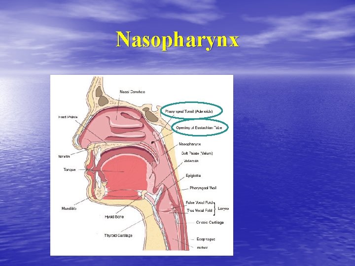
Nasopharynx
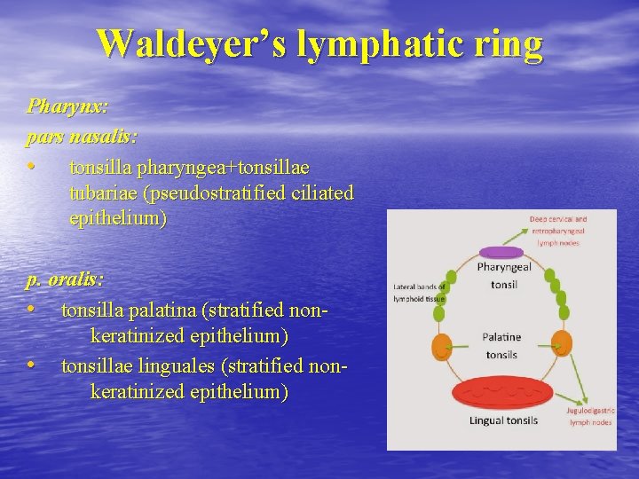
Waldeyer’s lymphatic ring Pharynx: pars nasalis: • tonsilla pharyngea+tonsillae tubariae (pseudostratified ciliated epithelium) p. oralis: • tonsilla palatina (stratified nonkeratinized epithelium) • tonsillae linguales (stratified nonkeratinized epithelium)
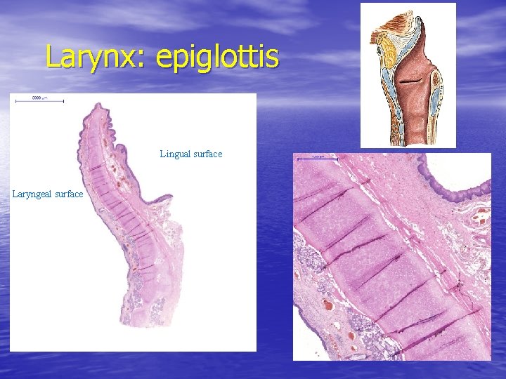
Larynx: epiglottis Lingual surface Laryngeal surface
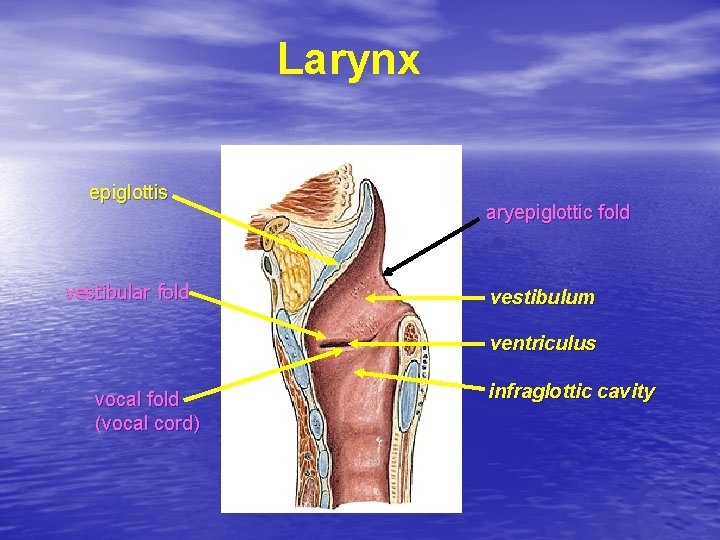
Larynx epiglottis vestibular fold aryepiglottic fold vestibulum ventriculus vocal fold (vocal cord) infraglottic cavity
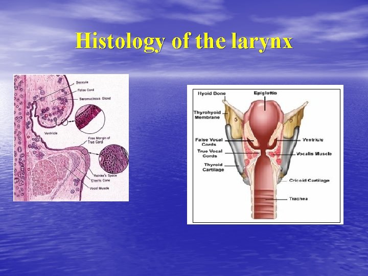
Histology of the larynx
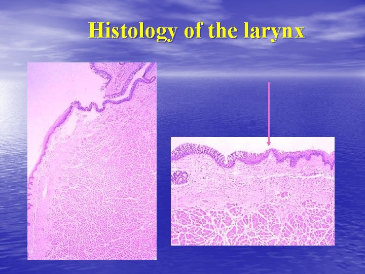
Histology of the larynx
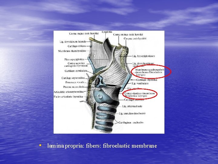
• lamina propria: fibers: fibroelastic membrane
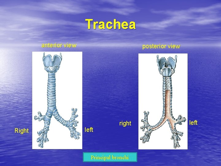
Trachea anterior view Right posterior view left right Principal bronchi left
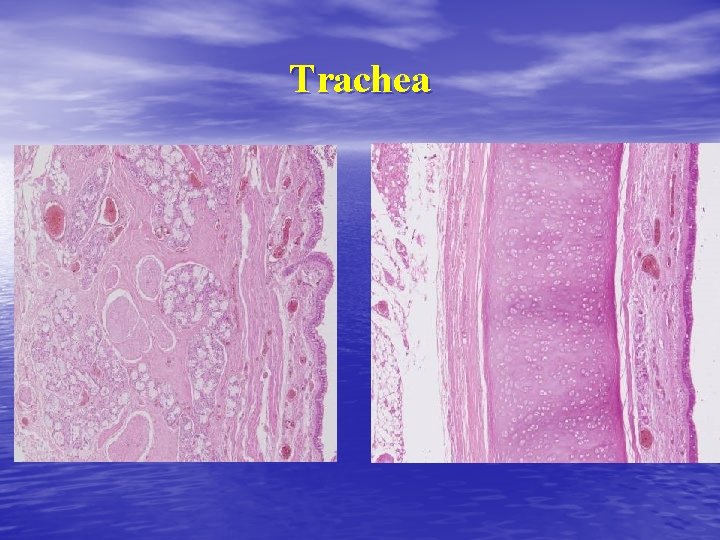
Trachea
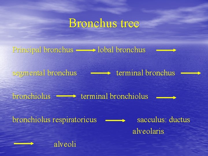
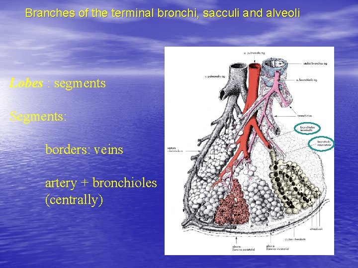
Bronchus tree Principal bronchus lobal bronchus segmental bronchus bronchiolus terminal bronchiolus respiratoricus alveoli sacculus: ductus alveolaris
Branches of the terminal bronchi, sacculi and alveoli Lobes : segments Segments: borders: veins artery + bronchioles (centrally)
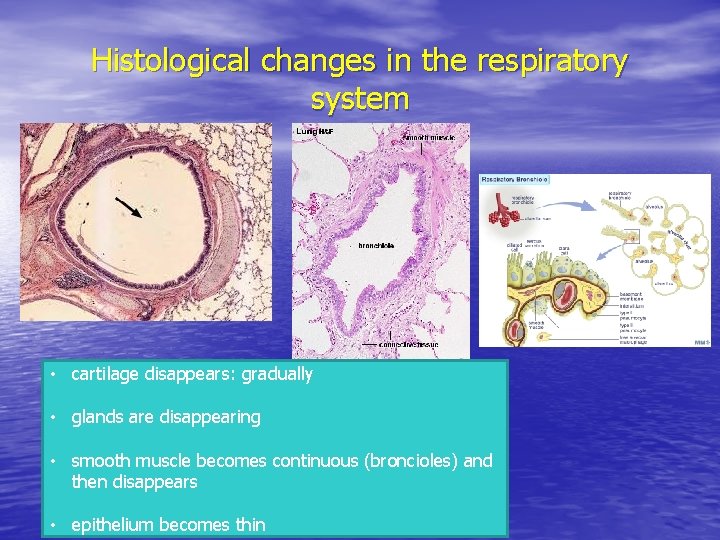
Histological changes in the respiratory system • cartilage disappears: gradually • glands are disappearing • smooth muscle becomes continuous (broncioles) and then disappears • epithelium becomes thin
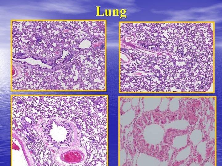
Lung
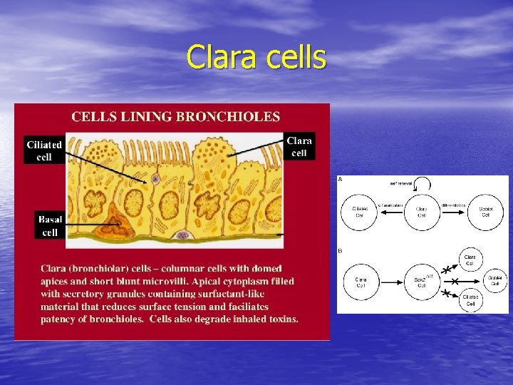
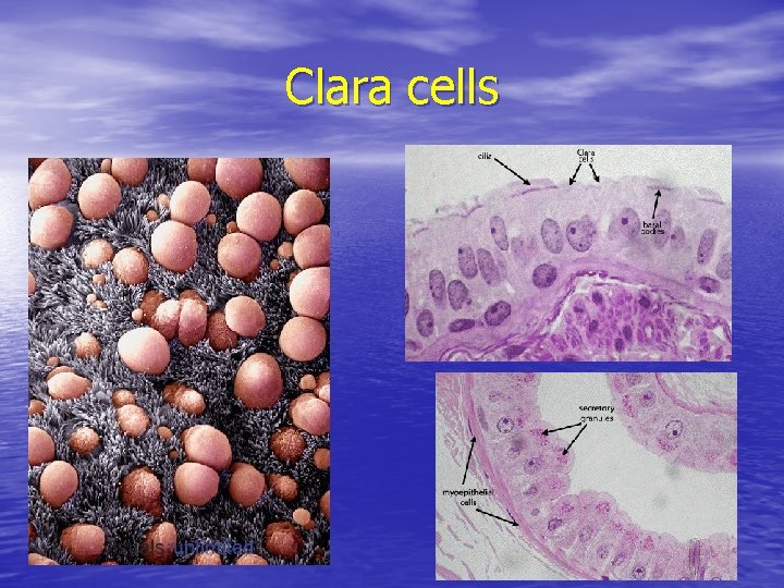
Clara cells
Clara cells
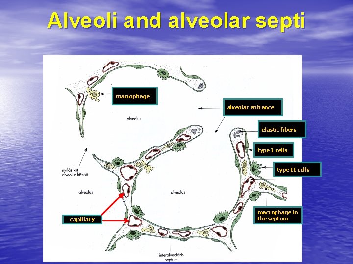
Alveoli and alveolar septi macrophage alveolar entrance elastic fibers type I cells type II cells capillary macrophage in the septum
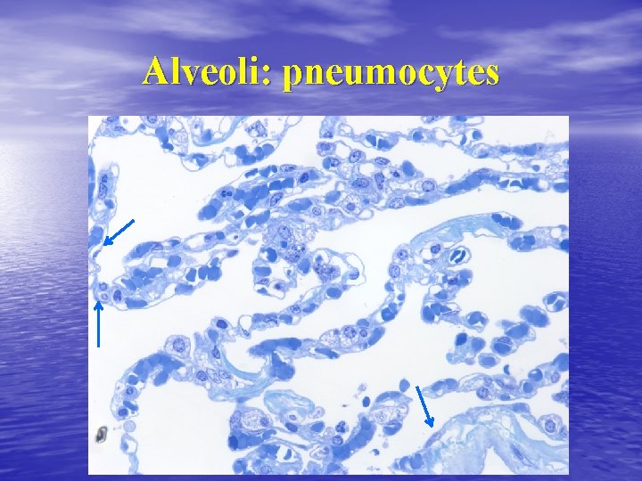
Alveoli: pneumocytes
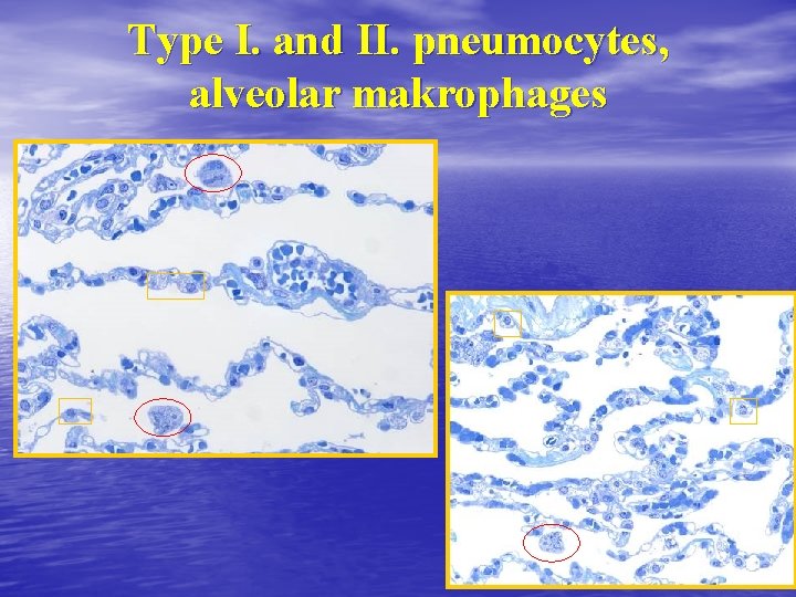
Type I. and II. pneumocytes, alveolar makrophages
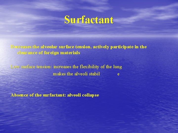
Surfactant Decreases the alveolar surface tension, actively participate in the clearance of foreign materials Low surface tension: increases the flexibility of the lung makes the alveoli stabil e Absence of the surfactant: alveoli collapse
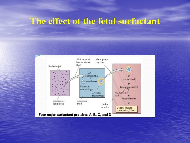
The effect ot the fetal surfactant
Bibliography • Snell RS, Clinical Anatomy, Little, Brown & Co, • • • Boston, 1995 Moore KL, Dalley AF: Clinically Oriented Anatomy, Lippincott, 1999 Sobotta: Atlas of Human Anatomy Röhlich: Szövettan