Histology of the Male Reproductive System Repro 5
 PROF. DR. FAUZIAH OTHMAN DEPT OF")


- Slides: 28
Histology of the Male Reproductive System (Repro 5) PROF. DR. FAUZIAH OTHMAN DEPT OF HUMAN ANATOMY
Contents • Histology of testes • Histology of vas deferens • Histology of seminal vesicle • Histology of prostate gland • Histology of the Bulbourethral gland
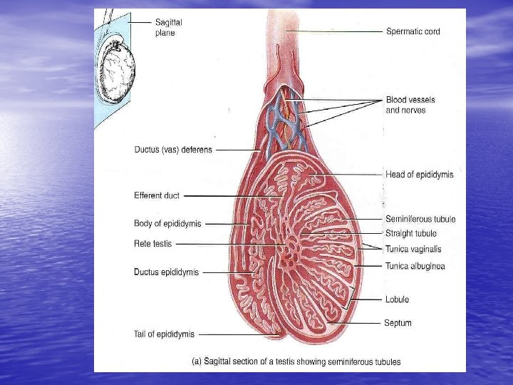
Histology of testes
• Tunica albuginea • dense white fibrous capsule • composed of dense irregular connective tissue, • forming septa that divide each testis into series internal compartments called lobules.
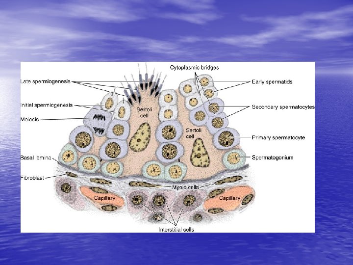
Spermatogenesis in seminiferous tubules
Seminiferous tubules • Stratified germinal epithelium • Surrounded by a layer CT with fibroblast and an inner basement membrane (bm). – Consist: Supporting (Sertoli cell) v Slender, elongated cells with irregular outlines that extend from the bm. v Primary spermatocytes v Spermatids
Spermatogenesis • Process where the spermatogenic cells in the • seminiferous tubules divide, differentiate, and produce sperm. 3 phases – Mitotic division of spermatogonia – Meiotic division of spermatocytes, somatic chromosome no. – spermatids – Spermiogenesis morphological transformation spermatids sperm.
Testes produce both testosterone and sperm • LH and FSH are produced by the pituitary gland. • LH binds to receptors on the interstitial cells (Leydig cells) and stimulate testosterone. • FSH stimulates Sertoli cells to synthesize and release ABP (androgen-binding protein) into seminiferous tubules to stimulate spermatogenesis. • ↑ concentration of testosterone essential for proper spermatogenesis.
Sertoli cells • Supportive cells in the seminiferous tubules located among the spermatogenic cells • Fx: physical support, protection and nutrition of the developing sperm (spermatids) • Phagocytosis of cytoplasm developing sperm (spermatids) • Produce and release ABP.
Seminiferous tubule, straight tubules, rete testis & ductuli efferentes
Ductuli efferentes of the ductus epididymis
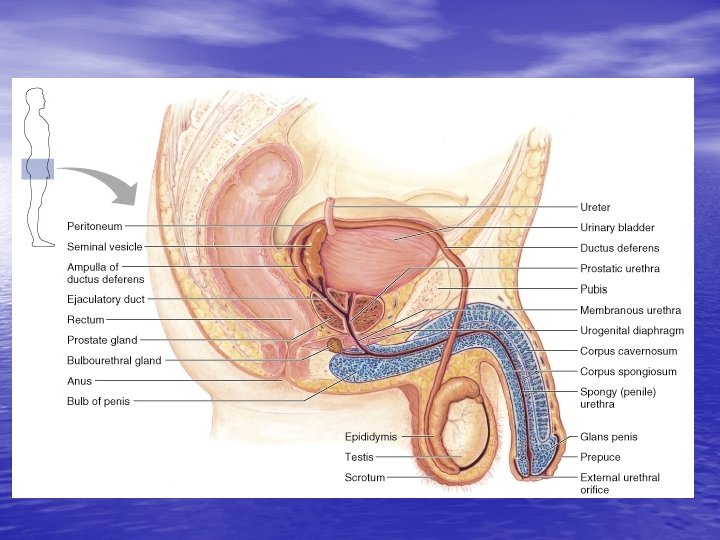
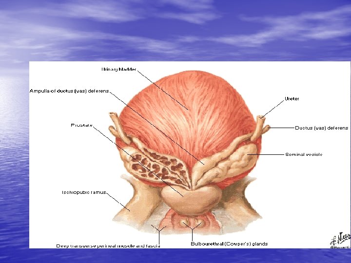
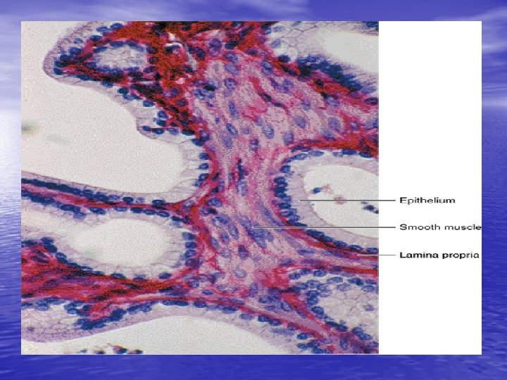
Histology of vas deferens
Vas deferens • A narrow and irregular lumen with longitudinal mucosal folds. • Psuedostratified columnar epithelium with stereocilia. • Thin lamina propria • consists of compact colagen fibers and fie net work of elastic fibers. • Thick muscularis • 3 SM layers – – – Inner longitudinal Middle circular Outer longitudinal • Adventitia. • Abundant of blood vessels
Ampulla of the vas deferens • Terminal portion of the vas deferens enlarges • into an ampulla Lumen of ampulla larger than vas deferens. • Numerous irregular branching mucosal folds • Deep glandular diverticula or crypts • Simple columnar or cuboidal epithelium. • Lamina propria • 3 layers of SM • Thin inner longitudinal • Thick middle circular • Thin outer longitudinal
Ampulla of the vas deferens
Accessory reproductive glands • Seminal vesicles • • • Yellowish viscous fluid high in fructose Energy source for sperm motility Produce most fluid found in semen • Prostate gland • Porduce thin, watery slightly acidic fluid • The enzyme fibrinolysin liquefies the semen after ejaculation. • Bulbourethral glands • Produce clear, viscid, mucus-like secretion during erotic stimulation • As lubricant for thr penile urethra.
Histology of seminal vesicle
Glandular epithelium of seminal vesicles normally varies low pseudostratified, low columnar or cuboidal.
Prostate gland & prostate urethra
• Prostate gland • Prostetic urethra – Psuedostratified epithelium • Glandular acini vary in size • Lumina of acini normally wide and irregular – Protrusion of epithelium-covered connective tissue folds – Proteinaceous secretions sometimes prostatic concretions. – Glandular epithelium simple-columnar or psuedot Fibromuscular stroma – Characteristic feature of prostate gland v SM bundles and CT fibers blend together in the stroma throughout the gland. v ratified, cells lightly stain, some region squamous or cuboidal.
Histology of prostate gland
Histology of the Bulbourethral gland