Histology of the Liver Dr Rehab Ahmed Associative

Histology of the Liver Dr Rehab Ahmed Associative professor Histology & Cell Biology Department

Liver Parenchyma cv
 LM structure Ø Shape: polyhedral cells large Ø Nucleus: large,")
THE HEPATOCYTE (liver cell) LM structure Ø Shape: polyhedral cells large Ø Nucleus: large, centrally located, spherical nucleus with prominent nucleolus. Ø Some hepatocytes are binucleated

The cytoplasm: is eosinophilic (due to the presence of numerous mitochondria and smooth endoplasmic reticulum) Sometimes vacuolated (due to abundant glycogen granules and fat droplets).

EM of Hepatocytes 1 - Mitochondria are abundant for energy requirement for metabolic activity (800 -1000/ cell). 2 - Rough endoplasmic reticulum is well developed for synthesis of plasma proteins & lipoproteins. 3 - Multiple Golgi complexes (50 units) (secretory activity of the cell). 4 - Smooth endoplasmic reticulum (detoxification & glycogen & cholestrol formation) s. ER

Ultrastructure of hepatocytes ØPeroxisomes For oxidation of fatty acids. N ØGlycogen granules L and lipid droplets. ØLysosmes & lipofuscin pigments r. ER
? 2. Intercellular g side; short")
1. Vascular side § Facing sinusoid; long microvilli (mv)? 2. Intercellular g side; short mv § Bounded by; tight junctions & desmosomes Bile canaliculus

Perisinusoidal space Space of Disse Where ? ! Between the endothelial lining of the sinusoids and the hepatocytes
 Definition: space that exists between the endothelial lining of")
Perisinusoidal space(Space of Disse ) Definition: space that exists between the endothelial lining of the sinusoids and the hepatocytes. Contents: (prevent the collapse of sinusoidal wall) 1. Plasma 2. Reticular fibres 3. Microvill. 4. Lipocytes or Ito cells =fat storing cells.

Perisinusoidal space of Disse Why ? ! Provides optimal conditions for Prevent the extensive collapse of metabolic sinusoidal wall. exchange between the blood plasma and hepatic cells.

Perisinusoidal space Space of Disse Contents ? ! Microvilli of Hepatocytes Ito cells Reticular fibers Plasma
 Vitamin A-storing Chronic inflamation Stellate shaped mesenchymmal cell")
Hepatic stellate cells (Ito cells ) Vitamin A-storing Chronic inflamation Stellate shaped mesenchymmal cell Myofibroblast Deposits collagen Liver fibrosis

Ø Few in number. ØSupport and prevent the collapse of sinusoidal wall.

Biliary tree

Bile formation & secretion Bile canaliculus
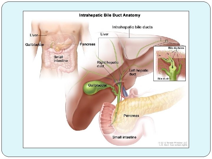

Bile canaliculi Bile ductules Canals of Hering Bile ducts Hepatic ducts Columnar cells Hepatocytes Cuboidal cells Cuboidal then columnar

Gall bladder ØHollow pear shaped organ attached to the lower surface of the liver. Right hepatic duct Left hepatic duct

Histology of gall bladder Mucosa Musculosa Adventitia or Serosa

Histology of gall bladder 1 - Mucosa ØHighly folded Ø Epithelium Simple columnar with apical microvilli (salt and water absorption) Ø Lamina propria

Histology of gall bladder 2 -Musculosa • Thin • Bundles of smooth muscle fibers (different directions) • Bundles separated by collagenous and elastic CT. 3 - Adventitia or Serosa

PANCREAS

THE PANCREAS Is a mexocrine gland

THE PANCREAS Stroma Parenchyma Lobulation

The Parenchyma of the Pancreas Exocrine part Compound tubuloalveolar Acini

The Pancreatic Acini. . LM • Serous • Oval • Without myoepithelial cells • Crowded • Larger than salivary glands

The Pancreatic Acini Narrow lumen Central acidophilia and peripheral deep basophilia

The Pancreatic Acini. . EM Typical protein secreting cells: Ør. ER ØProminent Golgi ØApical zymogen granules ZG r. ER
 ; flat squamous")
I. Intralobular Ducts Ø Centro acinar cell (inside lumen of acinus) ; flat squamous Ø Continues as intralobular intercalary duct lined by low cuboidal cells

The Pancreatic Duct System II- The Interlobular Ducts Seen in the septa between the lobules. Lined by simple cuboidal to simple columnar cells

The Pancreatic Duct System IV- The Main Pancreatic Duct • Larger duct with a wider lumen. • Lined by simple columnar cells with goblet cells (similar to the duodeum where it opens.

THANK YOU
- Slides: 33