Histology of the Liver Dr Rehab ahmed Abd

Histology of the Liver Dr. Rehab ahmed Abd el Moneim Histology & Cell Biology Department

Histology of the Liver I Dr Rehab Ahmed Abd el Moneim Associate Pr. in Histology & cell biology

Integrated Learning Objectives of this lecture ØDescribe stroma & parenchyma of liver. ØIdentify the dual blood supply of liver. ØCorrelate vascular distribution with hepatic lobulation ØDescribe histological structure of hepatic sinusoids. ØRecognize different parts of the biliary tree.
 Exocrine (Bile) Blood Duodenum (Ducts)")
Liver function Dual function Endocrine (Glucose and proteins) Exocrine (Bile) Blood Duodenum (Ducts)

Structural Organization of the Liver It is a compound tubular gland. It is formed of stroma and parenchyma.
 2 - Porta hepatis 3 - CT septa")
Stroma 1 - Capsule (Glisson's capsule) 2 - Porta hepatis 3 - CT septa (Pigs) 4 - Reticular fibers Classical hepatic lobules
 cv")
Liver Parenchyma (Hepatocytes) cv
 LM structure Ø Shape: large polyhedral cells Ø Nucleus: large,")
THE HEPATOCYTE (liver cell) LM structure Ø Shape: large polyhedral cells Ø Nucleus: large, centrally located, spherical nucleus with prominent nucleolus. Ø Some hepatocytes are binucleated

• The cytoplasm: is eosinophilic (due to the presence of numerous mitochondria and smooth endoplasmic reticulum) • Sometimes pale or vacuolated (due to abundant glycogen granules and fat droplets).

EM of Hepatocytes 1 - Mitochondria are abundant for energy requirement for metabolic activity (800 -1000/ cell). 2 - Rough endoplasmic reticulum is well developed for synthesis of plasma proteins & lipoproteins. 3 - Multiple Golgi complexes (50 units) (secretory activity of the cell). 4 - Smooth endoplasmic reticulum (detoxification & glycogen & cholestrol formation) s. ER

Ultrastructure of hepatocytes ØPeroxisomes For oxidation of fatty acids. N L ØGlycogen granules and lipid droplets. ØLysosmes & lipofuscin pigments r. ER
? 2. Intercellular g side; short")
1. Vascular side § Facing sinusoid; long microvilli (mv)? 2. Intercellular g side; short mv § Bounded by; tight junctions & desmosomes Bile canaliculus
 Rich in O 2")
Blood supply Portal vein Rich in nutrients Hepatic artery (25%) Rich in O 2

Branches of portal vein & hepatic artery inside liver Interlobar & interlobular br Portal tract Inlet br ? ?

All branches end in Hepatic sinusoids Triad in portal tract

cv 1 a s 2 s cv PT 3 s

Hepatic Sinusoids

Hepatic Sinusoids Lined by: 1 -Endothelial cells: Flat Fenestrated -NO diaphragm -Thin discont. basal lamina (WHY? ) 2 -Kupffer cells: Large branching Oval nuclei Phagocytic (organelles? )

Central vein Sub lobular veins
")
Central vein (C. V. )

Perisinusoidal space Space of Disse Where ? ! Between the endothelial lining of the sinusoids and the hepatocytes
 Definition: space that exists between the endothelial lining of the")
Perisinusoidal space(Space of Disse) Definition: space that exists between the endothelial lining of the sinusoids and the hepatocytes. Contents: (prevent the collapse of sinusoidal wall) 1. Plasma 2. Reticular fibres 3. Microvill. 4. Lipocytes or Ito cells =fat storing cells.

Perisinusoidal space of Disse Why ? ! Provides optimal Prevent the conditions for collapse of extensive sinusoidal metabolic wall. exchange between the blood plasma and hepatic

Perisinusoidal space Space of Disse Contents ? ! Microvilli of Hepatocyte s Ito cells Reticular fibers Plasma
 Vitamin A-storing Chronic inflamation Stellate shaped mesenchymmal cell")
Hepatic stellate cells (Ito cells ) Vitamin A-storing Chronic inflamation Stellate shaped mesenchymmal cell Myofibroblast Deposits collagen Liver fibrosis

Ø Few in number. ØSupport and prevent the collapse of sinusoidal wall.

Biliary tree

Bile formation & secretion Bile canaliculus
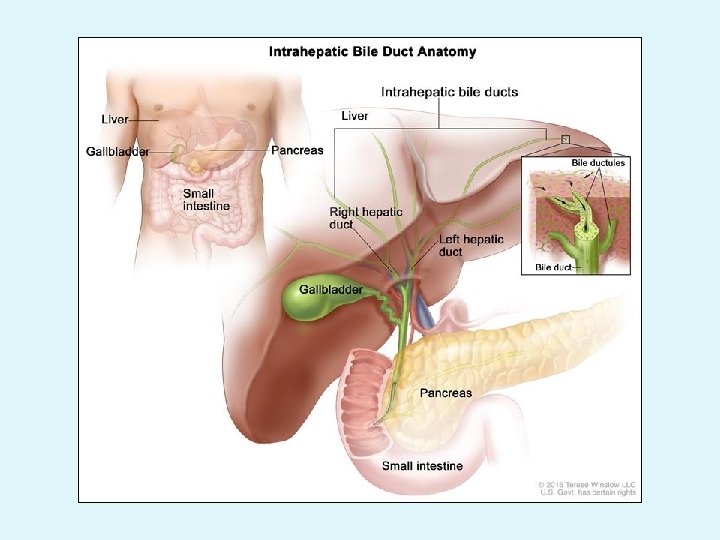

Bile canaliculi Bile ductules Canals of Hering Bile ducts Hepatic ducts Columnar cells Hepatocytes Cuboidal cells Cuboidal then columnar

Histology of the liver Lobes Lobules Hepatocytes

Liver Lobule • Liver has 50. 000 -100. 000 lobules. Functional unit

ØLobules are composed of rows of Hepatocytes arranged radially around a central vein. ØBlood sinusoids are between Hepatocytes. central vein hepatocytes sinusoids

What is the structural & functional unit of the liver? ? ! Liver lobule

Liver lobules Three classifications of liver lobulations: Structural ØClassic hepatic lobule Functional ØLiver acinus ØPortal lobule

ØIn each lobule identify the following: 1 - Shape 2 - Borders 3 - Functional description

1 - Classical hepatic lobule

Pigs

Quiz. . ? ?

What flows from 1 - What flows from 3 ? ? 3 -1? ?
 ØShape: shaped. Diamond- ØBorders: 4 angles; 2 CV")
2 - Hepatic acinus (Liver acinus) ØShape: shaped. Diamond- ØBorders: 4 angles; 2 CV of two neighbouring classical lobules; 2 portal tracts ØCenter: interlobular branches of hepatic art & portal vein
")
Hepatic acinus (Liver acinus)

• Functionally: determines the degree of oxygen & nutrients/toxins supply of cells. Zone III: Zone I: from • Farthest • Close tosupply, blood Zone II: close supply. to CV. Intermediate • • Rich Poorininoxygen&& nutrients. • • Oxidative Glycolysis metabolism & • protien First to synthesis. degenerate.

• • III- Portal lobule Shape: Triangular. Borders: Central veins of three lobules joined. Center: Portal canal. Function:

To sum up… A B 1 - CV 2 - sinusoids 9 - bile duct 10 - hepatic art 11 - PV

- Slides: 46