Histology of the Digestive System Same four layers


 changes as it")


 – Hexagonal solid made")


- Slides: 20
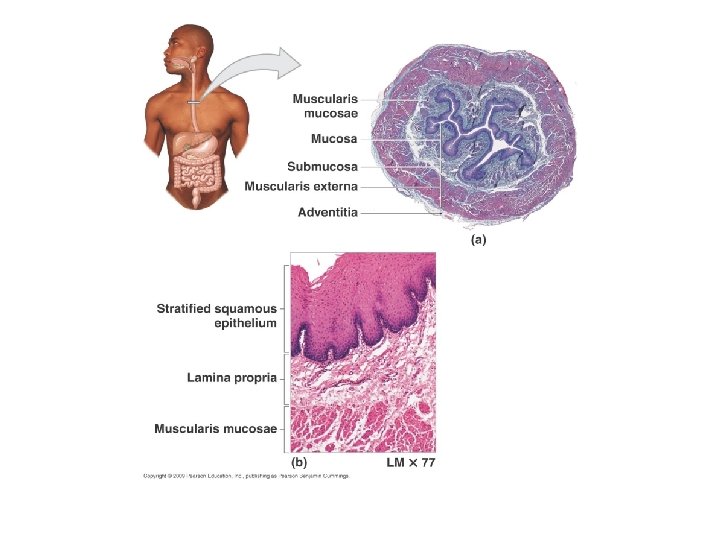
Histology of the Digestive System
Same four layers from esophagus to anal canal 1. 2. 3. 4. Mucosa Submucosa Muscularis externa Serosa from lumen (inside) out
The mucosa Three sub-layers 1. Lining epithelium absorbs nutrients, secretes mucus 2. Lamina propria Loose connective tissue with nourishing and absorbing capillaries , mucosaassociated lymphoid tissue 3. Muscularis mucosae local movements
The submucosa • Connective tissue containing major blood and lymphatic vessels and nerves • Many elastic fibers so gut can regain shape after food passes
The muscularis externa • Two layers of smooth muscle ? ? !!!!! Inner circular layer Outer longitudinal layer
The serosa ? ? ? adventitia ? ?
Esophagus • Epithelium: nonkeratinized stratified squamous epithelium • Muscle (muscularis externa) changes as it goes down – Superior 1/3 of esophagus: skeletal muscle (like pharynx) – Middle 1/3 mixture of skeletal and smooth muscle – Inferior 1/3 smooth muscle (as in stomach and intestines
Stomach Mucosa • Simple columnar epithelium: • Gastric pits opening into gastric glands – Mucus neck cells – Parietal cells • HCL • Intrinsic factor (for B 12 absorption) – Chief cells • Pepsinogen (activated to pepsin with HCL) • Stimulated by gastrin: a stomach hormone
• Submucosa Connective tissue. • Muscularis 3 layers: Outer > Longitudinal Middle > Circular Inner > Oblique
Mucosa ? ? ? Submucosa ? ? ? Muscularis ? ?
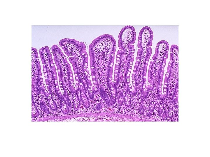
Small intestine designed for absorption -Structural modifications also increase absorptive area -Circular folds (plicae circulares) -Villi (fingerlike projections) 1 mm high – simple columnar epithelium: velvety -Microvilli
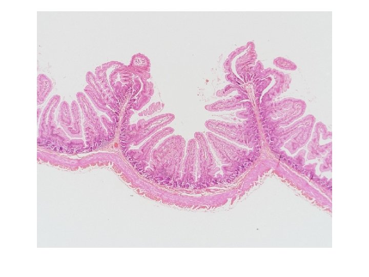
Large intestine • No villi – Fewer nutrients absorbed • “Columnar cells” • A lot of goblet cells for mucus • More lymphoid tissue – A lot of bacteria in stool
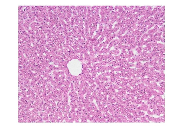
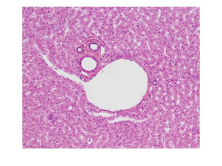
Liver histology • Liver lobules (about one million of them) – Hexagonal solid made of sheets of hepatocytes (liver cells) around a central vein – Corners of lobules have “portal triads”
• Portal triad – Portal arteriole – Portal venule • Branch of hepatic portal vein • Delivers substances from intestines for processing by hepatocytes – Bile duct • Carries bile away • Liver sinusoids – Large capillaries between plates of hepatocytes – Contribute to central vein and ultimately to hepatic veins and IVC • Kupffer cells – Liver macrophages – Old blood cells and microorganisms removed
Pancreas exocrine and endocrine