Histology of the blood vessels 2020 02 27

Histology of the blood vessels 2020 02 27 Dr. Nandor Nagy Semmelweis University, Budapest

Developing blood vessels in zebrafish embryo Nagy N et al, 2009 Developmental Biology 5 dpf Tg(fli-EGFP) RED: neurons GREEN: blood vessels
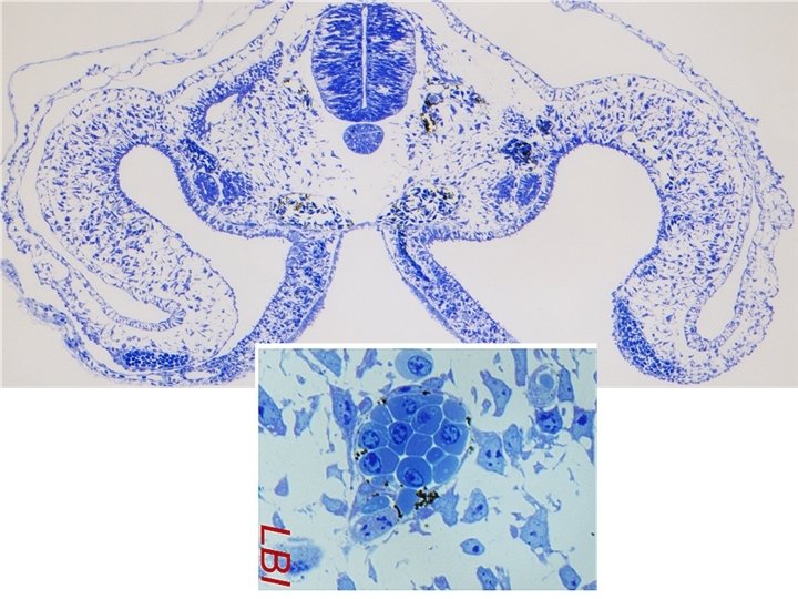

: Pericytes are fibroblast-like cells with extensive cytoplasmic processes that wrap around endothelial cells.

Histology of vessels 1. Elastic arteries 2. Muscular arteries 3. Arterioles 4. Capillaries 5. Venules 6. Veins Layers of the wall: 1. Tunica intima (endothelium) 2. Tunica media (smooth muscle / elastic fibers) 3. Tunica adventitia (loose connective tissue)

Elastic artery
 T. intima: endothelium, subendothelial layer T. media: 40 -70 fenestrated elastic")
Elastic artery (HE) T. intima: endothelium, subendothelial layer T. media: 40 -70 fenestrated elastic membrane, smooth muscle cell layers in between T. adventitia: connective tissue, vasa vasorum

Elastic artery „Roller blind”

vasa vasorum
 T. intima External")
Muscular artery (orcein staining; dyes extracted from several species of lichen) T. intima External elastic membrane Internal elastic membrane

Muscular artery http: //img 3. wikia. nocookie. net/__cb 20140529213818/vroniplag/de/images/3/35/Pew_02_diss. png Elsevier Kierszenbaum: Histology and Cell Biology: An introduction to pathology

Small artery membr. elastica int.

Arteriole http: //www. edu. upmc. fr/histologie/A/arteriole/pages/ariole 01. htm http: //micro. ronaldschulte. nl/esophagus/05_schema_arteriole. JPG
 (endothelium, lamina basalis, 1 layer of smooth muscle) 2")
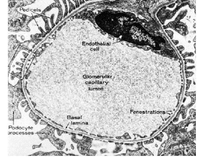
Microcirculation 1 Precapillary arteriole (sphincter) (endothelium, lamina basalis, 1 layer of smooth muscle) 2 Capillary 3 Postcapillary venule (endothelium, lamina basalis, pericyte)
 wall: only endothelium and lamina basalis (pericytes often surround the capillaries)")
Capillaries (toluidine blue) wall: only endothelium and lamina basalis (pericytes often surround the capillaries)

https: //quizlet. com/32301631/histology-5 -blood-circulatory-system-and-hemopoiesis-flash-cards/

Periferal vessels: vein valves, vessels of the limbs

Venules Elsevier, Ovalle & Nahirney: Netter’s Essential Histology

Small and middle sized veins http: //histohelp. files. wordpress. com/2011/09/screen-capture 2. png

Large vein T. intima 1. endothelium 2. subendothelial layer T. media 3. smooth muscle and connective tissue T. adventitia 4. longitudinal smooth muscle bundles, connective tissue 5. collagen fibres

Most vein are equiped with one-way valves to ptevent blood flowing in reverse direction!

Lymph vessels Arteriole Lymph vessel Venule

vessel type Tunica intima Tunica media Tunica adventitia fibrocytes, macrophages, elastic and collagen fibers, vasa vasorum, nerves Internal elastic membrane (IEM) elastic artery endothel, subendothel (IEM) fenestrated elastic lamellae / membranes smooth muscle muscular artery endothel, subendothel IEM smooth muscle (spiral) few elastic fibrocytes, collagen and elastic fibers, vasa fibers, circular collagen fibers (ext. vasorum, nerves elastic membr. ) small artery endothel, subendothel IEM 5 -10 layers of smooth muscle, collagen fibers arteriole, precapillary arteriole endothel (IEM) 2 -5 layers of smooth muscle pericyte, some collagen fibers precap. art. : 1 layer smooth muscle capillary endothel (continuous, fenetrated or discontinuous) pericyte postcapillary venule endothel pericyte venule endothel some smooth muscle cells some collagen, elastic and reticular fibers small vein endothel 2 -3 layers of smooth muscle loose conn. tiss. , thicker than the media, longitudinal smooth muscle, collagen and elastic fibers, middle sized vein endothel, subendothel (IEM) circular smooth muscle layers loose conn. tiss. , thicker than the media, longitudinal smooth muscle, collagen and elastic fibers, large vein endothel, subendothel (IEM) smooth muscle, varying thickness, collagen fibers loose conn. tiss. , thicker than the media, longitudinal smooth muscle, collagen and elastic fibers, vasa vasorum, nerves, lymph vessels thinner than the tunica media
- Slides: 25