HISTOLOGY OF ESOPHAGUS GASTRO ESOPHAGEAL JUNCTION By Dr

HISTOLOGY OF ESOPHAGUS &GASTRO ESOPHAGEAL JUNCTION By Dr. Sobia Ibrahim Assistant Professor Anatomy, KEMU

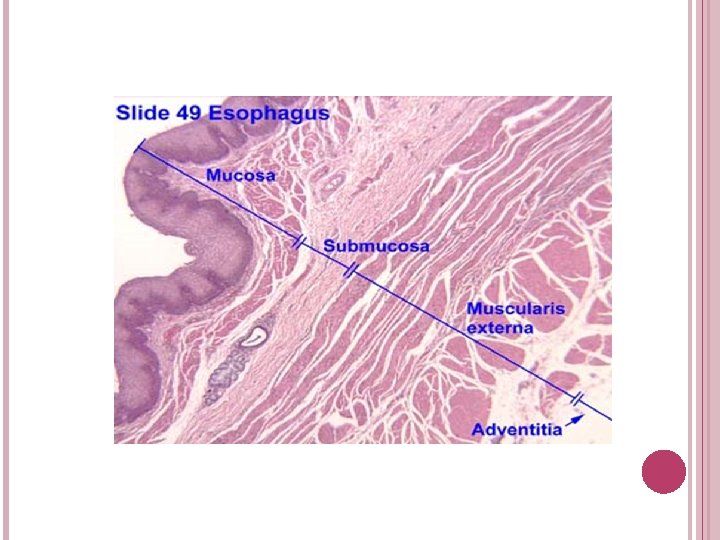
ESOPHAGUS Is muscular tube Extends from pharynx to stomach Wall has four layers: � Mucosa Epithelium Lamina propria Muscularis interna or muscularis mucosae � Submucosa � Muscularis externa � Adventitia / Serosa
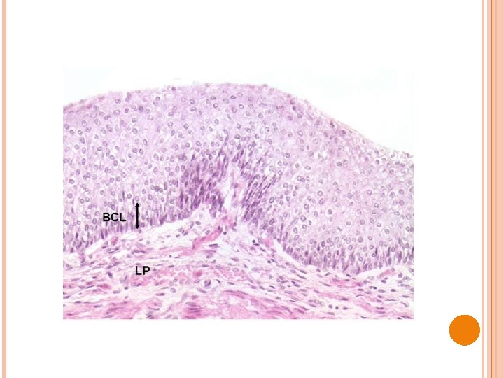

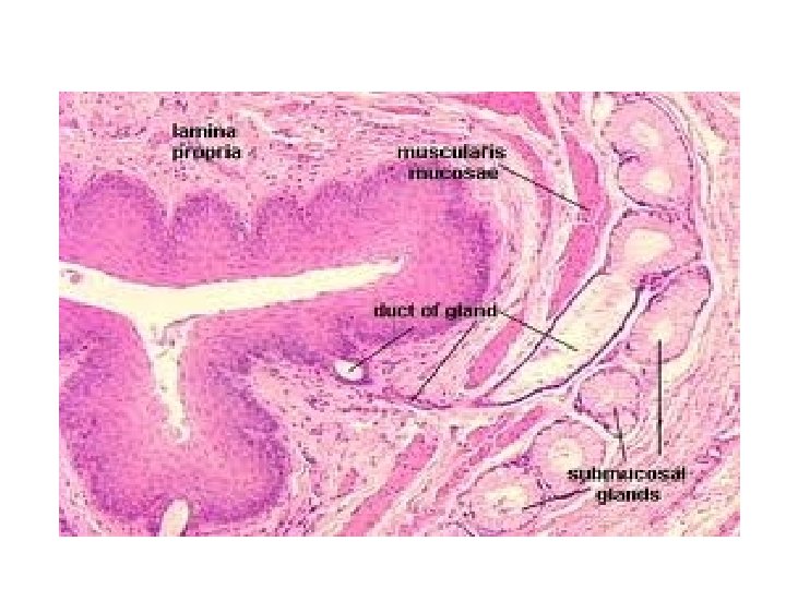
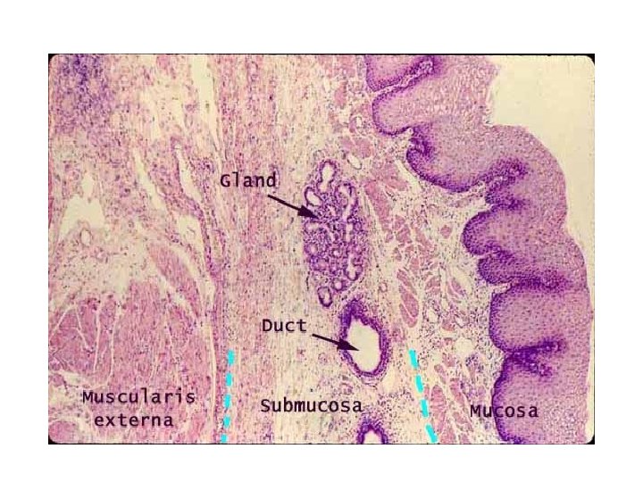
UPPER 1/3 OF ESOPHAGUS MUCOSA Epithelium: Stratified squamous non-keratinized � Lamina propria: scattered lymphocytes, few lymphatic nodules, mucous glands, blood vessels � Muscularis mucosae: � Thick, smooth muscles longitudinally arranged � q SUB-MUCOSA § Collagen & elastic fibres, mucous glands, blood vessels, nerves q MUSCULARIS EXTERNA § Skeletal (striated) muscles q ADVENTITIA § Collagen & elastic fibers, blood vessels

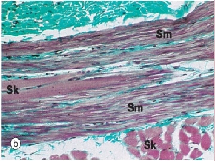
MIDDLE 1/3 OF ESOPHAGUS MUCOSA Epithelium: stratified squamos nonkeratinized Lamina propria: : scattered lymphocytes, few lymphatic nodules, mucous glands, blood vessels � Muscularis mucosae: � thick, smooth muscles longitudinally arranged � q § SUB-MUCOSA Collagen & elastic fibres, mucous glands, blood vessels, nerves q § § MUSCULARIS EXTERNA Skeletal (striated) muscles, Smooth muscles q § ADVENTITIA Collagen & elastic fibers, blood & lymphatic vessels

LOWER 1/3 OF ESOPHAGUS MUCOSA Epithelium: Simple columnar � Lamina propria: scattered lymphocytes, few lymphatic nodules, mucous glands, blood vessels � Muscularis mucosae: thick, smooth muscles longitudinally arranged q SUB-MUCOSA § Collagen & elastic fibres, mucous glands, blood vessels, nerves q MUSCULARIS EXTERNA § Smooth muscles q ADVENTITIA/ SEROSA § Collagen & elastic fibers, blood & lymphatic vessels

GASTROESOPHAGEAL REFLUX DISEASE It is associated with incompetent barriers at this junction; caused by hiatal hernia, decrease in lower esophageal sphincter tone. Results in reflux esophagitis as mucosal barriers are not sufficient to protect esophagus from acids, pepsin & bile causing heart burns/atypical chest pain

BARRETT’S ESOPHAGUS Reflux of gastric contents throu lower esophageal sphincter Gastric acid in esophagus Heart burn With time, metaplasia of epithelium (protective) Barrett’s esophagus High risk for carcinoma

STOMACH PARTS OF STOMACH Cardia Fundus Body Pylorus Rugae Three histological zones

CARDIOESOPHAGEAL JUNCTION

Esophageal cardiac junction

MUCOSA: 1: Epithelium changes from: Startified squamous non keratinised into simple columnar. 2: Lamina propria: esophageal cardiac glands but in stomach (cardiac, fundic and pyloric glands). They are tubular and deep. 3: Muscularis mucosae: thick longitudinal in esophagus but thin and two layered in stomach. Submucosa: esophageal glands proper disappear. No glands in submucosa of stomach. Muscularis externa: three layered in stomach an additional oblique layer is there


GASTROESOPHAGEAL REFLUX DISEASE • It is associated with incompetent barriers at this junction; caused by hiatal hernia, decrease in lower esophageal sphincter tone. • Results in reflux esophagitis as mucosal barriers are not sufficient to protect esophagus from acids, pepsin & bile • causing heart burns/atypical chest pain
- Slides: 19