Histology Circulatory System Part 2 Vena Cava The


pm or")
 Are most common in body consist the wall endothelial")

 The Endocardium is composed of a continuous endothelial lining, with")
 The myocardium is a variably-sized layer of cardiomyocytes that lies")
 The Epicardium of the heart is lined by a layer of")


- Slides: 18
Histology Circulatory System Part -2 -
Vena. Cava The inferior vena cava is the largest vein in the human body. It collects blood from veins serving the tissues inferior to the heart and returns this blood to the right atrium of the heart. There are two in humans, the inferior vena cava (carrying blood from the lower body) and the superior vena cava (carrying blood from the head, arms, and upper body). Although the vena cava is very large in diameter, its walls are incredibly thin due to the low pressure exerted by venous blood.
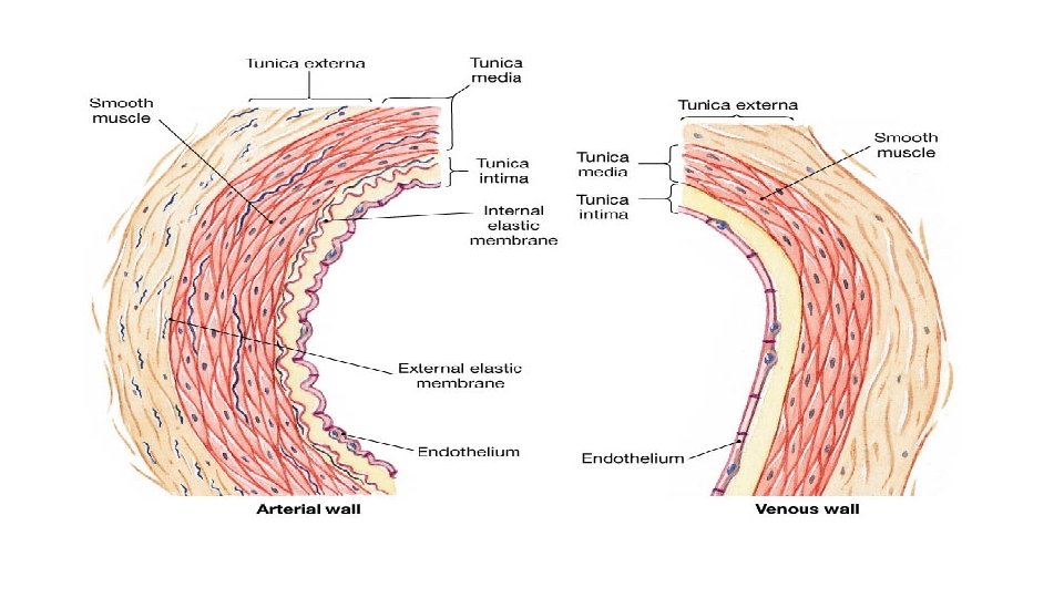
Comparison between a vein and artery Artery Vein 1. Thick wall 1. Thin wall 2. shape less deformed 2. shape flatted 3. tunica intina crinkled 3. Smooth 4. three distinct layers 4. layering indistinct 5. tunica media prominent 5. Weak
Capillaries are small, normally around 3 -4µm, but some capillaries can be 30 -40 µm in diameter. The largest capillaries are found in the liver. Capillaries connect arterioles to venules. They allow the exchange of nutrients and wastes between the blood and the tissue cells, together with the interstitial fluid. This exchange occurs by passive diffusion and by pinocytosis which means 'cell drinking.
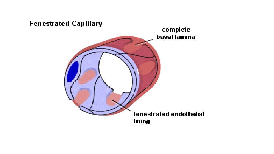
Types of capillaries 1 -Fenestratedcappillaries: Have the same structure of non fenestrated but several circular opening in their endothelium cell are closed by diaphragm. This type capillary found in kidney and endocrine gland.
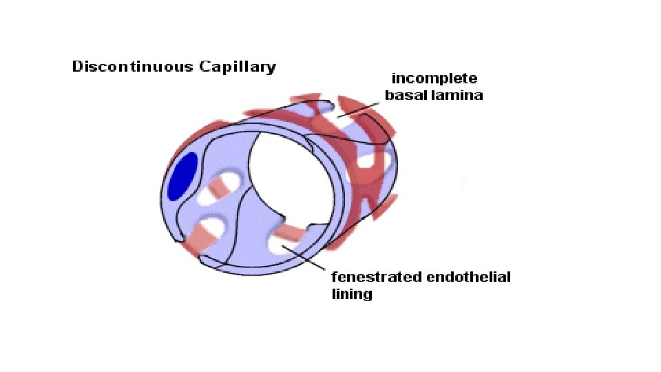
2 -Sinusoid or discontinuous capillary A large capillaries with diameter of (30 -40)pm or more. It consists of cells which are irregular in shape and discontinuous basal membrane. The endothelium is not connected by desmosome and associated with macrophagocytic or (Kupffer cells). This type is found in spleen, bone marrow, and liver.
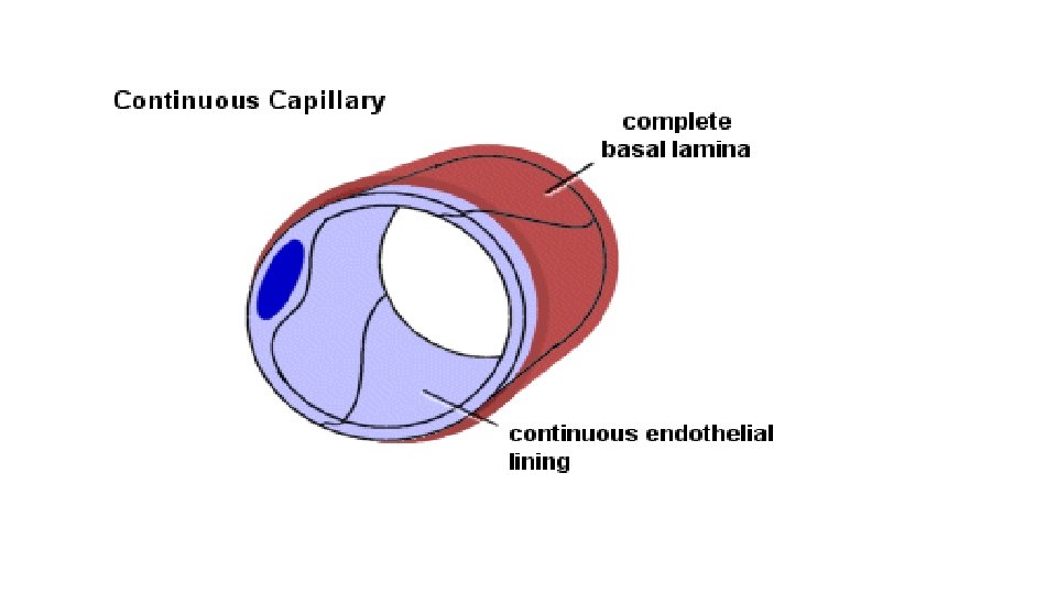
3 -Non fenestrated (Continuous capillaries) Are most common in body consist the wall endothelial cell resting on basal membrane and contain pericyte and pinocytotic vessel. This type capillary found in skin and muscle. Are formed by "continuous" endothelial cells and basal lamina. The endothelial cell and the basal lamina do not form openings, which would allow substances to pass the capillary wall without passing through both the endothelial cell and the basal lamina. Both endothelial cells and the basal lamina can act as selective filters in continuous capillaries.
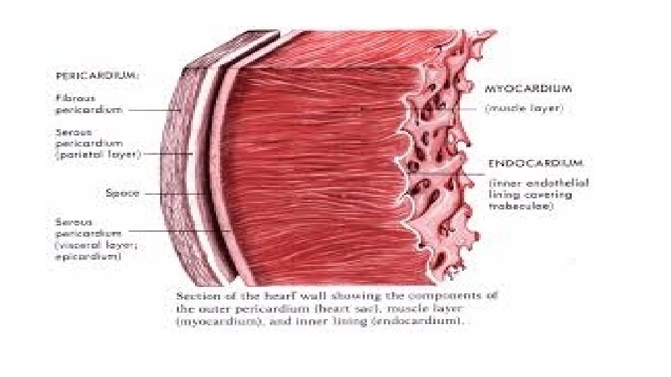
Heart The heart is a muscular organ in humans and other animals, which pumps blood through the blood vessels of the circulatory system. Blood provides the body with oxygen and nutrients, and also assists in the removal of metabolic wastes. The heart is located in the middle compartment of the mediastinum in the chest, the heart is a pump with four chambers and valves that maintain a one -way flow of blood. The wall of heart consists of three layers that are homologous to the three tunic of blood vessels.
Tunica Intima: ( Endocardium) The Endocardium is composed of a continuous endothelial lining, with attendant basement membrane, that lies on top of a layer of collagen. The endothelium of the heart is continuous with that of the aorta, vena cava, pulmonary artery, and pulmonary veins, Purkinje Fibers ( impulse conducting fibers , it is large modified of muscle cell).
Tunica Media: ( Myocardium) The myocardium is a variably-sized layer of cardiomyocytes that lies between the endocardium and epicardium and makes up the bulk of the heart's mass. Cardiomyocytes are arranged end to end, in a branching network of fibers and are attached to one another by specialized sections of membrane known as intercalated discs. The membrane at intercalated discs possess a large number of gap junctions which allow nearly free passage of ions between cardiomyocytes which is critical for cardiac action potential propagation.
Tunica Adventitia: (Epicardium) The Epicardium of the heart is lined by a layer of flat mesothelial cells that lie on top of a layer of collagenous tissue. The epicardium makes up the visceral pericardium and serves as a lubricated surface which allows for the free movement of the heart within the pericardium. The outer, parietal pericardium also possesses a layer of mesothelium but is not considered a histological component of the heart itself.
Heart also have four valves composed of connective tissue layered with endothelium on each side, valves consist of three layes: Spongiosa (loose collagen), fibrosa(dense of connective tissue), Ventricularis