Histology Anatomy of the Skeletal System Dr Nabil

Histology & Anatomy of the Skeletal System Dr. Nabil Khori MD. MSc. Ph. D
")
Connective Tissue: Bone (Osseous Tissue)

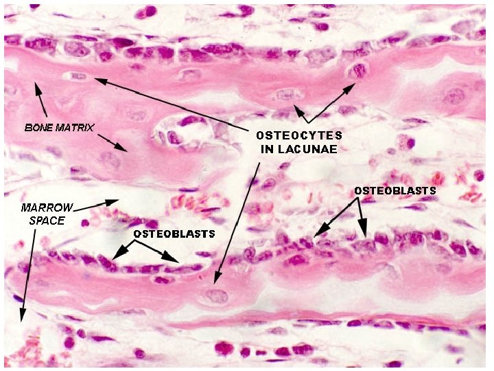
Bone Cells 1. Osteoblast and Osteocyte: Are mono-nucleate cells that are responsible for bone formation Specialized fibroblasts that produce protein matrix of osteoid, which is composed mainly of Type I collagen and prostaglandin hormone. derive from osteogenic stem cells (osteoprogenitor cell) that differentiate to form pre-osteoblast and then osteoblasts , when maturing form Osteocyte

2. Osteoclasts: Specialized cells that work in synchronization with osteoblasts to maintain the skeletal system. It contains 15 -20 closely packed oval-shaped nuclei Found in pits in the bone surface which are called resorption bays, or Howship's Lacunae. cytoplasm is homogeneous, "foamy" appearance. Are type of bone cell that removes bone tissue by removing its mineralized matrix and breaking up the organic bone

Osteoclasts

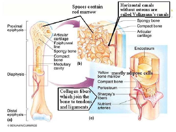
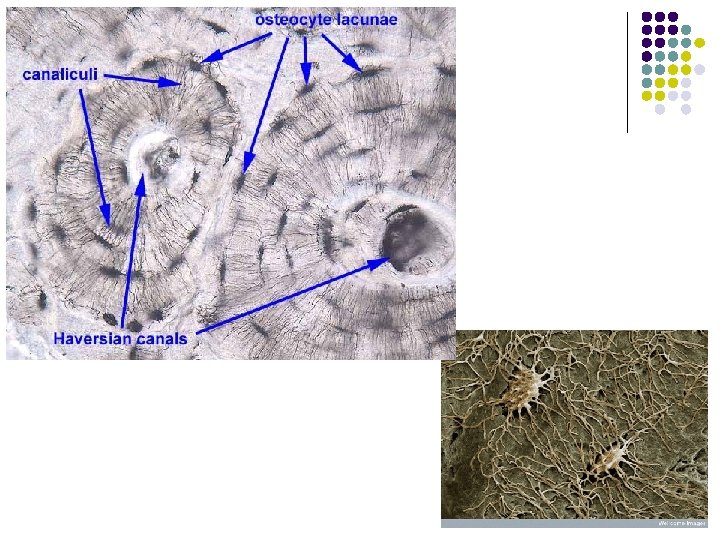
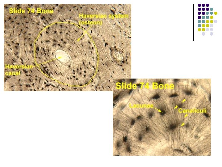
Haversion system

Bones of the skeleton l l l Axial skeleton Vertebral Column 26 Skull 22 Hyoid bone 1 Ribs and sternum 25 --------- l l l Appendicl. skeleton Upper Extrimities 64 Lower Exremities 62 ---------- l l Auditory ossicles 6 ----------total 206

Types of Bones: By Shape

Skeletal System The Axial Skeleton

The Axial Skeleton l Eighty bones segregated into three regions l l l Skull Vertebral column Bony thorax

Skull – Anterior View

Skull – Posterior View

Parietal Bones and Major Associated Sutures l Form most of the superior and lateral aspects of the skull

Interior Lateral View of the Skull

Occipital Bone and Its Major Markings

Inferior View of the Skull Base

Facial Bones l l Fourteen bones of which only the mandible and vomer are unpaired The paired bones are the maxillae, zygomatics, nasals, lacrimals, palatines, and inferior conchae

Skull – Anterior View

Maxillary Bones l Their major markings include palatine, frontal, and zygomatic processes, the alveolar margins, inferior orbital fissure, and the maxillary sinuses

Inferior View of the Skull Base
 that form the prominenc es of")
Zygomatic Bones l Irregularly shapes bones (cheekbon es) that form the prominenc es of the cheeks and the inferolatera l margins of the orbits

Ethmoid Bone l l l Most deep of the skull bones; lies between the sphenoid and nasal bones Forms most of the bony area between the nasal cavity and the orbits Major markings include the cribriform plate, crista galli, perpendicular plate, nasal conchae, and the ethmoid sinuses

Nasal Cavity

Nasal septum

Other Facial Bones l l l Nasal bones – thin medially fused bones that form the bridge of the nose Lacrimal bones – contribute to the medial walls of the orbit and contain a deep groove called the lacrimal fossa that houses the lacrimal sac Palatine bones – two bone plates that form portions of the hard palate, the posterolateral walls of the nasal cavity, and a small part of the orbits Vomer – plow-shaped bone that forms part of the nasal septum Inferior nasal conchae – paired, curved bones in the nasal cavity that form part of the lateral walls of the nasal cavity

The Orbital Cavity
 is the largest, strongest")
Mandible and Its Markings l l The mandible (lower jawbone) is the largest, strongest bone of the face Its major markings include the coronoid process, mandibular condyle, the alveolar margin, and the mandibular and mental foramina Figure 7. 8 a

Paranasal Sinuses l l l Mucosa-lined, air-filled sacs found in five skull bones – the frontal, sphenoid, ethmoid, and paired maxillary bones Air enters the paranasal sinuses from the nasal cavity and mucus drains into the nasal cavity from the sinuses Lighten the skull and enhance the resonance of the voice
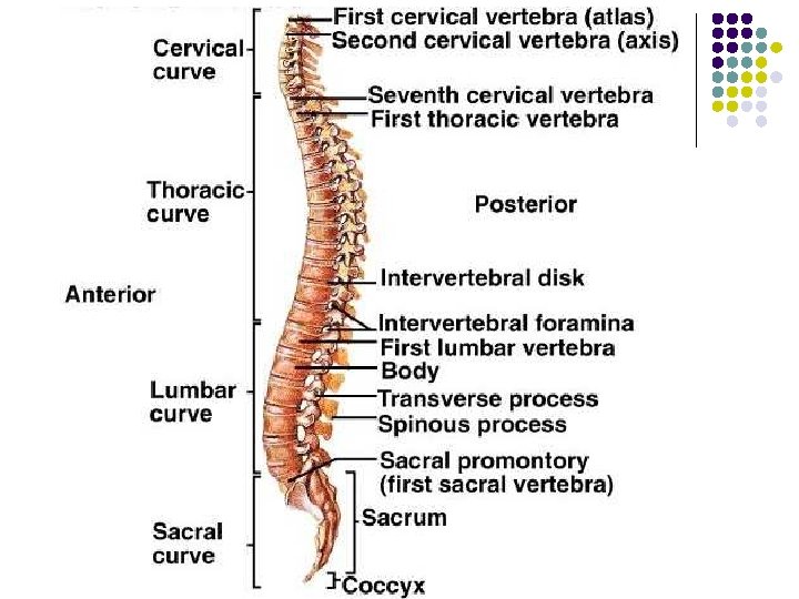

Curvature of vertebral column Cervical and lumbar : concave posterior, develop during the fetal period, due to deference in IV disc thickness cervical - infant hold head lumber - infant walk and assume upright position, prominent in female. Thoracic and sacral; primary, develop during fetal period, deference beteween ant and post parts of the vertebra
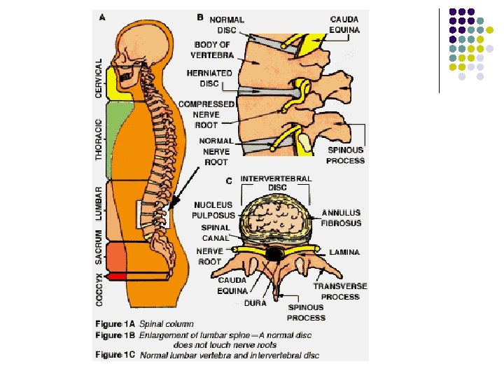

Abnormal curvature Kyphosis: abnormal increase in thoracic curv. Erosion of anterior vertebral part. Lordosis: (hollow back) anterior rotation of pelvis ubnormal increase in lumber curvature (pregnancy) Scoliosis: (Crooked or curved back) abnormal lateral curvature and rotation of the back (appears between ages of 10 -15)

General Structure of Vertebrae l l l Spinous process project posteriorly, and transverse processes project laterally Superior and inferior articular processes – protrude superiorly and inferiorly from the pediclelamina junctions Intervertebral foramina – lateral openings formed from notched areas on the superior and inferior borders of adjacent pedicles
 l l l Has no body and no spinous")
Cervical Vertebrae: Atlas (C 1) l l l Has no body and no spinous process Consists of anterior and posterior arches, and two lateral masses The superior surface of lateral masses articulate with the occipital condyles
 l The axis has a body, spine, and")
Cervical Vertebrae: The Axis (C 2) l The axis has a body, spine, and vertebral arches as do other cervical vertebrae

Thoracic Vertebrae

Lumbar Vertebrae

Sacrum and Coccyx

Sacrum and Coccyx

Bones of the Thorax: Sternum and Ribs

Structure of a Typical True Rib l Bowed, flat bone consisting of a head, neck, tubercle, and shaft
- Slides: 45